Unlocking the Medial Side of the Knee: A Surgical Anatomy Guide
Key Takeaway
Discover the latest medical recommendations for Unlocking the Medial Side of the Knee: A Surgical Anatomy Guide. The medial side of the knee is anatomically structured in three distinct layers. The outer layer consists of the deep fascia of the thigh, while the middle layer contains the superficial medial ligament, also known as the medial collateral ligament. The deepest layer comprises the true joint capsule and other reinforcing structures. This layered understanding is crucial for surgical approaches and diagnosis.
Applied Surgical Anatomy of the Medial Side of the Knee
Overview
Fig. 10-37).
---
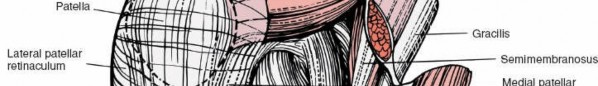
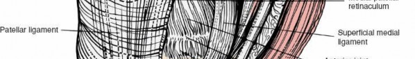
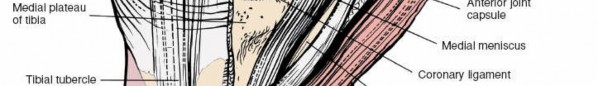

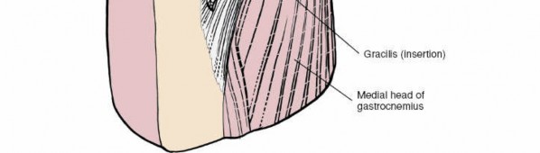 Figure 10-33 The sartorius and the medial patellar retinaculum (outer layer) have been resected to reveal the superficial medial ligament of the middle layer. The true joint capsule (deep layer) also is exposed.
 ---
---
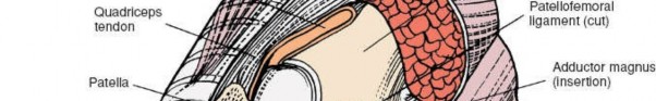
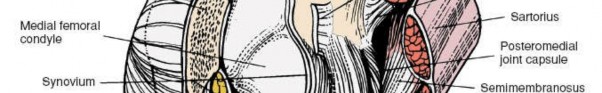
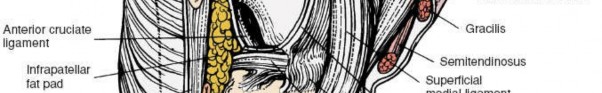
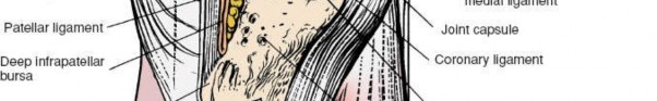
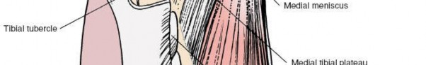
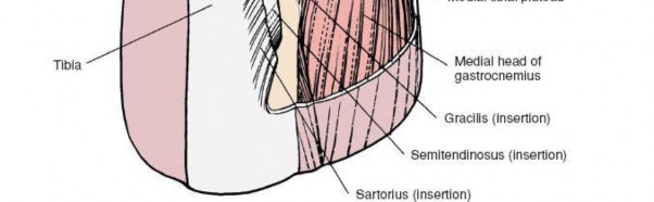 Figure 10-34 The joint cavity of the knee, with all the more superficial structures removed.
 ---
---
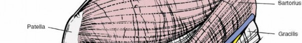
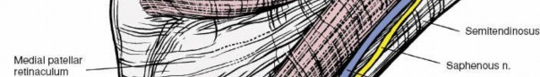
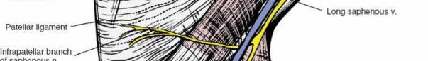
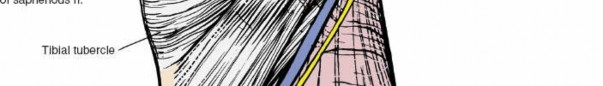
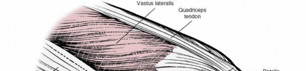
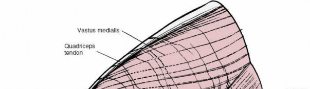
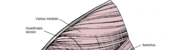
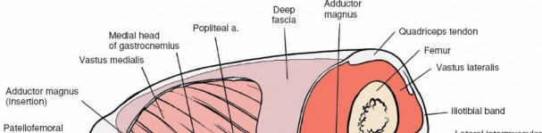
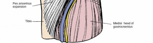 Figure 10-35 The outer layer of the medial aspect of the knee joint consists of the sartorius, the fascia of the thigh, and the medial patellar retinaculum.
 ---
---
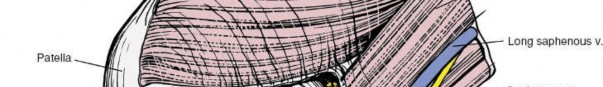
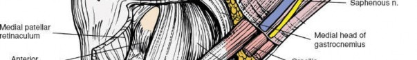
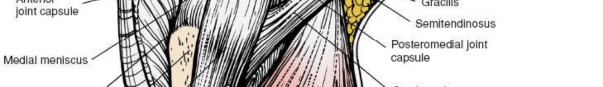
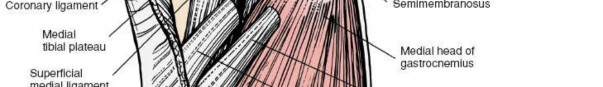
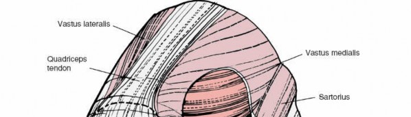
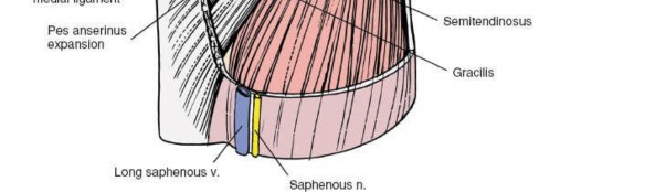 Figure 10-36 The outer layer has been resected to reveal the intermediate layer, consisting of the superficial medial ligament. Between the superficial and middle layers run the semitendinosus and gracilis muscles. The deep medial ligament (meniscofemoral ligament) of the deep layer is visible. The true joint capsule anterior to the superficial medial ligament also is visible.
 ---
---
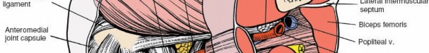
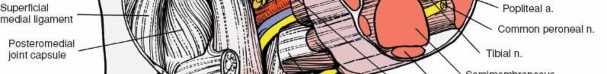
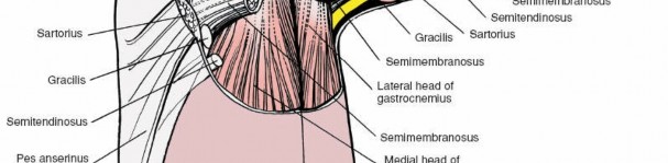
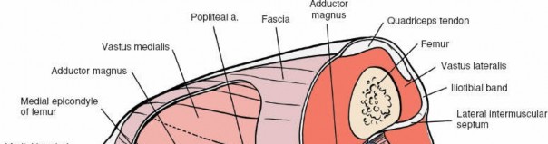
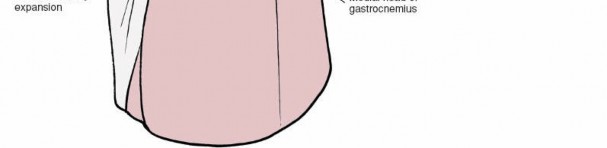 Figure 10-37 A more posteromedial view of the knee joint. The sartorius, the deep fascia of the outer layer, the gracilis, the semitendinosus, and the semimembranosus have been resected to reveal the superficial medial ligament (middle layer), the posteromedial joint capsule (deep layer), and the medial head of the gastrocnemius.
 ---
---
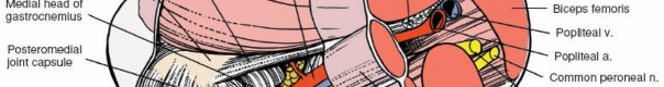
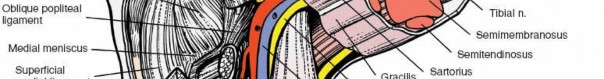
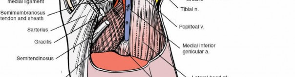
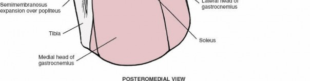 Figure 10-38 The medial head of the gastrocnemius has been resected to reveal the three expansions of the semimembranosus.
 ---
---
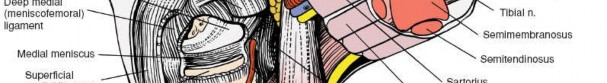
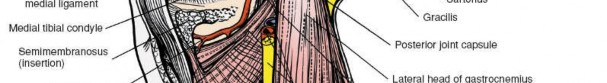
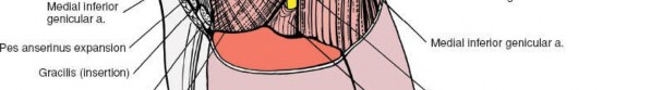
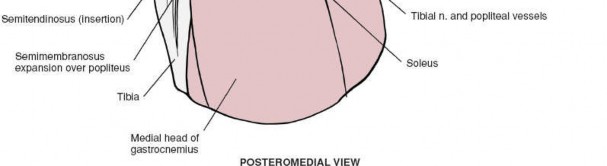 Figure 10-39 The posterior aspect of the superficial medial ligament (middle layer) has been excised to reveal the true joint capsule and its thickening, the deep medial ligament (the meniscofemoral ligament and the coronary ligaments). The posteromedial joint capsule has been excised to reveal the corner of the joint. The insertion of the semimembranosus and a portion of its expansion are visible. 2. Further posterior retraction brings the posteromedial corner of the joint into view. The cover consists of fibrous tissue derived from the semimembranosus muscle (the
middle layer
), which has fused with the true joint capsule (the
deep layer;
see 2.
Approach for medial meniscectomy
1. Incising the medial patellar retinaculum exposes the true capsule of the joint, which is very thin at this point. 2. The true capsule of the joint, incised with the synovium, allows access to the anteromedial portion of the joint (seeFigs. 10-33 and 10-34). 3.
Medial parapatellar approach to the knee
1. The joint is dissected through the same fascial layers as in the approach for the medial meniscus.
Special Anatomic Points ** Three muscles, the sartorius, semitendinosus, and gracilis, insert into the upper part of the subcutaneous surface of the tibia. Each muscle has a different nerve supply: The sartorius is innervated by the femoral nerve, the semitendinosus by the sciatic nerve, and the gracilis by the obturator nerve. In addition, each muscle crosses both the hip and the knee. The actions of the three muscles are duplicated by other, more powerful, muscles. At their pelvic origins, the three attach to three points on the bony pelvis that are separated as widely as the pelvis allows: The anterior-superior iliac spine (sartorius), the ischial tuberosity (semitendinosus), and the inferior pubic ramus (gracilis). With these origins and insertions, the muscles are arranged ideally to stabilize the pelvis on the leg. The sartorius, semitendinosus, and gracilis insert into the subcutaneous surface of the tibia at a point called the
pes anserinus
(goose foot). Acting together, they not only flex the knee, but also internally rotate the tibia.
Figure 10-39 The posterior aspect of the superficial medial ligament (middle layer) has been excised to reveal the true joint capsule and its thickening, the deep medial ligament (the meniscofemoral ligament and the coronary ligaments). The posteromedial joint capsule has been excised to reveal the corner of the joint. The insertion of the semimembranosus and a portion of its expansion are visible. 2. Further posterior retraction brings the posteromedial corner of the joint into view. The cover consists of fibrous tissue derived from the semimembranosus muscle (the
middle layer
), which has fused with the true joint capsule (the
deep layer;
see 2.
Approach for medial meniscectomy
1. Incising the medial patellar retinaculum exposes the true capsule of the joint, which is very thin at this point. 2. The true capsule of the joint, incised with the synovium, allows access to the anteromedial portion of the joint (seeFigs. 10-33 and 10-34). 3.
Medial parapatellar approach to the knee
1. The joint is dissected through the same fascial layers as in the approach for the medial meniscus.
Special Anatomic Points ** Three muscles, the sartorius, semitendinosus, and gracilis, insert into the upper part of the subcutaneous surface of the tibia. Each muscle has a different nerve supply: The sartorius is innervated by the femoral nerve, the semitendinosus by the sciatic nerve, and the gracilis by the obturator nerve. In addition, each muscle crosses both the hip and the knee. The actions of the three muscles are duplicated by other, more powerful, muscles. At their pelvic origins, the three attach to three points on the bony pelvis that are separated as widely as the pelvis allows: The anterior-superior iliac spine (sartorius), the ischial tuberosity (semitendinosus), and the inferior pubic ramus (gracilis). With these origins and insertions, the muscles are arranged ideally to stabilize the pelvis on the leg. The sartorius, semitendinosus, and gracilis insert into the subcutaneous surface of the tibia at a point called the
pes anserinus
(goose foot). Acting together, they not only flex the knee, but also internally rotate the tibia.
 ---
---
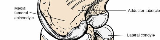
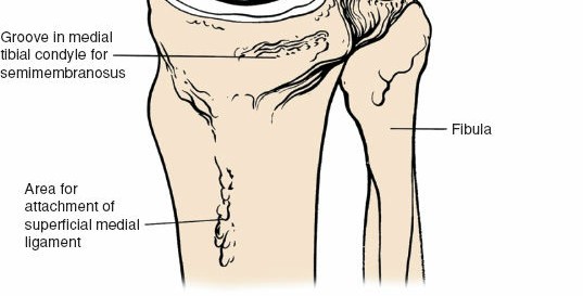 Figure 10-40 Osteology of the posteromedial aspect of the knee joint.
You Might Also Like