Open Reduction and Internal Fixation of Tibial Plateau Fractures: A Master Surgical Guide
Key Takeaway
Open reduction and internal fixation (ORIF) of tibial plateau fractures aims to restore articular congruity, mechanical alignment, and joint stability. This comprehensive guide details the surgical approaches, biomechanical principles, and step-by-step techniques for managing lateral, medial, and bicondylar fractures. Emphasizing soft tissue management, meniscal preservation, and rigid subchondral support, these evidence-based protocols ensure optimal functional recovery and minimize posttraumatic arthritis in complex intraarticular knee injuries.
Comprehensive Introduction and Patho-Epidemiology
Tibial plateau fractures represent a highly complex and heterogenous spectrum of intraarticular injuries that consistently challenge the orthopedic trauma surgeon. The fundamental objectives of intervention are to restore joint congruity, re-establish axial alignment, and impart mechanical stability, all while meticulously preserving the fragile surrounding soft tissue envelope. The proximal tibia serves as the critical anatomical junction for load transmission across the knee joint. When this structural integrity is compromised, the resultant altered joint kinematics and pathological contact stresses inexorably lead to accelerated cartilage degradation and debilitating post-traumatic osteoarthritis (PTOA) if left unaddressed.
The epidemiology of tibial plateau fractures demonstrates a classic bimodal distribution, reflecting two distinct mechanisms of injury and patient populations. In the younger demographic, predominantly males in their third and fourth decades of life, these fractures are typically the result of high-energy trauma, such as motor vehicle collisions, motorcycle accidents, or falls from significant heights. These high-energy mechanisms impart massive axial loads combined with severe varus or valgus moments, resulting in highly comminuted, displaced bicondylar fractures (Schatzker Types V and VI) with profound soft tissue devastation, including fracture blisters, acute compartment syndrome, and open wounds.
Conversely, the second peak occurs in the elderly population, predominantly females over the age of sixty, secondary to low-energy mechanisms such as simple falls from a standing height. In this cohort, osteopenia or frank osteoporosis renders the subchondral bone highly susceptible to failure under minimal physiological loading. These injuries frequently manifest as pure depression fractures (Schatzker Type III) or split-depression fractures of the lateral plateau (Schatzker Type II). Despite the lower energy imparted, the compromised bone stock in this demographic presents a unique surgical challenge, frequently necessitating the use of fixed-angle locking plate technology and extensive structural bone grafting to prevent postoperative subsidence and catastrophic hardware failure.
The pathophysiology of these injuries extends far beyond the osseous disruption. The initial impact causes a massive release of inflammatory cytokines within the joint space, initiating a cascade of chondrocyte apoptosis and extracellular matrix degradation. Furthermore, the violent displacement of the bony fragments frequently results in concomitant soft tissue trauma, including meniscal avulsions, cruciate ligament ruptures, and collateral ligament sprains. A comprehensive understanding of this patho-epidemiology is paramount, as the surgeon must not only address the bony architecture but also respect the profound biological insult to the entire knee organ, tailoring the surgical approach and timing to mitigate further iatrogenic trauma.
Detailed Surgical Anatomy and Biomechanics
A profound mastery of the surgical anatomy and biomechanics of the proximal tibia is the absolute prerequisite for executing safe and effective open reduction and internal fixation (ORIF). The proximal tibia expands from the diaphyseal shaft into the metaphyseal flare, culminating in the medial and lateral articular plateaus. The medial plateau is anatomically larger, concave in the sagittal and coronal planes, and composed of highly dense subchondral bone. Biomechanically, it is the primary weight-bearing surface of the knee, supporting approximately 60% of the physiological load during the normal gait cycle. Due to its robust architecture, fractures of the medial plateau generally require significantly higher energy to occur and are highly correlated with concomitant ligamentous disruptions and knee subluxations.
In stark contrast, the lateral plateau is convex, sits slightly higher anatomically than its medial counterpart, and possesses a significantly thinner, less dense subchondral bone plate. This structural vulnerability makes the lateral plateau highly susceptible to depression and split-depression fractures when subjected to combined axial and valgus loading. Furthermore, both plateaus exhibit a natural posterior slope of approximately 7 to 10 degrees relative to the longitudinal axis of the tibial shaft. Anatomical restoration of this posterior slope is a critical surgical objective; failure to restore the slope alters anterior cruciate ligament (ACL) tension and knee kinematics, leading to chronic instability and restricted range of motion.
The soft tissue envelope surrounding the proximal tibia dictates the surgical approaches and the timing of intervention. The anteromedial surface of the tibia is subcutaneous, covered only by skin and a thin fascial layer, making it highly vulnerable to necrosis following high-energy trauma and extensive surgical dissection. The pes anserinus (comprising the conjoined tendons of the sartorius, gracilis, and semitendinosus) inserts onto the proximal anteromedial tibia and must be carefully mobilized during medial approaches. Laterally, Gerdy’s tubercle serves as the insertion site for the iliotibial (IT) band, while the fibular head anchors the lateral collateral ligament (LCL) and the biceps femoris tendon.
Neurovascular proximity adds a layer of extreme peril to proximal tibial surgery. The common peroneal nerve courses intimately around the fibular neck, placing it at high risk during posterolateral approaches, fibular osteotomies, or the application of lateral external fixation pins. Posteriorly, the popliteal artery bifurcates at the distal border of the popliteus muscle into the anterior tibial artery, posterior tibial artery, and peroneal artery. The anterior tibial artery passes through the interosseous membrane in close proximity to the posterior tibial cortex, making it exquisitely vulnerable to injury from errant posterior drill bits, over-penetrating screws, or displaced posteromedial coronal shear fragments.
Exhaustive Indications and Contraindications
The decision-making process regarding the operative versus non-operative management of tibial plateau fractures is nuanced, requiring a careful synthesis of patient-specific factors, fracture morphology, and the integrity of the soft tissue envelope. The overarching goal is to achieve a stable, congruent joint that permits early range of motion, but this must never be pursued at the expense of catastrophic soft tissue complications.
Operative intervention is definitively indicated for fractures demonstrating significant articular incongruity, axial malalignment, or clinical instability. While historical literature frequently cited a rigid threshold of 2 to 3 millimeters of articular step-off as an absolute indication for surgery, modern orthopedic trauma philosophy emphasizes a more dynamic assessment. A 2-millimeter step-off in a high-demand, 25-year-old athlete warrants aggressive anatomical restoration, whereas the same step-off in a low-demand, 85-year-old non-ambulatory patient may be managed conservatively. Furthermore, any condylar widening exceeding 5 millimeters, which indicates significant disruption of the peripheral meniscal rim and capsular attachments, strongly necessitates operative reduction and buttressing to restore meniscal function and hoop stresses.
Absolute contraindications to definitive ORIF are primarily dictated by the biological state of the limb and the patient. Active surgical site infection, profound soft tissue compromise (e.g., massive fracture blisters, skin necrosis, or unresolved acute compartment syndrome), and severe peripheral vascular disease preclude immediate internal fixation. In such scenarios, the "span, scan, and plan" protocol using a joint-spanning external fixator is the mandatory standard of care. Relative contraindications include extreme medical comorbidities rendering the patient unfit for anesthesia, profound osteoporosis where hardware purchase is impossible (necessitating primary arthroplasty consideration), or non-ambulatory baseline status where the risks of surgery outweigh the functional benefits.
| Parameter | Indications for Operative Management (ORIF) | Contraindications to Immediate ORIF |
|---|---|---|
| Articular Congruity | Articular depression or step-off > 2-3 mm (patient-dependent). | Acceptable alignment (< 2mm step-off) in low-demand patients. |
| Axial Alignment | Metaphyseal-diaphyseal varus/valgus deformity > 5 degrees. | Pre-existing severe osteoarthritis (consider primary TKA). |
| Joint Stability | Clinical varus/valgus instability > 10 degrees in near extension. | Non-ambulatory baseline functional status. |
| Soft Tissue Envelope | Open fractures (requires immediate I&D, staged fixation). | Unresolved fracture blisters, massive edema, acute compartment syndrome. |
| Polytrauma Status | Polytrauma requiring early mobilization for pulmonary toilet. | Hemodynamic instability (Damage Control Orthopedics required). |
| Vascular Status | Concomitant popliteal artery injury requiring repair/stabilization. | Severe, uncorrectable peripheral vascular disease (ischemic limb). |
Pre-Operative Planning, Templating, and Patient Positioning
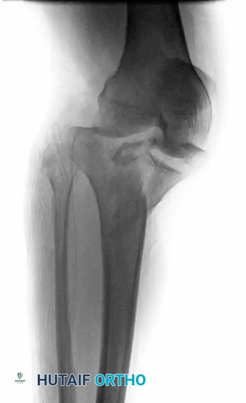
Meticulous preoperative planning is the cornerstone of successful tibial plateau fracture management. The evaluation begins with high-quality, orthogonal plain radiographs, including an anteroposterior (AP), lateral, and bilateral oblique views. A 10-degree caudal tilt AP view (plateau view) is frequently utilized to account for the native posterior slope of the tibia, providing a true tangential projection of the articular surface. While plain films provide a gross overview of the fracture pattern and overall alignment, they profoundly underestimate the degree of articular depression and the complexity of metaphyseal comminution.
Consequently, a preoperative computed tomography (CT) scan with fine cuts (1mm or less) and multiplanar 2D (coronal and sagittal) and 3D reconstructions is the absolute gold standard and is mandatory for all tibial plateau fractures scheduled for operative intervention. The CT scan precisely delineates the location of the fracture apex, the presence and size of coronal shear fragments (particularly the critical posteromedial fragment), and the exact topography of the articular impaction. Digital templating is then performed utilizing the CT and plain film data. The surgeon must meticulously plan the sequence of reduction, the trajectory of subchondral raft screws, the required volume of bone graft substitute, and the precise selection and positioning of the osteosynthesis plates (e.g., lateral periarticular plates, posteromedial buttress plates).
Patient positioning and operating room setup must be executed with precision to facilitate unhindered fluoroscopic imaging and surgical access. The patient is placed supine on a fully radiolucent operating table. A bump is placed beneath the ipsilateral gluteus to correct the natural external rotation of the lower extremity, ensuring the patella points directly towards the ceiling. A non-sterile pneumatic tourniquet is applied to the proximal thigh; however, in cases of high-energy trauma, exsanguination via Esmarch bandage is avoided to prevent the mobilization of deep vein thromboses. Tourniquet inflation is generally reserved for critical portions of the procedure to minimize ischemic time and mitigate the risk of postoperative compartment syndrome.
The fluoroscopic C-arm is positioned on the contralateral side of the table, entering perpendicular to the operative limb. The monitor should be placed directly in the surgeon's line of sight. Prior to the surgical incision, the surgeon must confirm that perfect AP and lateral fluoroscopic images of the knee can be obtained without interference from the table base or the contralateral leg. The operative limb is prepped and draped freely, allowing for full flexion and extension. A sterile bump or triangle is frequently utilized to maintain the knee in varying degrees of flexion during the approach and reduction maneuvers, relaxing the posterior neurovascular structures and the gastrocnemius muscle.
Step-by-Step Surgical Approach and Fixation Technique
The Anterolateral Approach and Articular Elevation
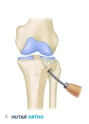
For fractures involving the lateral condyle (Schatzker Types I, II, and III), the anterolateral approach provides the workhorse exposure. The incision begins 3 to 5 cm proximal to the joint line, centered over Gerdy's tubercle, and extends distally along the anterolateral border of the tibia. Deep dissection involves incising the iliotibial band in line with its fibers and elevating the anterior compartment musculature extraperiosteally from the lateral tibial flare. To access the joint space, a submeniscal arthrotomy is performed by sharply incising the coronary ligament. Tagging sutures are placed in the peripheral margin of the lateral meniscus, allowing it to be retracted superiorly, providing direct, unparalleled visualization of the lateral articular surface.
Once exposed, the lateral split fragment is gently hinged open like a book, utilizing the intact periosteal hinge distally. This maneuver exposes the impacted central and posterior articular fragments. A cortical window may be created distally if the lateral wall is intact. Using a curved bone tamp, the depressed articular segments are elevated en masse, taking great care to preserve the remaining subchondral bone attachments.
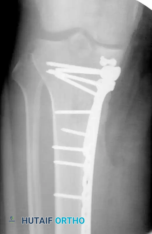
Following anatomical elevation to the level of the native joint line (confirmed visually and fluoroscopically), the resultant metaphyseal void is densely packed with a structural bone graft substitute, such as calcium phosphate cement, which provides immediate compressive strength and prevents subsidence. The articular surface is temporarily held with K-wires, followed by the placement of a subchondral raft of 3.5-mm or 4.0-mm screws. Finally, a precontoured lateral locking plate is applied to buttress the lateral cortex and neutralize axial and valgus forces.
The Posteromedial Approach
Fractures involving a posteromedial coronal shear fragment (Schatzker IV or the medial component of Schatzker V/VI) cannot be adequately addressed through an anteromedial approach. The apex of these fractures lies posterior to the mid-coronal plane, necessitating a dedicated posteromedial approach to apply an anti-glide or buttress plate directly over the fracture apex. The patient remains supine, but the hip is externally rotated and the knee slightly flexed (a "figure-of-four" position can be utilized).
An incision is made along the posteromedial border of the proximal tibia. The saphenous nerve and vein are identified and protected. Dissection proceeds between the medial head of the gastrocnemius (retracted posteriorly) and the pes anserinus (retracted anteriorly or distally). This exposes the posteromedial tibial crest. The fracture is debrided of hematoma, and reduction is achieved using a pointed reduction forceps applied from the anterior tibial crest to the posteromedial fragment.
Fixation is achieved using an under-contoured 3.5-mm T-plate or a dedicated posteromedial locking plate. The plate is positioned to act as a buttress against the distal apex of the fracture, neutralizing the vertical shear forces. Proximal screws are placed carefully to avoid intraarticular penetration and to prevent collision with laterally placed hardware in bicondylar fracture patterns.
Arthroscopically Assisted Reduction and Internal Fixation (ARIF)
For less comminuted lateral plateau fractures (Schatzker I, II, and III), ARIF offers a minimally invasive alternative that significantly reduces soft tissue morbidity. Standard anterolateral and anteromedial arthroscopic portals are established. The hemarthrosis is evacuated, allowing direct, magnified visualization of the articular surface and the identification of concomitant intraarticular pathology, such as meniscal tears, which are addressed concurrently.
Crucially, fluid management must be strictly controlled. A dry arthroscopy technique or gravity flow without a high-pressure pump is mandatory to prevent massive fluid extravasation through the metaphyseal fracture lines into the fascial compartments of the calf, which carries a catastrophic risk of iatrogenic compartment syndrome. The depressed articular fragments are elevated percutaneously using a bone tamp introduced through a small metaphyseal cortical window, guided by fluoroscopy and direct arthroscopic visualization. Once reduced, the fracture is stabilized with percutaneous subchondral screws or a minimally invasive plate osteosynthesis (MIPO) technique.
Management of Bicondylar Fractures (Schatzker V & VI)
Bicondylar fractures represent the zenith of surgical complexity in this anatomical region. Historically, extensive single-incision exposures (e.g., the Y-shaped or Mercedes-Benz incision) with massive dual plating led to catastrophic wound necrosis and deep infection rates exceeding 30%. Modern management dictates a staged, tissue-preserving approach. Initial management consists of a spanning external fixator to restore length, alignment, and rotation while allowing the soft tissue envelope to recover over 7 to 14 days.
Definitive fixation typically employs a dual-incision technique: a standard anterolateral approach and a separate posteromedial approach. A wide skin bridge (minimum of 7 cm) must be maintained between the two incisions to preserve the delicate angiosomal vascular supply to the anterior tibial skin. The more comminuted or displaced column (frequently the medial side) is reduced and provisionally fixed first to establish a stable cornerstone. The contralateral side is then reconstructed against this stable foundation. In cases of profound metaphyseal comminution or persistent soft tissue compromise, the surgeon may opt for a hybrid construct, utilizing a lateral locking plate combined with a fine-wire circular external fixator (e.g., Ilizarov or Taylor Spatial Frame) to minimize periosteal stripping.
Complications, Incidence Rates, and Salvage Management
Despite meticulous surgical technique and adherence to biological principles, the management of tibial plateau fractures is fraught with a high incidence of both early and late complications. The severity of the initial trauma, combined with the tenuous nature of the proximal tibial soft tissue envelope, creates a precarious environment for healing. Surgeons must maintain a high index of suspicion and employ aggressive, preemptive strategies to mitigate these risks.
Early complications are predominantly related to the soft tissues and the acute physiological response to trauma. Acute compartment syndrome (ACS) is the most devastating early complication, occurring in up to 10-15% of high-energy bicondylar fractures (Schatzker V/VI). Diagnosis is primarily clinical, characterized by pain out of proportion to the injury and pain with passive stretch of the involved compartments. If suspected, emergent four-compartment fasciotomies are mandatory; delayed diagnosis leads to irreversible myonecrosis, ischemic contractures, and potential amputation. Surgical site infection (SSI) remains a significant challenge, with deep infection rates ranging from 2% in low-energy fractures to over 20% in severe, open bicondylar injuries. Management of deep SSI requires aggressive serial irrigation and debridement, hardware retention if the construct is stable and the fracture is healing, and targeted intravenous antibiotic therapy guided by intraoperative cultures.
Late complications are primarily mechanical and biological failures of the reconstructed joint. Post-traumatic osteoarthritis (PTOA) is the most common long-term complication, developing in 20% to 45% of patients within 5 to 10 years post-injury. PTOA is driven by residual articular incongruity, uncorrected axial malalignment, meniscal loss, and the initial chondrocyte apoptosis triggered by the traumatic impact. Hardware failure, including screw breakage or plate pullout, typically occurs secondary to premature weight-bearing or failure to adequately fill metaphyseal voids with structural graft, leading to articular subsidence. Nonunion and malunion are less common but occur in cases of severe comminution or deep infection.
Salvage management for end-stage PTOA or profound structural failure frequently necessitates Total Knee Arthroplasty (TKA). However, TKA following a tibial plateau fracture ORIF is technically demanding. The surgeon must navigate compromised soft tissues, retained hardware, distorted anatomical landmarks, and metaphyseal bone loss. Staged hardware removal and culture of the medullary canal may be required prior to arthroplasty to rule out indolent infection. In cases of severe infection, massive bone loss, or extensor mechanism failure, knee arthrodesis may be the only viable salvage option to provide a stable, painless, though stiff, lower extremity.
| Complication | Estimated Incidence | Primary Etiology / Risk Factors | Prevention and Salvage Management |
|---|---|---|---|
| Compartment Syndrome | 5% - 15% (High-energy) | Massive tissue trauma, ARIF fluid extravasation. | Prevention: Staged protocol, gravity fluid. Salvage: Emergent 4-compartment fasciotomy. |
| Deep Surgical Infection | 2% - 20% (Pattern dependent) | Early ORIF through compromised soft tissues, open fractures. | Prevention: "Span, scan, plan", meticulous handling. Salvage: Serial I&D, IV antibiotics, eventual hardware removal. |
| Peroneal Nerve Palsy | 1% - 5% | Posterolateral approaches, traction injury, fibular neck fractures. | Prevention: Direct visualization, avoiding excessive traction. Salvage: AFO bracing, tendon transfers (late). |
| Articular Subsidence | 10% - 25% | Premature weight-bearing, inadequate metaphyseal void filling. | Prevention: Dense structural grafting, strict NWB protocol. Salvage: Revision ORIF (early) or TKA (late). |
| Post-Traumatic OA | 20% - 45% | Residual step-off, meniscal excision, initial cartilage impact. | Prevention: Anatomic reduction, meniscal repair. Salvage: Corrective osteotomy (young) or TKA (elderly). |
Phased Post-Operative Rehabilitation Protocols
The postoperative rehabilitation protocol following ORIF of a tibial plateau fracture is a delicate, tightly choreographed balance. The surgeon and physical therapist must facilitate early joint mobilization to nourish the articular cartilage and prevent arthrofibrosis, while simultaneously enforcing strict weight-bearing restrictions to protect the fragile osteosynthesis construct from catastrophic mechanical failure. Communication between the surgical team and the rehabilitation specialists is paramount to ensure patient compliance and optimize functional outcomes.
Phase I: Maximum Protection and Early Motion (Weeks 0 to 6)
Immediately postoperatively, the knee is placed in a hinged knee brace locked in full extension to protect the soft tissues and provide stability during transfers. However, early motion is initiated within 24 to 48 hours. Continuous passive motion (CPM) machines or active-assisted range of motion (AAROM) exercises are utilized to promote synovial fluid diffusion, which is critical for chondrocyte nutrition and healing. The goal is to achieve 90 degrees of flexion by week 4 and full range of motion by week 6. Weight-bearing is strictly restricted to non-weight-bearing (NWB) or toe-touch weight-bearing (TTWB, < 20 lbs) using crutches or a walker. Crucial Exception: If an extensive meniscal repair or a concomitant collateral ligament repair was performed, motion may be restricted or delayed for 3 to 4 weeks to protect the repair site.
Phase II: Progressive Loading and Strengthening (Weeks 6 to 12)
At the 6-week mark, orthogonal radiographs are obtained to assess for the maintenance of reduction, the absence of hardware failure, and the early signs of bridging callus. If clinical and radiographic progression is satisfactory, weight-bearing is gradually advanced. Patients typically progress to 50% partial weight-bearing (PWB) and begin weaning from the hinged knee brace. Rehabilitation focuses on closed kinetic chain exercises, isometric quadriceps strengthening, and patellar mobilization to prevent patella infera. Aquatic therapy is highly beneficial during this phase, allowing for gait mechanics training in a reduced-gravity environment.
Phase III: Full Weight-Bearing and Functional Restoration (Weeks 12 to 24+)
By 10 to 12 weeks, assuming radiographic consolidation of the fracture, patients are cleared for full weight-bearing (FWB) as tolerated. The focus of physical therapy shifts towards aggressive muscular strengthening, proprioceptive retraining, and functional restoration. Advanced balance exercises, stationary cycling, and progressive resistance training are incorporated. Return to high-impact sports or heavy manual labor is generally not permitted until 6 to 9 months postoperatively, and only after the patient demonstrates symmetrical quadriceps strength and dynamic stability on functional testing. Patients must be counseled that maximal medical improvement frequently takes up to 12 to 18 months, and some degree of residual stiffness or aching is common.
Summary of Landmark Literature and Clinical Guidelines
The evolution of tibial plateau fracture management is deeply rooted in landmark orthopedic literature and continuously refined clinical guidelines. Historically, the Schatzker classification system, introduced in 1979, provided the foundational framework for understanding these fractures based on two-dimensional radiographic patterns. Schatzker divided the fractures into six types, guiding the initial surgical approach (Types I-III lateral, Type IV medial, Types V-VI bicondylar). However, as high-resolution CT imaging became ubiquitous, the limitations of the Schatzker system in addressing complex, multi-planar comminution became apparent.
To address this, Luo et al. (2010) introduced the CT-based Three-Column Concept, a paradigm-shifting classification that divides the tibial plateau into lateral, medial, and posterior columns. This conceptual framework revolutionized preoperative planning, explicitly highlighting the frequency and importance of the posterior coronal shear fragment (the posterior column), which is often missed on plain radiographs and cannot be adequately stabilized through traditional anterior approaches. The Three-Column Concept dictates that fixation must be customized to address each involved column independently, leading to the widespread adoption of posteromedial and posterolateral approaches.
Furthermore, the timing and strategy of soft tissue management have been profoundly influenced by the work of Egol et al. and the Canadian Orthopaedic Trauma Society (COTS). The COTS multicenter randomized controlled trial (Moore et al.) comparing dual plating to circular external fixation for severe bicondylar fractures demonstrated that while both techniques yield similar long-term functional outcomes, dual plating through a single extensile incision carried an unacceptably high rate of deep infection. This landmark study cemented the modern standard of care: utilizing a staged "span, scan, and plan" protocol with delayed, dual-incision ORIF or fine-wire circular fixation to respect the compromised soft tissue envelope and minimize catastrophic postoperative complications.
