Hip Dysplasia Treatment: Outcomes, Recovery, & Preventing Complications
Key Takeaway
Learn more about Hip Dysplasia Treatment: Outcomes, Recovery, & Preventing Complications and how to manage it. Hip dysplasia treatment typically involves Pavlik harnesses for infants or closed reduction and casting for young children. Early detection significantly improves hip dysplasia treatmentoutcomesrecoverycomplications, often leading to predictable results and normal hip development. If detected later, treatment becomes more complex, yielding less predictable outcomes and potentially prolonged recovery or requiring surgical intervention to prevent further complications.
Introduction & Epidemiology
Developmental Dysplasia of the Hip (DDH) represents a spectrum of conditions ranging from mild acetabular dysplasia to irreducible dislocation of the femoral head. It is characterized by an abnormal relationship between the femoral head and the acetabulum, leading to instability, subluxation, or complete dislocation. The incidence of DDH varies globally, with reported rates between 1-3 per 1,000 live births, though milder forms of dysplasia may be higher. Females are predominantly affected (4:1 ratio), and risk factors include breech presentation, first-born status, oligohydramnios, and a positive family history. Early detection is paramount, as it significantly influences treatment outcomes and mitigates the long-term sequelae of secondary osteoarthritis.
When DDH is detected at birth through clinical examination (Ortolani, Barlow maneuvers) or screening ultrasound, non-operative management with a harness or brace, such as the Pavlik harness, is typically highly successful. This early intervention leverages the plasticity of the infant hip, promoting concentric reduction and remodeling of the dysplastic acetabulum. If the hip is not dislocated at birth, or if mild dysplasia is initially missed, the condition may not be noticed until the child begins walking, often presenting with gait abnormalities, limb length discrepancy, or delayed motor milestones. At this stage, treatment becomes more complex, requiring more invasive interventions with less predictable long-term results and an increased risk of complications.
The management strategy for DDH evolves with the child's age, the degree of instability, and the severity of acetabular and femoral head maldevelopment. Primary acetabular dysplasia without hip instability, or residual acetabular dysplasia following successful treatment for instability, may initially warrant observation. Continuous monitoring of acetabular development, particularly the Acetabular Index (AI) and femoral head coverage, is crucial. If the AI continues to improve and the hip remains concentrically reduced, observation can be continued. However, if hip subluxation develops, the AI fails to improve over a 12-month period, or if significant migration of the femoral head is noted, operative treatment is indicated to prevent progressive deformity and early degenerative joint disease.
Surgical Anatomy & Biomechanics
Successful management of DDH, whether non-operative or operative, hinges on a thorough understanding of the unique surgical anatomy and biomechanics of the pediatric hip. The hip joint in infants and children is characterized by its dynamic growth and remodeling potential, which diminishes with age.
Bony Anatomy:
*
Acetabulum:
In infants, the acetabulum is predominantly cartilaginous, particularly the triradiate cartilage, which is a key growth center contributing to acetabular depth and orientation. The bony acetabulum contributes to the anterior, posterior, and superior walls. The acetabular index (AI), measured from the horizontal line connecting the triradiate cartilages to the lateral lip of the acetabulum, is a critical radiographic parameter for assessing acetabular development. A normal AI is typically less than 30 degrees at birth and decreases with age. Dysplasia is characterized by an increased AI, indicating a shallow, oblique acetabulum.
*
Femoral Head and Neck:
The femoral head is entirely cartilaginous at birth, with ossification commencing around 4-6 months. Its spherical shape is essential for concentric reduction. In DDH, the femoral head may be hypoplastic, flattened, or exhibit altered anteversion due to chronic dislocation or subluxation. The femoral neck-shaft angle (coxa valga) and excessive femoral anteversion are common compensatory or associated deformities, which contribute to instability and require addressing during surgical correction.
*
Pelvis:
The overall pelvic configuration can also be affected, with some dysplastic hips exhibiting a more vertical orientation of the ilium.
Soft Tissue Anatomy:
*
Capsule:
In chronically dislocated hips, the capsule often becomes redundant and stretched laterally but constricts medially, forming an hourglass constriction that can impede reduction. The inferior aspect of the capsule may thicken, contributing to the "limbus."
*
Ligamentum Teres:
This intra-articular ligament can hypertrophy and elongate in dislocated hips, acting as an obstacle to reduction.
*
Labrum:
The acetabular labrum, a fibrocartilaginous ring, deepens the acetabulum. In DDH, it can be everted, inverted, or hypertrophied, creating a barrier to concentric reduction.
*
Psoas Tendon:
The iliopsoas tendon passes anterior to the hip joint. In DDH, it can become tight and act as a tether, pulling the femoral head anteriorly and superiorly, hindering reduction or contributing to redislocation. Adductor longus and magnus muscles are also frequently contracted.
Biomechanics:
*
Acetabular Remodeling:
The primary goal of DDH treatment is to achieve and maintain a stable, concentric reduction. This allows for normal growth and remodeling of the cartilaginous acetabulum, which is stimulated by the presence of a well-centered femoral head. The optimal window for harnessing this remodeling potential is typically before 6 months of age.
*
Joint Stability:
Hip stability is a complex interplay of bony congruence, capsuloligamentous integrity, and muscle forces. In DDH, the shallow acetabulum and abnormal femoral head morphology compromise this stability, leading to increased joint stress, articular cartilage degeneration, and eventual osteoarthritis.
*
Contact Pressures:
A dysplastic hip often results in a reduced weight-bearing surface area, concentrating forces on a smaller portion of the articular cartilage. This elevated stress is a major contributor to premature degenerative changes. Surgical intervention aims to normalize these contact pressures by improving acetabular coverage and joint congruence.
*
Muscular Imbalance:
Chronic dislocation can lead to muscle imbalances, with contracture of adductors, iliopsoas, and hip flexors, while abductors and extensors become relatively weak or elongated. This imbalance can further contribute to instability and gait abnormalities.
Indications & Contraindications
The decision-making process for DDH treatment is multifactorial, considering the patient's age, the severity of dysplasia, the stability of the hip, and the response to prior interventions.
Non-Operative Indications:
*
Infants < 6 months:
*
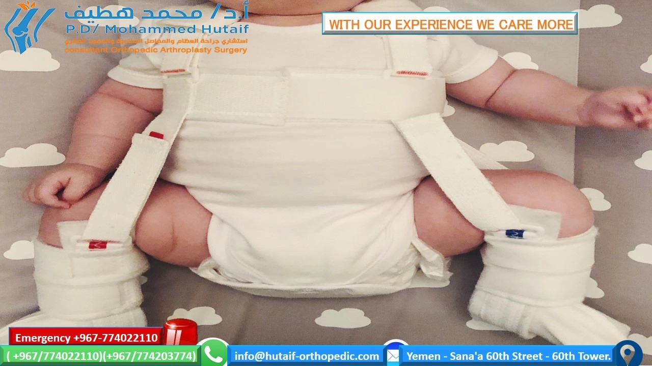
Pavlik Harness:
The initial treatment for primary acetabular dysplasia and reducible subluxation or dislocation. Requires the hip to be reducible and stable within the harness's safe zone (flexion > 90°, abduction 40-60°). Contraindications include irreducible hips, teratologic dislocations, hyperlaxity (leading to posterior dislocation), or age > 6 months (where the success rate decreases significantly due to less cartilaginous plasticity and greater soft tissue contracture).
*
Children 6-18 months (or up to 24 months):
*
Closed Reduction and Cast Immobilization:
Indicated when Pavlik harness fails or is no longer appropriate due to age. Requires general anesthesia, often with adductor tenotomy and/or psoas release to facilitate reduction. Success is predicated on achieving a stable reduction that can be maintained in a hip spica cast. Pre-reduction traction may be employed for several weeks to stretch soft tissues.
*
Residual Acetabular Dysplasia (post-treatment or mild primary):
*
Observation:
As long as the acetabular index continues to improve and the hip remains concentrically reduced. Regular radiographic monitoring is essential.
*
Neuromuscular Patients:
*
Observation:
For patients with a migration index less than 25% and abduction greater than 45 degrees, indicating reasonable stability and function.
Operative Indications:
*
Failed Non-Operative Management:
Persistent instability, redislocation, or unacceptably high Acetabular Index despite adequate bracing or closed reduction.
*
Older Children (typically >18-24 months):
*
Open Reduction:
Required when closed reduction is unsuccessful due to significant soft tissue obstruction (e.g., inverted labrum, hypertrophied ligamentum teres, constricted capsule, interposition of iliopsoas tendon). Often combined with a pelvic osteotomy and/or femoral osteotomy.
*
Persistent Acetabular Dysplasia (after age 2-4 years, or failed observation):
* If hip subluxation develops, the acetabular index fails to improve over a 12-month period, or if the Wiberg Lateral Center-Edge angle remains critically low (e.g., < 19° at skeletal maturity).
*
Pelvic Osteotomies:
Indicated to improve acetabular coverage and reorient the acetabulum.
*
Redirectional Osteotomies (e.g., Salter innominate osteotomy, Triple osteotomy, Bernese periacetabular osteotomy - PAO):
Used when the native acetabulum has sufficient volume but is maloriented. Generally preferred in older children/adolescents with open triradiate cartilage (Salter, Triple) or closed triradiate cartilage (PAO).
*
Reshaping/Augmenting Osteotomies (e.g., Pemberton pericapsular osteotomy, Dega osteotomy):
Used to deepen and reshape a deficient acetabulum, typically in younger children with open triradiate cartilage.
*
Significant Femoral Deformity:
*
Femoral Varus Derotation Osteotomy (VDRO):
Indicated for excessive femoral anteversion (>40-50°) and/or coxa valga (neck-shaft angle >145°) that contributes to instability or limits abduction. Often performed concurrently with open reduction and/or pelvic osteotomy.
*
Neuromuscular Patients:
*
Surgical Treatment:
For migration indexes over 50%, or progressive subluxation/dislocation, which can lead to pain, perineal hygiene issues, and functional limitations. This can include femoral or pelvic osteotomies.
Contraindications for Specific Surgical Procedures:
*
Pelvic Osteotomies:
*
Salter Osteotomy:
Contraindicated with a closed triradiate cartilage.
*
Pemberton/Dega Osteotomy:
Contraindicated with closed triradiate cartilage, as these osteotomies go through it.
*
PAO:
While often used for closed triradiate cartilage, meticulous planning is required to avoid growth disturbance if the triradiate cartilage is still open.
*
General Surgical Contraindications:
* Severe medical comorbidities precluding safe anesthesia.
* Active infection.
* Severe neurological impairment where surgical benefits are unlikely to outweigh risks.
Table: Operative vs. Non-Operative Indications in DDH
| Category | Non-Operative Indications | Operative Indications |
|---|---|---|
| Age | < 6 months: Reducible hip, Pavlik harness | > 18-24 months: Irreducible dislocations (Open Reduction) |
| 6-18 months: Closed reduction, spica cast | Any age: Failed closed reduction/harness treatment | |
| Hip Stability | Reducible subluxation/dislocation, stable in harness/cast | Irreducible dislocation (requires Open Reduction) |
| Stable dysplasia without signs of progression | Recurrent subluxation/dislocation | |
| Acetabular | Improving Acetabular Index (AI), adequate femoral coverage | Persistent dysplasia (elevated AI, poor coverage), AI not improving |
| Morphology | Severe acetabular deficiency (requires Pelvic Osteotomy - Salter, Pemberton, Dega, PAO) | |
| Femoral | Normal femoral anteversion/neck-shaft angle | Excessive femoral anteversion or coxa valga (requires Femoral Osteotomy) |
| Obstacles to | None (or easily managed with minor soft tissue release) | Significant soft tissue interposition (limbus, psoas, capsule) |
| Reduction | ||
| Neuromuscular | Migration index < 25%, abduction > 45° | Migration index > 50%, progressive subluxation, symptomatic dislocation |
| Prognosis | High likelihood of normal hip development with non-op management | Low likelihood of successful non-op management; high risk of OA without surgery |
Pre-Operative Planning & Patient Positioning
Meticulous pre-operative planning is critical for optimizing outcomes and minimizing complications in DDH surgery.
Imaging:
*
Plain Radiographs:
Anteroposterior (AP) pelvis and frog-leg lateral views are standard. Key measurements include the Acetabular Index (AI), Shenton's line, Hilgenreiner's line, Perkin's line, and Wiberg's Lateral Center-Edge (LCE) angle (for older children/adolescents). For older children approaching skeletal maturity, a false profile view may be useful to assess anterior coverage.
*
Ultrasound:
Crucial for initial diagnosis and monitoring in infants, especially before ossification of the femoral head (typically up to 6 months). Static (Graf classification) and dynamic assessments (Barlow, Ortolani maneuvers) evaluate stability and acetabular morphology.
*
MRI:
Indicated for evaluating soft tissue obstacles to reduction (e.g., inverted labrum, hypertrophied ligamentum teres, constricted capsule, psoas impingement), especially in older infants or failed closed reductions. It also assesses femoral head viability (pre-existing AVN) and triradiate cartilage status.
*
CT Scan:
Valuable for 3D assessment of complex deformities, particularly for planning osteotomies, assessing joint congruity post-reduction, and evaluating residual dysplasia or early AVN. Often used for post-reduction spica cast assessment if concerns about concentricity persist.
Templating and Surgical Strategy:
* Based on imaging, the specific surgical approach (open reduction vs. osteotomy) and osteotomy type are determined.
* For osteotomies, precise templating helps determine the amount of correction needed (e.g., angle of acetabular fragment redirection, amount of femoral derotation/varus).
* Planning for potential adjunctive procedures like adductor tenotomy, psoas release, or capsulorrhaphy.
* Discussion with the anesthesiology team regarding blood loss management, pain control, and anesthetic technique.
Patient and Family Education/Consent:
* Comprehensive discussion with parents regarding the surgical procedure, expected outcomes, potential complications (especially AVN), post-operative care (spica cast), and long-term prognosis.
* Informed consent must detail all risks and benefits.
Equipment Preparation:
* Specific instruments for pediatric hip surgery, including small osteotomes, drills, wires, plates, and screws suitable for pediatric bone.
* Image intensifier (fluoroscopy) for intra-operative confirmation of reduction and osteotomy fixation.
* Appropriate size spica cast materials.
Patient Positioning:
*
Closed Reduction with Spica Cast:
Patient is typically supine on a radiolucent table. Traction may be applied pre-operatively or during the procedure. After reduction, the patient is often moved to a spica cast table (e.g., Gigli or similar frame) to facilitate cast application in the desired position of flexion and abduction.
*
Open Reduction/Pelvic Osteotomy (Anterior Approach - e.g., Smith-Petersen):
Patient is positioned supine on a radiolucent table. A bump is placed under the ipsilateral hip to allow slight external rotation. The entire lower extremity should be prepped and draped free to allow full range of motion, particularly internal and external rotation, for intra-operative stability assessment.
*
Pelvic Osteotomy (Posterior Approach - e.g., Bernese PAO):
Typically lateral decubitus position, allowing access to the posterior ilium.
*
Femoral Osteotomy:
Patient is usually supine with a bump under the ipsilateral hip. The leg is prepped and draped free.
Detailed Surgical Approach / Technique
The surgical technique for DDH is highly individualized based on age, severity, and specific deformities.
1. Closed Reduction with Adductor Tenotomy
This is often performed in children aged 6-18 months.
*
Anesthesia:
General anesthesia.
*
Adductor Tenotomy:
A small transverse skin incision (approximately 1-2 cm) is made in the groin crease overlying the adductor longus tendon. The tendon is identified and released percutaneously or with a small open incision. Some surgeons also perform a psoas tenotomy via a similar medial approach if indicated.
*
Arthrogram (Optional but Recommended):
Following tenotomy, an arthrogram may be performed by injecting contrast into the hip joint under fluoroscopic guidance. This allows visualization of the cartilaginous femoral head and acetabulum, identifying potential obstacles to reduction (e.g., inverted labrum, hypertrophied ligamentum teres, pulvinar fat pad, constricted capsule forming an hourglass shape).
*
Reduction Maneuvers:
Gentle, controlled maneuvers are used to achieve reduction. This typically involves gradual flexion, gentle traction, and controlled abduction and internal rotation (reverse Ortolani). Forceful reduction must be avoided to minimize the risk of Avascular Necrosis (AVN). The "safe zone" of stability (the arc of motion between redislocation) is identified.
*
Post-Reduction Assessment:
Fluoroscopy confirms concentric reduction. An arthrogram can also confirm a stable, concentric reduction with good femoral head coverage.
*
Spica Cast Application:
A hip spica cast is applied to maintain the reduction in the optimal "human position" (typically 90-100 degrees of hip flexion and 30-45 degrees of abduction), avoiding extreme positions that increase AVN risk. The cast is usually applied for 6-12 weeks.
2. Open Reduction (Anterior Approach - Modified Smith-Petersen)
Indicated when closed reduction fails due to irreducible obstacles or in older children.
*
Incision:
A bikini or oblique incision is made, starting from the anterior superior iliac spine (ASIS) and extending distally and medially towards the sartorius muscle.
*
Internervous Plane:
The interval between the sartorius (supplied by femoral nerve) and the tensor fascia lata (supplied by superior gluteal nerve) is developed. The sartorius is retracted medially, and the rectus femoris is identified. The lateral femoral cutaneous nerve should be protected along the medial border of the sartorius. The ascending branch of the lateral circumflex femoral artery and vein, which crosses the field, should be ligated or cauterized.
*
Rectus Femoris Release:
The direct and reflected heads of the rectus femoris are often released from their origins to facilitate exposure and avoid tension during reduction.
*
Capsulotomy:
A T-shaped or inverted T-shaped capsulotomy is performed. The anterior limb of the capsule is incised longitudinally, and then transversely. Meticulous care is taken to avoid damage to the labrum and femoral head articular cartilage.
*
Obstacle Excision:
Intra-articular obstacles hindering reduction are systematically addressed:
*
Inverted Labrum:
The everted or inverted labrum, which forms a major block to reduction, is carefully excised or repositioned if possible.
*
Ligamentum Teres:
Often hypertrophied, it is typically excised.
*
Pulvinar:
Hypertrophied pulvinar fat pad is resected to deepen the acetabulum.
*
Constricted Capsule:
The medial constricting portion of the capsule (hourglass constriction) is released to allow the femoral head to seat concentrically. The psoas tendon, if tight, is released at its insertion.
*
Reduction:
The hip is gently reduced under direct visualization.
*
Capsulorrhaphy:
Once reduced, a meticulous capsulorrhaphy is performed to tighten the redundant capsule and enhance stability. The anterior capsule is imbricated and sutured.
*
Stability Assessment:
The hip's stability is assessed through a gentle range of motion, ensuring it remains reduced in a functional arc. The "safe zone" is confirmed.
*
Closure:
Deep fascia and subcutaneous layers are closed, followed by skin closure.
*
Spica Cast Application:
A hip spica cast is applied to maintain the reduction. A single-leg spica is usually sufficient if the hip is stable.

Figure 1: Intraoperative view during open reduction of developmental dysplasia of the hip, illustrating the exposure of the femoral head and acetabulum after capsulotomy and removal of soft tissue obstacles.
3. Pelvic Osteotomies
Performed to correct acetabular deficiency and improve coverage.
*
Salter Innominate Osteotomy:
*
Indications:
Children 18 months to 8 years with a mobile, concentrically reduced hip, but persistent acetabular dysplasia (shallow acetabulum with sufficient volume, but poor orientation). Requires open triradiate cartilage.
*
Technique:
An oblique incision from ASIS towards the iliac crest. The gluteal muscles are reflected subperiosteally. A full-thickness osteotomy of the ilium is performed just above the ASIS down to the greater sciatic notch. The distal fragment, containing the acetabulum, is then redirected (rotated inferolaterally) to improve anterior and lateral coverage. A triangular bone graft (typically from the posterior ilium) is inserted into the osteotomy gap, and fixation is achieved with two or three Kirschner wires (K-wires).
*
Pemberton Pericapsular Osteotomy:
*
Indications:
Children 18 months to 8 years with true acetabular deficiency (anterior and lateral coverage deficit). Requires open triradiate cartilage.
*
Technique:
Similar approach to Salter. An osteotomy is created superior to the anterior inferior iliac spine (AIIS) and extends posteriorly, stopping just proximal to the triradiate cartilage. The outer table is preserved to act as a hinge. The acetabular fragment is then rotated inferomedially to deepen the acetabulum, and a bone graft (often from the inner table of the ilium) is inserted. Fixation typically with K-wires or a single screw.
*
Dega Osteotomy:
*
Indications:
Similar to Pemberton, focusing on lateral and posterior coverage. Requires open triradiate cartilage.
*
Technique:
Similar approach to Pemberton, but the osteotomy begins more posteriorly and extends towards the AIIS, maintaining the integrity of the anterior column. The goal is to improve posterior and lateral acetabular coverage by rotating the fragment around a hinge near the anterior limb of the triradiate cartilage. Bone graft is inserted, and fixation is with K-wires.
*
Triple Innominate Osteotomy (Sutherland, Steel):
*
Indications:
Adolescents with open triradiate cartilage, significant acetabular dysplasia, and a concentrically reduced hip.
*
Technique:
Involves three separate osteotomies: iliac, pubic, and ischial. This allows for complex 3D reorientation of the acetabulum. The fragment is then fixed with screws and/or plates.
*
Bernese Periacetabular Osteotomy (PAO):
*
Indications:
Adolescents and young adults with symptomatic hip dysplasia and closed triradiate cartilage.
*
Technique:
A complex procedure involving periacetabular osteotomies that detach the entire acetabular rim and a portion of the innominate bone, allowing for multiplanar correction. It preserves the posterior column and the weight-bearing integrity of the pelvis. Fixation is typically with screws. This procedure allows for correction of acetabular version and inclination without significantly changing the pelvic volume.
4. Femoral Varus Derotation Osteotomy (VDRO)
Performed to correct excessive femoral anteversion and/or coxa valga.
*
Indications:
Excessive femoral anteversion (>40-50°) and/or coxa valga (neck-shaft angle >145°) contributing to instability, impingement, or gait abnormalities. Often performed concurrently with open reduction and/or pelvic osteotomy.
*
Technique:
A lateral approach to the proximal femur is made. A transverse or oblique osteotomy is performed in the subtrochanteric region. A wedge of bone may be resected to achieve varus correction, and the distal fragment is internally rotated to correct anteversion. Fixation is typically with a plate and screws (e.g., blade plate, dynamic hip screw, pediatric locking plate).
Following any of these procedures, a hip spica cast is generally applied for 6-12 weeks to protect the reduction and allow for bone healing.
Complications & Management
Despite meticulous surgical technique, complications can occur in DDH treatment, impacting long-term outcomes. The risk of specific complications varies significantly with patient age, the severity of dysplasia, and the chosen intervention.
Table: Common Complications, Incidence, and Salvage Strategies
| Complication | Incidence (Approximate) | Salvage Strategy / Management |
|---|---|---|
| Avascular Necrosis (AVN) of Femoral Head | 5-30% (higher with open reduction, older age, forceful reduction) |
Early:
Observation, protected weight-bearing.
Late: Containment procedures (pelvic/femoral osteotomies) to improve coverage; microfracture/drilling for focal lesions; in severe cases, arthrodesis or total hip arthroplasty (THA) in adulthood. |
| Redislocation / Subluxation | 5-15% (higher with severe dysplasia, inadequate initial reduction/fixation) |
Early:
Re-reduction (closed or open) and re-casting; revision osteotomy; adjust fixation.
Late: Pelvic/femoral osteotomies to improve stability; consider arthroscopy for intra-articular pathology. |
| Nerve Palsy (Femoral, Sciatic, LFCN) | 1-5% (often transient) |
Prevention:
Careful positioning, avoid excessive traction/stretch.
Management: Observation (most resolve spontaneously); nerve conduction studies/EMG; surgical exploration if no recovery. |
| Infection | 1-3% |
Superficial:
Oral antibiotics, wound care.
Deep: Surgical debridement, IV antibiotics, implant removal if necessary. |
| Bleeding / Hematoma | 2-5% | Careful hemostasis intra-op; drains; hematoma evacuation if symptomatic. Transfusion if significant blood loss. |
| Leg Length Discrepancy (LLD) | 5-10% (can be developmental or iatrogenic) |
Minor:
Shoe lift.
Significant: Epiphysiodesis of the longer limb; limb lengthening procedures for the shorter limb (e.g., guided growth, external fixator). |
| Stiffness / Loss of Motion | 5-10% | Aggressive physical therapy; anti-inflammatory medications; rarely, manipulation under anesthesia or arthroscopic capsular release. |
| Over/Undercorrection of Dysplasia | 5-10% |
Under-correction:
Revision pelvic/femoral osteotomy, further non-operative management if mild.
Over-correction: Rare, may require reverse osteotomy or observation. |
| Growth Arrest / Physeal Damage | < 1% (specific to osteotomies through physis) |
Prevention:
Careful surgical planning to avoid growth plates, especially triradiate cartilage.
Management: Observation, corrective osteotomies for limb length discrepancy or angular deformity. |
| Hardware-Related Complications | 5-10% (prominence, breakage, migration) | Symptomatic hardware removal; revision surgery for inadequate fixation. |
| Residual Deformity / Early Osteoarthritis | Long-term risk, especially with AVN or persistent dysplasia | Surveillance; symptomatic management; consider reconstructive surgery (e.g., redirectional osteotomy, PAO) or joint replacement (THA) in adulthood. |
Detailed Discussion of Key Complications:
-
Avascular Necrosis (AVN) of the Femoral Head: This is the most dreaded and devastating complication, significantly impacting long-term outcomes.
- Etiology: Interruption of the blood supply to the femoral epiphysis, often due to forceful reduction maneuvers, extreme positioning in a cast (excessive abduction), increased intra-articular pressure, or direct surgical trauma to epiphyseal vessels.
- Risk Factors: Older age at reduction, severe pre-existing dislocation, repeated reduction attempts, forceful reduction, extreme spica cast positions.
- Classification: Kalamchi and MacEwen classification (Grade I-IV) describes the extent of epiphyseal involvement and subsequent remodeling.
- Management: Varies based on severity. Mild AVN may resolve with minimal deformity. Severe AVN can lead to femoral head collapse, coxa plana, coxa magna, premature physeal arrest, and progressive osteoarthritis. Treatment often focuses on containment (improving acetabular coverage over the compromised head) with pelvic or femoral osteotomies, or bracing, to optimize remodeling. In adulthood, severe residual deformity may necessitate total hip arthroplasty.
-
Redislocation / Subluxation: Can occur early or late.
- Early: Often due to inadequate initial reduction, missed intra-articular obstacles, or improper cast application.
- Late: May be secondary to inadequate initial capsulorrhaphy, progression of residual acetabular or femoral dysplasia, or growth of the patient beyond the scope of the initial correction.
- Management: Requires careful investigation (radiographs, CT, MRI) to identify the cause. Re-reduction (closed or open) and re-casting are often necessary. If structural deficiencies persist, revision pelvic or femoral osteotomies are indicated.
-
Nerve Palsy:
- Femoral Nerve Palsy: Most common, typically transient, due to prolonged hip flexion in the spica cast causing nerve stretch or compression. Presents as quadriceps weakness.
- Sciatic Nerve Palsy: Less common, usually associated with forceful posterior reduction or extreme external rotation.
- Lateral Femoral Cutaneous Nerve (LFCN) Palsy: Common with anterior surgical approaches, causing numbness or dysesthesia in the anterolateral thigh. Usually a transient sensory deficit.
- Management: Prevention is key (avoid extreme positions, careful surgical dissection). Most cases of femoral and sciatic nerve palsy resolve spontaneously within weeks to months. Physical therapy is initiated. Surgical exploration is rarely indicated unless there is persistent, severe deficit or an identifiable cause like a hematoma.
-
Leg Length Discrepancy (LLD): Can result from growth disturbances of the proximal femur secondary to AVN or physeal damage during osteotomy. Also, from over-correction or under-correction of femoral or pelvic deformities.
- Management: For minor LLD, shoe lifts are used. For significant LLD, surgical interventions such as epiphysiodesis of the longer limb or limb lengthening of the shorter limb (e.g., using an external fixator or intramedullary nail) may be considered, usually near skeletal maturity.
Post-Operative Rehabilitation Protocols
Post-operative rehabilitation is crucial for achieving optimal outcomes, maintaining reduction, promoting healing, and restoring function. Protocols vary based on the specific procedure performed, patient age, and surgeon preference.
1. Immediate Post-Operative Period (Spica Cast Immobilization):
*
Duration:
Typically 6-12 weeks, depending on the procedure and stability achieved.
* Closed reduction and adductor tenotomy: 6-12 weeks.
* Open reduction with capsulorrhaphy: 6-12 weeks.
* Pelvic/Femoral osteotomies: 8-12 weeks for bone healing.
*
Cast Type:
Usually a one-and-a-half hip spica cast (immobilizing the operative hip and both lower extremities) or a single hip spica (immobilizing only the operative hip and leg), depending on the desired stability.
*
Cast Care:
*
Monitoring:
Regular neurovascular checks of the toes. Ensure no signs of cast pressure sores (redness, blistering, pain, foul odor).
*
Hygiene:
Specialized diapering techniques to prevent cast soiling. Sponge baths.
*
Positioning:
Frequent repositioning to prevent pressure areas and encourage circulation.
*
Family Education:
Comprehensive instruction for parents on cast care, signs of complications, and safe handling.
*
Weight-Bearing:
Strictly non-weight-bearing (NWB) on the operative side during cast immobilization. Transport in a wagon or stroller.
2. Cast Removal and Early Mobilization:
*
Radiographic Assessment:
After cast removal, plain radiographs are obtained to confirm maintenance of reduction and assess osteotomy healing.
*
Initiation of Motion:
Gentle, passive range of motion exercises are initiated, avoiding extreme positions or forceful movements. The goals are to prevent contractures and gradually restore hip motion.
*
Weight-Bearing Progression:
*
Touch-down weight-bearing (TDWB) or Partial weight-bearing (PWB):
Often initiated with crutches or a walker, typically for 4-6 weeks after cast removal, depending on bone healing and stability.
*
Full Weight-Bearing (FWB):
Progressed gradually as pain allows and muscle strength improves, usually within 2-3 months post-cast removal.
*
Physical Therapy (PT):
Essential for strengthening hip musculature (abductors, extensors, core stabilizers) and restoring proprioception.
*
Initial Focus:
Gentle stretching (flexors, adductors), isometric strengthening.
*
Progressive:
Isotonic exercises, balance training, gait training.
3. Long-Term Rehabilitation and Activity Progression:
*
Ongoing PT:
May continue for several months to a year, focusing on achieving full, pain-free range of motion, muscle strength, and functional mobility.
*
Activity Restrictions:
*
Avoid high-impact activities:
Running, jumping, contact sports are typically restricted for 6-12 months post-surgery, or until full healing and strength are confirmed.
*
Return to Sport:
Gradual return to sports is allowed based on clinical and radiographic assessment, muscle strength testing, and the patient's comfort level.
*
Monitoring:
Regular clinical and radiographic follow-up is necessary to monitor for signs of residual dysplasia, AVN, or other complications. Long-term surveillance into skeletal maturity and adulthood is paramount.
Summary of Key Literature / Guidelines
The management of DDH has evolved considerably, guided by extensive research and consensus from leading orthopedic societies.
-
Early Diagnosis is Paramount:
The overriding theme in the literature is the critical importance of early diagnosis, preferably in the neonatal period. Studies consistently show that DDH detected and treated in infancy (especially with a Pavlik harness) has significantly higher success rates (up to 95%) and a lower incidence of complications compared to treatment initiated in older children. This is largely due to the unique remodeling potential of the infant hip.
- Evidence: Loder et al. (2018) in JBJS Reviews emphasize the effectiveness of screening programs and early harness treatment.
-
Pavlik Harness as First-Line Treatment:
For reducible hips in infants younger than 6 months, the Pavlik harness remains the gold standard. Its success hinges on careful application and parent education to ensure proper wear and monitoring for signs of failure or complications (e.g., femoral nerve palsy, inferior dislocation).
- Guidelines: The American Academy of Orthopaedic Surgeons (AAOS) and Pediatric Orthopaedic Society of North America (POSNA) guidelines strongly advocate for Pavlik harness use for reducible DDH in infants.
-
Transition to Closed Reduction and Spica Casting:
For infants 6-18 months or those failing Pavlik harness, closed reduction under general anesthesia with adductor tenotomy and spica cast immobilization is the next step. The literature highlights the need for gentle reduction techniques to minimize the risk of AVN, often with pre-reduction traction.
- Evidence: Cooperative studies from the International Hip Dysplasia Institute emphasize the balance between adequate reduction and avoiding excessive force.
-
Open Reduction for Irreducible Hips or Older Children:
When closed reduction fails or for older children (typically >18-24 months) with irreducible dislocations, open reduction becomes necessary. The goal is to remove all intra-articular obstacles, achieve concentric reduction, and perform a capsulorrhaphy.
- Evidence: Studies by Schoenecker et al. (1995) and others underscore the importance of meticulous surgical technique to remove obstacles and achieve stable reduction, often combined with osteotomies.
-
Role of Pelvic and Femoral Osteotomies:
-
Pelvic Osteotomies:
The literature provides strong support for pelvic osteotomies (Salter, Pemberton, Dega, Triple, PAO) to address residual or primary acetabular dysplasia, improving femoral head coverage and normalizing joint mechanics. The choice of osteotomy is highly dependent on patient age, the severity and type of acetabular deficiency, and the status of the triradiate cartilage.
- Salter Osteotomy: Widely reported for its efficacy in reorienting a shallow but well-formed acetabulum in younger children.
- PAO: Considered the gold standard for symptomatic hip dysplasia in adolescents and young adults with closed triradiate cartilage due to its ability to achieve multiplanar correction while preserving the posterior column. Studies by Ganz et al. (1988) and subsequent follow-ups have demonstrated excellent long-term outcomes with PAO.
-
Femoral Osteotomies:
Varus derotation osteotomies (VDRO) are often necessary to correct excessive femoral anteversion or coxa valga that contribute to instability, impingement, or gait abnormalities.
- Evidence: Research by Kitoh et al. (2009) and others demonstrate the importance of correcting femoral geometry concurrently with acetabular procedures to optimize hip biomechanics and prevent redislocation.
-
Pelvic Osteotomies:
The literature provides strong support for pelvic osteotomies (Salter, Pemberton, Dega, Triple, PAO) to address residual or primary acetabular dysplasia, improving femoral head coverage and normalizing joint mechanics. The choice of osteotomy is highly dependent on patient age, the severity and type of acetabular deficiency, and the status of the triradiate cartilage.
-
Avascular Necrosis (AVN) as a Major Determinant of Long-Term Outcome:
AVN of the femoral head remains the most significant complication. Extensive research focuses on minimizing its incidence through careful reduction techniques, avoiding extreme cast positions, and early recognition.
- Evidence: Tönnis and Heinecke (1993) and subsequent authors have elucidated the risk factors and classifications of AVN, highlighting its long-term implications for premature osteoarthritis.
-
Long-Term Outcomes:
While early treatment dramatically improves outcomes, long-term studies reveal that even successfully treated DDH can lead to premature osteoarthritis in adulthood, especially if residual dysplasia or AVN occurs. This necessitates lifelong surveillance.
- Evidence: Studies by Murphy et al. (1990) and Cooperman et al. (2003) have provided crucial insights into the natural history of DDH and the long-term results of various treatments, underscoring the importance of achieving near-normal radiographic parameters (e.g., LCE angle > 25° at maturity) to mitigate the risk of late osteoarthritis.
- Current Controversies: Ongoing debates include the optimal age for specific pelvic osteotomies, the precise indications for combined femoral and pelvic procedures, and the role of arthroscopy in detecting and addressing intra-articular obstacles to reduction. The debate surrounding universal ultrasound screening versus selective screening based on risk factors also continues.
In conclusion, the current literature emphasizes a stepwise, age-appropriate approach to DDH treatment, prioritizing non-operative methods in infancy, progressing to closed reduction, and ultimately to open reduction with corrective osteotomies for persistent deformities. Meticulous surgical planning, precise execution, and diligent post-operative care are all critical to mitigating complications and ensuring the best possible long-term outcomes for patients with DDH.
