Management of Acute Septic Arthritis: A Comprehensive Surgical Guide
Key Takeaway
Acute septic arthritis is an orthopedic emergency requiring immediate intervention to prevent irreversible articular cartilage destruction. Management relies on Nade’s three essential principles: adequate joint drainage, targeted antibiotic therapy, and joint rest in a stable position. This comprehensive guide details evidence-based diagnostic protocols, empirical and definitive antibiotic regimens, and step-by-step surgical techniques—including arthroscopic lavage and open arthrotomy—to ensure optimal joint preservation and functional recovery in affected patients.
INTRODUCTION AND CORE PRINCIPLES
Acute septic arthritis represents a profound orthopedic emergency. The rapid accumulation of purulent exudate within a closed joint space initiates a catastrophic cascade of articular cartilage destruction, subchondral bone loss, and irreversible joint dysfunction. The foundational management of this condition remains anchored in Nade’s three essential principles:
1. The joint must be adequately and promptly drained.
2. Appropriate systemic antibiotics must be administered to neutralize the systemic effects of sepsis and eradicate the pathogen.
3. The joint must be rested in a stable, functional position to minimize mechanical trauma to vulnerable cartilage.
Pathophysiology of Cartilage Destruction
Prompt drainage and the evacuation of purulent synovial fluid are paramount for the preservation of articular cartilage. The biomechanical and biochemical degradation of the joint occurs rapidly. Experimental rabbit models by Daniel et al. demonstrated that prompt joint lavage significantly delays the loss of collagen matrix. Furthermore, Smith et al. established a critical timeline for joint destruction:
* 8 Hours: Enzymatic degradation of the cartilage matrix begins following bacterial inoculation.
* 48 Hours: A devastating 40% loss of glycosaminoglycans (GAGs) occurs, severely compromising the compressive stiffness of the cartilage.
* Several Days: Irreversible collagen breakdown commences, leading to permanent structural failure.
Clinical Pearl: Time is cartilage. The 8-hour window before enzymatic degradation begins highlights the necessity of treating suspected septic arthritis with the same urgency as a compartment syndrome or a dysvascular limb.
DIAGNOSTIC PROTOCOLS AND SYNOVIAL FLUID ANALYSIS
If an infected joint is suspected, diagnostic aspiration using a large-bore needle (18-gauge or larger) must be performed before the initiation of any antibiotic therapy. Careful, sterile skin preparation (e.g., chlorhexidine gluconate) is mandatory to prevent iatrogenic inoculation.
Synovial Fluid Evaluation
The aspirated fluid must be immediately dispatched for:
* Gram staining (yields immediate, albeit low-sensitivity, guidance)
* Aerobic and anaerobic cultures
* Total leukocyte (WBC) count with differential
* Crystal analysis (to rule out concomitant or isolated gout/pseudogout)
Interpreting Cell Counts:
Typically, a synovial leukocyte count exceeding 50,000/mm³ is highly indicative of infectious arthritis. However, rigid adherence to this threshold can lead to missed diagnoses. McCutchan and Fisher reported that up to 50% of patients with culture-proven infectious arthritis presented with initial synovial fluid leukocyte counts of 28,000/mm³ or less. This blunted inflammatory response is particularly prevalent in immunocompromised patients, those with rheumatoid arthritis, or those on systemic corticosteroids.
Regardless of the total leukocyte count, a differential demonstrating greater than 90% polymorphonuclear neutrophils (PMNs) is a highly sensitive indicator of bacterial infection.
The Culture-Negative Dilemma
The orthopedic literature reports a surprisingly high rate of culture-negative septic arthritis, ranging from 18% to 48% in adults. In the pediatric population, the diagnostic challenge is even more pronounced. Lyon and Evanich demonstrated that 70% of children presenting with classic clinical findings of septic arthritis yielded negative synovial fluid cultures.
Surgical Warning: In pediatric patients, aggressive surgical and medical treatment must be instituted based on clinical presentation and inflammatory markers (CRP, ESR), regardless of whether a causative organism is successfully cultured.
PHARMACOLOGIC MANAGEMENT: ANTIBIOTIC THERAPY
Initial antibiotic treatment is strictly empirical, guided by the patient’s age, comorbidities, and specific risk factors. Empirical therapy must be maintained until definitive culture and sensitivity results dictate a targeted approach. If cultures remain negative, empirical therapy should be continued for the full duration of treatment.
Empirical Antibiotic Selection
- Neonates: High risk for Staphylococcus aureus, Enterobacteriaceae, and Group B streptococci. Recommended regimen: Nafcillin combined with cefotaxime or gentamicin.
- Children (< 5 years): S. aureus, Haemophilus influenzae type b (decreasing due to vaccination), and Streptococci. Recommended regimen: Nafcillin or cefuroxime.
- Children (≥ 5 years) & Adults (Unlikely STD): Predominantly S. aureus and Streptococci. Recommended regimen: Nafcillin (or Vancomycin if MRSA is prevalent) and cefotaxime.
- Adolescents & Adults (Possible STD): High suspicion for Neisseria gonorrhoeae. Recommended regimen: Ceftriaxone or cefotaxime.
- Prosthetic Joints / Post-Procedural: High risk for Staphylococcus epidermidis, S. aureus, and Gram-negative bacilli (including Pseudomonas). Recommended regimen: Vancomycin combined with ceftazidime, aztreonam, or ciprofloxacin.
Definitive Antibiotic Therapy
Once susceptibilities are available, therapy is narrowed:
* MSSA: Nafcillin or Cefazolin.
* MRSA: Vancomycin or Teicoplanin.
* Streptococcus pyogenes: Benzylpenicillin or Cefazolin.
* Neisseria gonorrhoeae: Ceftriaxone.
* Pseudomonas aeruginosa: Piperacillin/Mezlocillin plus an aminoglycoside, or Ceftazidime.
Duration of Therapy
The duration of antimicrobial therapy is dictated by the pathogen's virulence and the host's response:
* Brief Therapy (< 2 weeks): Infections caused by H. influenzae, Neisseria, or Streptococcus generally respond rapidly.
* Prolonged Therapy (4 to 6 weeks): Required for S. aureus and Gram-negative bacilli. Extended courses are also mandatory for deep joint infections (hip, shoulder), immunocompromised hosts, or cases demonstrating a sluggish clinical response.
SURGICAL MANAGEMENT STRATEGIES
While the necessity of early drainage is universally accepted, the optimal modality—serial aspiration, arthroscopic lavage, or open arthrotomy—remains a subject of debate. Excellent outcomes have been reported with all three techniques, and clinical comparisons remain largely inconclusive.
The Gonococcal Exception: Gonococcal arthritis is uniquely responsive to systemic antibiotics and rarely requires surgical drainage unless a large, loculated effusion persists.
Indications for Specific Modalities
- Serial Aspiration: Appropriate for superficial joints (elbow, ankle, wrist) diagnosed early. If clinical parameters (pain, swelling, fever) do not improve and repeat aspiration fails to show a decreasing synovial leukocyte count within 24 to 48 hours, immediate escalation to surgical drainage is required.
- Arthroscopic Drainage: The gold standard for large, accessible joints (knee, shoulder, ankle). It allows for comprehensive visualization, thorough lavage with large volumes of fluid, and targeted synovectomy or debridement of fibrinous loculations with minimal morbidity.
- Open Surgical Drainage (Arthrotomy): Mandatory for deep joints (hip) where aspiration or arthroscopy is technically prohibitive or carries high risk. It is also indicated when arthroscopic management fails, or in the presence of extensive extra-articular soft tissue extension.
STEP-BY-STEP SURGICAL APPROACHES
1. The Hip: Open Arthrotomy
Septic arthritis of the hip is an absolute indication for emergent open arthrotomy, particularly in pediatric patients where elevated intra-articular pressure rapidly leads to avascular necrosis of the femoral head.
- Positioning: Supine on a radiolucent table. The entire hemipelvis and lower extremity are prepped and draped free to allow manipulation.
- Approach: An anterior (Smith-Petersen) or anterolateral (Watson-Jones) approach is preferred.
- Superficial Dissection: For the anterior approach, utilize the internervous plane between the sartorius (femoral nerve) and tensor fasciae latae (superior gluteal nerve).
- Deep Dissection: Retract the rectus femoris medially and the gluteus medius laterally to expose the anterior hip capsule.
- Capsulotomy: Perform a generous T-shaped or cruciate capsulotomy. Crucial Step: Do not close the capsule. It must be left open to prevent re-accumulation of purulent fluid and subsequent tamponade of the retinacular vessels.
- Lavage: Irrigate the joint copiously with 6 to 9 liters of sterile saline.
- Closure: Place a large-bore suction drain down to, but not inside, the joint capsule. Close the fascial layers, subcutaneous tissue, and skin loosely.
2. The Knee: Arthroscopic Lavage and Debridement
The knee is the most commonly affected joint and is highly amenable to arthroscopic management.
- Positioning: Supine with a lateral post or leg holder. A tourniquet is applied but inflated only if visualization is severely compromised by bleeding.
- Portal Placement: Standard anterolateral and anteromedial portals are established. A superomedial or superolateral outflow portal is highly recommended to manage the large volumes of irrigation required.
- Diagnostic Round: Systematically evaluate the suprapatellar pouch, medial/lateral gutters, and intercondylar notch.
- Debridement: Septic joints often contain thick, fibrinous exudate that clogs standard arthroscopic cannulas. Use a motorized shaver (4.5 mm or 5.5 mm) to aggressively resect fibrinous loculations and perform a limited synovectomy to break up compartments.
- Irrigation: Perform high-volume lavage (minimum 9 to 12 liters of saline).
- Closure: Insert a closed-suction drain through one of the portals into the suprapatellar pouch. Close the portals with non-absorbable sutures.
Pitfall: Failure to adequately clear the posterior compartments of the knee. If preoperative imaging suggests posterior loculations, posteromedial and posterolateral portals must be established to ensure complete eradication of the nidus.
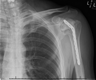
3. The Shoulder: Arthroscopic vs. Open Drainage
- Arthroscopic Approach: Utilizes standard posterior viewing and anterior working portals. Excellent for early infections. Allows thorough irrigation of the glenohumeral joint and subacromial space.
- Open Approach (Deltopectoral): Indicated for delayed presentations with thick purulence or osteomyelitis of the proximal humerus. The subscapularis is tenotomized or peeled to access the joint. As with the hip, the capsule is left partially open over a drain to prevent re-accumulation.
POSTOPERATIVE PROTOCOL AND REHABILITATION
The postoperative phase is as critical as the surgical intervention. Adjuvant therapies such as systemic salicylate administration or continuous passive motion (CPM), while theoretically beneficial for cartilage nutrition, have not demonstrated statistically significant improvements in long-term outcomes.
Phase I: Protection and Inflammation Control (Days 1-7)
- Splinting: The joint should be initially splinted in a position of function to prevent contractures (e.g., knee in full extension, ankle in neutral, wrist in slight extension).
- Monitoring: The patient must be closely observed for resolution of systemic inflammatory signs (fever, tachycardia) and local signs (erythema, swelling). Serial CRP levels should be tracked; a failure of CRP to halve every 48-72 hours post-drainage suggests retained purulence requiring re-operation.
Phase II: Early Mobilization (Weeks 1-3)
- As the acute infection resolves and drain output ceases, rigid splinting is discontinued.
- Isometric Exercises: Initiated immediately to prevent profound muscle atrophy, which occurs rapidly in the presence of joint effusion (arthrogenic muscle inhibition).
- Active Range of Motion (ROM): Gentle, active, and active-assisted ROM exercises are begun to restore joint kinematics and promote cartilage nutrition through synovial fluid diffusion.
Phase III: Residual Deformity Management
Patients treated for delayed or severe infectious arthritis often present with varying degrees of joint contracture or deformity.
* Conservative Correction: Treatment with dynamic splints, serial casting, and sustained passive stretching may be required to overcome soft tissue contractures.
* Surgical Reconstruction: In the residual stage—where the infection has completely subsided but the joint is left with severe deformity, ankylosis, or secondary osteoarthritis—treatment is directed at functional restoration (e.g., corrective osteotomy, arthrodesis, or staged arthroplasty).
> Surgical Warning: When undertaking reconstructive procedures in a previously septic joint, the surgeon must maintain a high index of suspicion for latent infection. Preoperative aspiration, intraoperative frozen sections for PMN counts, and extended prophylactic antibiotic coverage are mandatory to prevent catastrophic reactivation of the dormant pathogen.
You Might Also Like