Posterior Hip Approach: Optimize the Aspect of the Trochanter
Key Takeaway
Here are the crucial details you must know about Posterior Hip Approach: Optimize the Aspect of the Trochanter. The posterior approach is the most common surgical method for hip joint access, offering easy visualization and preserving abductor power. Ideal for total hip replacements and revisions, its curved incision is centered on the posterior aspect of the greater trochanter. This technique ensures safe, quick access for multiple hip procedures, though it carries a potential dislocation risk.
As an Academic Orthopedic Surgeon and Medical Educator, I present a comprehensive review of the posterior hip approach, with a particular emphasis on optimizing the management of the greater trochanter and its associated musculature for enhanced patient outcomes.
Introduction and Epidemiology
The posterior approach to the hip, often referred to as the Southern approach and popularized by Moore, has long been a cornerstone in orthopaedic surgery for its versatility and efficacy in accessing the hip joint. Its widespread adoption stems from several key advantages that contribute to its popularity among surgeons for both primary and revision hip arthroplasty, as well as a spectrum of trauma and reconstructive procedures.
A significant benefit of the posterior approach is the relative ease, safety, and rapidity of joint exposure. It typically requires minimal surgical assistance, making it a practical choice in various operating theatre environments. Critically, this approach largely spares the abductor mechanism of the hip—comprising the gluteus medius and minimus—which translates to preserved abductor power in the immediate postoperative period, facilitating earlier mobilization and rehabilitation. Furthermore, the posterior approach offers unparalleled visualization of the femoral shaft, rendering it particularly advantageous for complex revision total hip arthroplasty (THA) cases where femoral component removal or revision is anticipated.
However, the posterior approach is not without its specific considerations. The access to the joint inherently involves division of the posterior capsule and, by tradition, the short external rotator muscles. Consequently, if a prosthetic dislocation occurs, it typically manifests with the classic mechanism of hip flexion, internal rotation, and adduction. Historical data suggest a potentially higher dislocation rate compared to certain anterior approaches, particularly when used in specific vulnerable populations, such as elderly, bedridden patients with femoral neck fractures who frequently adopt a flexed and adducted posture. Nevertheless, modern modifications involving meticulous capsular and short external rotator repair have significantly mitigated this risk, leading to comparable dislocation rates with other contemporary approaches in experienced hands. The critical aspect of optimizing outcomes lies in understanding and strategically managing the integrity of the posterior soft tissues and the greater trochanteric attachments.
The utility of the posterior approach spans a broad range of orthopaedic conditions:
1. Hemiarthroplasty for femoral neck fractures, a common application in elderly populations.
2. Total hip replacement, encompassing both primary and complex revision surgeries.
3. Open reduction and internal fixation of posterior acetabular fractures, where direct visualization is paramount.
4. Dependent drainage of hip sepsis, leveraging gravity for effective fluid evacuation.
5. Removal of loose bodies from the posterior compartment of the hip joint.
6. Pedicle bone grafting procedures requiring posterior access.
7. Open reduction of irreducible or unstable posterior hip dislocations.
The enduring relevance of the posterior approach in contemporary orthopaedic practice underscores the importance of a thorough understanding of its anatomical basis, surgical technique, and strategies for complication avoidance and management.
Surgical Anatomy and Biomechanics
A profound understanding of the surgical anatomy and biomechanics of the hip joint is fundamental to executing the posterior approach safely and effectively, particularly concerning the critical aspect of the greater trochanter.
Regional Anatomy Overview
The posterior approach navigates through distinct anatomical layers to expose the hip joint. The primary structures encountered include:
- Skin and Subcutaneous Tissue: The incision traverses the skin and subcutaneous fat, typically extending to the fascia lata.
- Fascia Lata and Gluteus Maximus: The fascia lata is incised, and the underlying gluteus maximus muscle is either split bluntly along its fibers or incised sharply. The gluteus maximus, innervated by the inferior gluteal nerve, is a powerful extensor and external rotator of the hip. Blunt splitting minimizes damage to its fibers and preserves its function.
- Deep External Rotators: This critical layer lies deep to the gluteus maximus and superficial to the posterior capsule. It consists of the piriformis, superior gemellus, obturator internus, inferior gemellus, and quadratus femoris. These muscles originate from the sacrum and ischium and insert onto the greater trochanter of the femur. Their primary function is external rotation of the hip; piriformis also acts as an abductor when the hip is flexed.
- The piriformis inserts into the superior facet of the greater trochanter. It is a key landmark, serving as a guide to the sciatic nerve.
- The superior and inferior gemelli flank the obturator internus tendon, which inserts into the medial surface of the greater trochanter.
- The quadratus femoris is the most inferior of the short external rotators, inserting into the quadrate tubercle and intertrochanteric crest. The nerve to the quadratus femoris typically courses anterior to the gemelli and obturator internus.
- Posterior Hip Capsule: Deep to the short external rotators lies the posterior capsule, a strong fibrous structure that provides static stability to the hip joint. It is reinforced by the ischiofemoral ligament and the zona orbicularis. Division of this capsule is essential for joint access.
Neurovascular Structures
Several neurovascular structures are at risk during the posterior approach:
- Sciatic Nerve: This is the most critical structure to protect. The sciatic nerve typically exits the pelvis below the piriformis muscle, running deep to the gluteus maximus and superficial to the deep external rotators. It lies medial to the proximal femur and is particularly vulnerable to direct trauma, excessive traction, or entrapment from hematoma or scar tissue. Its course varies relative to the piriformis, with variations occurring in a small percentage of individuals. Intraoperative identification and protection are paramount.
 - Superior Gluteal Nerve and Artery: These structures lie superior to the piriformis and innervate/supply the gluteus medius and minimus. They are generally superior to the dissection field but can be injured with overly extensive proximal retraction or dissection.
- Inferior Gluteal Nerve and Artery: These structures emerge inferior to the piriformis and supply the gluteus maximus. While the gluteus maximus is typically split, care must be taken to avoid injuring these vessels and nerve, particularly near the inferior border of the gluteus maximus.
- Medial Circumflex Femoral Artery: This artery typically runs anterior to the quadratus femoris. It is the main blood supply to the femoral head, particularly the retinacular branches. Injury during quadratus femoris release can compromise blood supply to the remaining femoral head (e.g., in hemiarthroplasty) or lead to significant hemorrhage.
The Greater Trochanter and its Biomechanical Significance
The greater trochanter is a crucial anatomical landmark and biomechanical fulcrum. It serves as the insertion point for the gluteus medius and minimus (on its lateral and superior facets) and the short external rotators (on its medial and superior aspects).
- Gluteus Medius and Minimus: These muscles are the primary abductors of the hip. Their attachments to the superior and lateral aspects of the greater trochanter are generally preserved in the posterior approach, which contributes to the rapid recovery of abductor strength post-surgery.
- Short External Rotators: These muscles, particularly the piriformis and obturator internus with the gemelli, insert onto the superior and medial aspects of the greater trochanter. Their detachment is a characteristic step of the posterior approach. The "optimization of the aspect of the trochanter" directly pertains to the meticulous management of these tendinous attachments. While their division is necessary for exposure, their careful repair during closure is paramount for restoring posterior hip stability and minimizing the risk of dislocation. These muscles also contribute to dynamic stability, and their absence or poor repair can theoretically influence functional outcomes, although the direct clinical impact beyond dislocation risk is debated.
Biomechanical Implications
The biomechanical rationale behind the posterior approach hinges on preserving the hip abductor mechanism. By splitting the gluteus maximus and detaching the short external rotators, the major abductors (gluteus medius and minimus) remain largely undisturbed. This contrasts with certain anterolateral approaches that may involve tenotomy or osteotomy of the abductor complex, potentially leading to a temporary or persistent abductor lurch.
However, the intentional division of the posterior capsule and short external rotators necessitates a compensatory strategy to maintain joint stability. Meticulous reattachment of these structures forms a critical component of modern posterior approach techniques, aiming to restore the natural posterior buttress against dislocating forces. The integrity of this repair significantly impacts the postoperative stability profile of the reconstructed hip.
Indications and Contraindications
The posterior hip approach offers broad utility across various orthopaedic pathologies. Careful patient selection and recognition of specific contraindications are crucial for optimizing outcomes.
Indications for the Posterior Hip Approach
The posterior approach is widely applicable due to its excellent exposure and versatility.
* Hemiarthroplasty: Particularly favored for displaced femoral neck fractures in elderly patients.
* Total Hip Arthroplasty Primary: A highly prevalent approach for elective THA due to good exposure, low abductor morbidity, and reproducibility.
* Total Hip Arthroplasty Revision: Especially useful when femoral component revision is anticipated, offering excellent visualization of the femoral shaft.
* Open Reduction and Internal Fixation (ORIF) of Posterior Acetabular Fractures: Provides direct access to the posterior column and posterior wall of the acetabulum.
* Dependent Drainage of Hip Sepsis: Allows for gravitational drainage of infected joint fluid.
* Removal of Loose Bodies from the Hip Joint: Particularly for bodies located in the posterior compartment.
* Open Reduction of Posterior Hip Dislocations: When closed reduction fails or is unstable.
* Femoral Head Osteonecrosis (FHO): For core decompression or arthroplasty.
* Non-union of Femoral Neck Fractures: Requiring arthroplasty.
* Certain Tumour Resections: Of the proximal femur or posterior acetabulum.
Contraindications for the Posterior Hip Approach
While versatile, specific situations may render the posterior approach less ideal or contraindicated.
* Absolute Contraindications:
* Active infection within the joint when the goal is primary arthroplasty (though it is indicated for drainage of sepsis).
* Severe posterior soft tissue compromise or extensive scarring from previous surgery or trauma that would preclude safe dissection or closure.
* Relative Contraindications:
* Patients with a high intrinsic risk of posterior dislocation (e.g., neurological disorders with spasticity, severe cognitive impairment, or specific anatomical variations that predispose to posterior instability).
* Extensive anterior acetabular pathology requiring primary anterior exposure.
* Significant surgeon preference or expertise with an alternative approach.
Summary of Operative and Non-Operative Indications
The following table summarizes common conditions addressed by the posterior hip approach and categorizes their typical management pathways.
| Indication Category | Specific Condition | Operative/Non-Operative | Rationale/Comments |
|---|---|---|---|
| Arthroplasty | Primary Total Hip Arthroplasty (THA) | Operative | Versatile, excellent exposure for both acetabulum and femur, preserves abductors. |
| Revision Total Hip Arthroplasty (THA) | Operative | Superior visualization of the femoral shaft for component extraction/insertion. | |
| Hemiarthroplasty (Femoral Neck Fx) | Operative | Common for displaced femoral neck fractures in older adults; straightforward and reproducible. | |
| Trauma | Posterior Acetabular Fracture | Operative | Direct access to posterior column and wall for anatomical reduction and stable fixation. |
| Posterior Hip Dislocation | Operative (if closed reduction fails/unstable) | For irreducible dislocations or to address associated injuries (e.g., labral tears, loose bodies). | |
| Infection | Hip Sepsis | Operative (drainage) | Allows for dependent drainage of purulent fluid and debridement of infected tissue. |
| Other Reconstructive | Loose Bodies in Hip Joint | Operative | Direct visualization for retrieval of intra-articular loose bodies. |
| Pedicle Bone Grafting Procedures | Operative | Provides access for various reconstructive bone grafting techniques. | |
| Femoral Head Osteonecrosis | Operative (arthroplasty or core decompression) | For advanced stages of FHO requiring arthroplasty or for core decompression in early stages. |
Pre Operative Planning and Patient Positioning
Thorough preoperative planning and meticulous patient positioning are foundational for a safe and efficient posterior hip approach, directly impacting surgical outcomes and the ability to optimize the aspect of the greater trochanter effectively.
Preoperative Planning
- Patient Assessment:
- Medical Co-morbidities: Comprehensive review of systemic health, particularly cardiovascular, pulmonary, renal, and endocrine status. Optimization of chronic conditions (e.g., diabetes, hypertension) is crucial.
- Neurological Status: Baseline assessment of sciatic nerve function to document any pre-existing deficits.
- Skin Integrity: Inspection of the operative hip and surrounding area for rashes, infections, or open wounds that might contraindicate surgery.
- Previous Surgeries: Detailed history of prior hip surgeries, including previous approaches, hardware, or complications. This informs incision planning and potential scar tissue management.
- Anticoagulation/Antiplatelet Agents: Strict protocols for discontinuation and bridging based on institutional guidelines and patient risk.
- Imaging Review:
- Standard Radiographs: Anteroposterior (AP) pelvis, lateral hip (frog-leg and/or cross-table) are essential for assessing joint morphology, osteophyte burden, bone quality, and any existing hardware.
- Computed Tomography (CT) Scan: Indicated for complex acetabular fractures, significant femoral deformity, or revision surgery to assess bone stock, implant loosening, and precise anatomical relationships.
- Magnetic Resonance Imaging (MRI): Useful for evaluating soft tissue pathology, avascular necrosis, or occult fractures.
- Templating: Crucial for preoperative planning of implant size, component position, leg length, and offset. Digital templating is now standard practice, allowing for precise estimation of reaming depth, cup inclination/anteversion, and femoral stem size.
- Risk Assessment and Prophylaxis:
- Venous Thromboembolism (VTE) Prophylaxis: Implementation of mechanical and/or pharmacological prophylaxis protocols based on patient risk factors and established guidelines.
- Antibiotic Prophylaxis: Administration of intravenous broad-spectrum antibiotics within one hour of incision, consistent with institutional protocols.
- Blood Management: Assessment of anemia, cross-matching blood products if anticipated blood loss is high.
- Anesthesia Consultation: Discussion with the anesthesia team regarding patient-specific risks, choice of anesthesia (general, spinal, epidural, regional blocks), and postoperative pain management strategies.
Patient Positioning
The patient is positioned in the true lateral decubitus position on the operating table, with the affected limb uppermost. This position is critical for providing optimal access to the posterior aspect of the hip and femur.
- Stability: The patient must be securely positioned using a beanbag or sacral posts to prevent inadvertent rolling. The torso should be stabilized by an anterior post at the sternum and a posterior post at the sacrum. The head and neck are supported in a neutral position.
- Limb Alignment:
- The affected (upper) limb is typically positioned with the hip slightly flexed (approximately 30-45 degrees), neutral rotation, and slight adduction. This combination helps to relax the gluteal muscles and bring the greater trochanter and posterior capsule into a more accessible plane.
- The contralateral (lower) limb is flexed at the hip and knee to create a stable base and allow the affected limb to drape freely without undue pressure. A pillow is placed between the knees to prevent contact and protect neurovascular structures.
- Padding of Bony Prominences: This is a crucial step, particularly in elderly or frail patients, to prevent pressure sores or nerve palsies.
- The lateral malleolus, fibular head, and knee of the bottom leg must be meticulously padded.
- The contralateral iliac crest, sacrum, and greater trochanter should also be protected.
- Draping: The limb is draped free to allow full range of motion during the surgical procedure (e.g., for dislocation and reduction maneuvers). The entire circumference of the affected thigh and pelvis is prepared and draped to provide a wide sterile field.


Appropriate positioning ensures surgical efficiency, minimizes patient morbidity, and sets the stage for optimal exposure and management of the greater trochanter and surrounding structures.
Detailed Surgical Approach and Technique
The posterior hip approach, while often described as the Kocher-Langenbeck for trauma, is typically a modified gluteus maximus splitting approach for arthroplasty. The meticulous execution of each step, particularly regarding the greater trochanter, is paramount for stability and functional outcomes.
Incision and Initial Dissection
- Skin Incision: A curvilinear or straight incision, typically 10-15 cm in length for primary arthroplasty, is centered over the greater trochanter. It extends proximally and slightly posteriorly towards the posterior superior iliac spine (PSIS) and distally along the line of the femoral shaft. For acetabular trauma, a longer, more posterior Kocher-Langenbeck incision may be used.
 - Subcutaneous Dissection: The subcutaneous tissue is incised down to the fascia lata. Skin flaps are raised sufficiently to expose the underlying gluteus maximus and the distal aspect of the iliotibial band.
- Fascia Lata Incision: The fascia lata is incised in line with the skin incision, distal to the greater trochanter, exposing the underlying gluteus maximus.
- Gluteus Maximus Splitting: The fibers of the gluteus maximus are bluntly split in the line of their fibers, which run obliquely from the sacrum to the IT band. This internervous plane is between the superior gluteal nerve (supplying gluteus medius/minimus, tensor fascia lata) and the inferior gluteal nerve (supplying gluteus maximus). This blunt dissection minimizes muscle trauma and preserves its innervation. The muscle edges are retracted with self-retaining retractors.
Exposure of the Short External Rotators and Sciatic Nerve Protection
- Identification of Deep Structures: Deep to the gluteus maximus, the layer of short external rotators and the sciatic nerve come into view. The piriformis muscle is typically the most superior of the short external rotators, inserting onto the superior aspect of the greater trochanter.
- Sciatic Nerve Protection: This is the most critical step for neurological preservation. The sciatic nerve usually emerges inferior to the piriformis and courses distally, lying deep to the gluteus maximus and superficial to the short external rotators. It is meticulously identified, protected, and gently retracted anteriorly and medially with a broad blunt retractor (e.g., a Charnley or bent Hohmann retractor). Care must be taken to avoid excessive traction or direct compression.


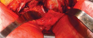
Optimization of the Aspect of the Trochanter: Short External Rotator Management
This is the core of "optimizing the aspect of the trochanter" in the posterior approach. The goal is to obtain sufficient exposure while preserving the potential for robust repair to enhance postoperative stability.
- Sequential Release: The short external rotators (piriformis, superior gemellus, obturator internus, inferior gemellus, and quadratus femoris) are sequentially released from their insertions on the greater trochanter.
- The piriformis tendon is usually released first, either with a cuff of bone or sharply at its insertion. This often marks the superior border of the tendinous insertions.
- The gemelli and obturator internus tendon complex are then released, typically as a single unit.
- The quadratus femoris is released last, distally. Care must be taken during the quadratus femoris release to identify and ligate/cauterize the ascending branch of the medial circumflex femoral artery, which lies on its anterior surface.
- Preserving a Tendon Cuff for Repair: To facilitate later repair, it is critical to leave a sufficient tendinous cuff attached to the greater trochanter. This cuff can be tagged with heavy non-absorbable sutures (e.g., #2 FiberWire, #5 Ethibond) for easier retrieval and repair at the end of the procedure. Alternatively, the tendons can be released sharply from the bone, and drill holes can be made in the trochanter to pass sutures through, allowing the tendon-muscle unit to be reattached directly to bone.
 - Capsulotomy: Once the short external rotators are detached and reflected, the posterior capsule of the hip joint is exposed. A T-shaped, H-shaped, or circumflex capsulotomy can be performed. The key is to create sufficient exposure for joint dislocation while planning for a robust capsular repair. A strong peripheral cuff of capsule should be left for reattachment. Tags are placed on the capsular flaps for later repair.

Joint Dislocation and Arthroplasty Procedures
- Dislocation: With the capsule opened and short rotators released, the hip joint is dislocated. This typically involves internally rotating, adducting, and flexing the hip. A posterior force on the femur helps to bring the femoral head out of the acetabulum.
- Femoral Head Resection: The femoral neck is resected at the planned level using an oscillating saw, guided by preoperative templating and intraoperative assessment.
- Acetabular Preparation: The acetabulum is exposed. The labrum and remaining cartilage are removed. Sequential reaming is performed to achieve the desired size and shape for the acetabular component, aiming for 40-45 degrees of inclination and 15-25 degrees of anteversion. The acetabular component is then implanted, ensuring secure fixation (press-fit or cemented).
- Femoral Preparation: The femoral canal is prepared using rasps or broaches, ensuring correct version and fit. The trial femoral stem and head are inserted.
- Trial Reduction: A trial reduction is performed to assess stability, leg length, and offset. Dynamic assessment through a full range of motion helps confirm stability, particularly in flexion, adduction, and internal rotation, which are the dislocating maneuvers for a posterior approach. Adjustments to implant size, neck length, or component position are made as needed.
Soft Tissue Repair and Wound Closure
This is where the "optimization of the aspect of the trochanter" is fully realized, directly influencing postoperative stability.
- Capsular Repair: The posterior capsule is meticulously repaired using strong, non-absorbable sutures. This can involve an imbrication technique, effectively tightening the capsule and reinforcing the posterior soft tissue envelope.
- Short External Rotator Repair: The released short external rotator tendons, ideally with the pre-placed sutures, are now reattached. They are either sutured back to their original insertion sites on the greater trochanter or advanced slightly and sutured to the posterior aspect of the capsule and the remaining tendinous cuff. This repair creates a robust posterior soft tissue barrier, significantly reducing the risk of posterior dislocation. The quality of this repair is a direct determinant of the need for strict postoperative hip precautions.
- Gluteus Maximus and Fascia Lata Closure: The gluteus maximus fibers are allowed to fall back into place, and the fascia lata is closed with strong absorbable sutures.
- Subcutaneous and Skin Closure: The subcutaneous layers are closed, and the skin is closed with staples or sutures. A drain may be placed in the deep wound, though this is often surgeon-dependent.

By meticulously managing the release and repair of the short external rotators and posterior capsule, the surgeon effectively optimizes the aspect of the trochanter, leveraging its anatomical position to create a stable, functional hip reconstruction with minimal long-term morbidity.
Complications and Management
Despite its effectiveness, the posterior hip approach, like all surgical procedures, carries potential complications. A comprehensive understanding of these risks, their incidence, and appropriate management strategies is vital for all orthopaedic surgeons.
General Surgical Complications
These are not specific to the posterior approach but are inherent risks of major orthopaedic surgery:
* Deep Vein Thrombosis (DVT) and Pulmonary Embolism (PE): Incidence significantly reduced with modern prophylaxis protocols.
* Surgical Site Infection (SSI): Can range from superficial to deep periprosthetic joint infection (PJI).
* Bleeding/Hematoma Formation: Requiring potential transfusion or evacuation.
* Anesthetic Complications: Related to general or regional anesthesia.
* Medical Complications: Myocardial infarction, stroke, renal failure, etc.
Specific Complications of the Posterior Hip Approach
- Dislocation:
- Incidence: Historically higher than with anterolateral approaches, often quoted at 1-5% for primary THA, but significantly reduced with modern soft tissue repair techniques to rates comparable with other approaches. Revision THA carries a higher risk.
- Predisposing Factors: Inadequate posterior soft tissue repair (capsule, short external rotators), component malposition (excessive anteversion of femoral component, excessive retroversion/verticality of acetabular component), patient non-compliance with precautions, neurological disorders, prior dislocation.
- Management:
- Acute Dislocation: Closed reduction under sedation/anesthesia. Assess stability after reduction.
- Recurrent Dislocation: Non-operative measures include bracing and physical therapy to strengthen periarticular muscles. Operative management may involve revision of malpositioned components, exchange to a constrained acetabular liner, femoral offset correction, capsular augmentation, or repeat meticulous repair of the posterior soft tissue envelope.
- Sciatic Nerve Palsy:
- Incidence: Rare, typically 0.1-2%.
- Predisposing Factors: Direct trauma (surgical instrument, suture entrapment), excessive traction during limb lengthening or dislocation maneuvers, compression from hematoma, cement extrusion.
- Management:
- Acute Postoperative: Immediate investigation (clinical assessment, electrophysiological studies).
- Observation: Many cases are traction-related neuropraxias and resolve spontaneously over weeks to months.
- Exploration: Indicated if there is suspicion of direct nerve transection, severe compression (e.g., from hematoma or cement), or no signs of recovery after a reasonable period.
- Rehabilitation: Foot drop brace if tibialis anterior is affected, physical therapy.
- Heterotopic Ossification (HO):
- Incidence: Clinical HO affecting function is less common (5-10%), but radiological evidence can be higher.
- Predisposing Factors: Male gender, ankylosing spondylitis, previous HO, hypertrophic osteoarthritis, diffuse idiopathic skeletal hyperostosis (DISH), history of traumatic brain injury.
- Management:
- Prophylaxis: Nonsteroidal anti-inflammatory drugs (NSAIDs) such as Indomethacin, or single-dose postoperative radiation therapy for high-risk patients.
- Excision: For symptomatic HO that restricts motion after skeletal maturity.
- Periprosthetic Fracture:
- Incidence: Approximately 0.1-1% intraoperatively, slightly higher postoperatively.
- Predisposing Factors: Osteoporosis, poor bone quality, tight femoral canal fit, impaction during stem insertion, revision surgery, stress shielding.
- Management: Dependent on fracture type (Vancouver classification for femoral, Paprosky for acetabular), stability of implants, and bone quality. May involve internal fixation (plates, cables, wires) with implant retention, or revision of the femoral or acetabular components.
- Abductor Insufficiency / Trochanteric Bursitis:
- Incidence: Less common than with direct lateral approaches that violate the gluteal insertion, but can occur if the gluteus medius/minimus are inadvertently damaged or if trochanteric pain develops.
- Predisposing Factors: Direct injury to gluteal tendons, trochanteric bursitis from irritation, retained hardware.
- Management: Physical therapy, anti-inflammatory medications, corticosteroid injections for bursitis. Rarely, revision surgery for severe abductor deficiency (e.g., abductor repair or transfer).
- Wound Healing Issues:
- Incidence: Relatively low.
- Predisposing Factors: Obesity, poor vascularity, smoking, diabetes, excessive tension on skin edges.
- Management: Local wound care, debridement if necrotic, antibiotics if infected. Rarely, flap coverage.
Table of Common Complications
| Complication | Incidence (approximate) | Predisposing Factors | Salvage Strategies |
|---|---|---|---|
| Dislocation | 1-5% (Primary THA) | Inadequate soft tissue repair, component malposition, patient non-compliance, neurological conditions | Closed reduction, bracing, revision (liner, components), constrained articulation, soft tissue repair augmentation |
| Sciatic Nerve Palsy | 0.1-2% | Direct trauma, excessive traction, hematoma, leg lengthening | Observation, bracing, neurolysis (rarely), exploration |
| Periprosthetic Fracture | 0.1-1% | Osteoporosis, impaction during stem insertion, revision surgery, stress shielding | ORIF (plates, wires), stem revision (Vancouver classification) |
| Deep Periprosthetic Infection | <1% | Co-morbidities, prolonged surgery, poor wound healing | Debridement and implant retention (DAIR), one/two-stage exchange arthroplasty, Girdlestone resection |
| Heterotopic Ossification | 5-10% (clinical) | Ankylosing spondylitis, previous HO, male gender, hyperostosis | Prophylaxis (NSAIDs, radiation), excision if symptomatic |
| DVT/Pulmonary Embolism | Low with prophylaxis | Advanced age, obesity, previous VTE, prolonged immobility | Anticoagulation, IVC filter (rarely) |
| Abductor Insufficiency | <1% (specific to approach) | Inadvertent gluteal damage, severe trochanteric bursitis | Physical therapy, anti-inflammatory medication, abductor repair/transfer |
Post Operative Rehabilitation Protocols
Postoperative rehabilitation following a posterior hip approach is crucial for achieving optimal functional outcomes, minimizing complications, and ensuring long-term prosthetic survivorship. The protocol is tailored to the individual patient, surgeon preference, implant stability, and the quality of the posterior soft tissue repair.
Phase I: Acute Postoperative (Days 1-7)
The primary goals of this phase are pain management, early mobilization, and adherence to hip precautions.
- Weight-Bearing Status: Generally, patients are encouraged to bear weight as tolerated (WBAT) immediately with uncemented components. Patients with cemented components are often full weight-bearing (FWB) immediately. Specific restrictions may be in place for complex cases (e.g., extensive revision, periprosthetic fracture fixation, or compromised bone quality).
- Hip Precautions: This is paramount for the posterior approach to prevent dislocation. Patients are instructed to avoid:
- Hip flexion beyond 90 degrees.
- Internal rotation past neutral.
- Adduction past midline.
- Specific instructions include using an elevated toilet seat, avoiding crossing legs, and sleeping on the back with an abductor pillow, particularly in the initial weeks. The duration and stringency of these precautions are directly influenced by the quality of the intraoperative posterior capsular and short external rotator repair. A meticulously repaired posterior soft tissue envelope may allow for less restrictive precautions.
- Early Mobilization:
- Bed Mobility: Assisted bed transfers and repositioning.
- Transfers: Supervised sit-to-stand and commode transfers.
- Gait Training: Initiation of ambulation with assistive devices (walker, crutches) with emphasis on proper gait mechanics and hip precaution adherence.
- Pain Management: Multimodal analgesia including oral opioids, NSAIDs (if not contraindicated), acetaminophen, and nerve blocks.
- Exercises: Gentle ankle pumps, quadriceps sets, gluteal sets to maintain muscle tone and promote circulation.
Phase II: Subacute/Intermediate (Weeks 2-6)
This phase focuses on gradual progression of strength, range of motion, and functional independence.
- Progressive Weight Bearing: If not already, progression to full weight-bearing as tolerated. Weaning from assistive devices as strength and balance improve.
- Increased Range of Motion: Gradual increase in active and passive range of motion within the limits of hip precautions.
- Strengthening Exercises:
- Isometric: Continued gluteal and quadriceps sets.
- Isotonic: Progressive resistance exercises for hip abductors, extensors, and flexors (within precautions). Exercises may include supine hip abduction, knee flexion, hip extension (prone or standing), and mini-squats.
- Core Strengthening: Essential for overall trunk stability and balance.
- Proprioception and Balance: Standing balance exercises, single-leg stance.
- Functional Training: Stair climbing, car transfers, dressing techniques while adhering to precautions.
- Education: Reinforcement of hip precautions and education on activity modification.
Phase III: Advanced/Return to Activity (Weeks 7-12 and beyond)
The final phase aims for restoration of full functional strength, endurance, and return to desired activities.
- Discontinuation of Precautions: Typically, hip precautions are relaxed or discontinued around 6-12 weeks, depending on the surgeon's preference, patient's progress, and the stability achieved by soft tissue repair. This decision is made after clinical and sometimes radiographic assessment.
- Advanced Strengthening: Progression to more challenging resistance exercises, incorporating functional movements. Focus on building endurance and power.
- Cardiovascular Conditioning: Stationary cycling, swimming, elliptical trainer.
- Return to Activity: Gradual return to light recreational activities (e.g., walking, golf). High-impact activities or contact sports are generally discouraged long-term for THA patients.
- Long-term Education: Patient education on activity modification, recognizing warning signs of complications, and importance of regular follow-up with the orthopaedic surgeon.
Key Considerations for Rehabilitation:
- Individualization: Protocols must be tailored to the patient's age, baseline function, comorbidities, and the specific surgical findings (e.g., quality of soft tissue repair).
- Communication: Close collaboration between the surgeon, physical therapist, and patient is crucial for optimal outcomes.
- Pain Management: Effective pain control facilitates participation in therapy.
The success of the posterior approach is not solely reliant on the surgical technique but equally on the rigorous and compliant execution of the postoperative rehabilitation protocol, particularly concerning the maintenance of hip stability and the restoration of muscular function.
Summary of Key Literature and Guidelines
The posterior hip approach has an extensive history in orthopaedic literature, with its efficacy, safety profile, and evolutionary modifications subject to continuous scrutiny and refinement. A review of key literature and guidelines highlights its enduring role and the factors influencing its outcomes.
Foundational Studies and Historical Context
The posterior approach gained prominence through the work of Moore in the mid-20th century. Early descriptions, often referred to as the "Southern approach" or modified Kocher-Langenbeck, established its utility for exposing the hip joint. Initially, a major concern with this approach was the relatively high dislocation rate, particularly in the era before routine capsular and short external rotator repair. These historical challenges spurred research into surgical modifications and postoperative management strategies.
Comparison with Other Approaches
Modern literature frequently compares the posterior approach to other contemporary approaches, primarily the direct anterior (DAA) and anterolateral approaches, across various outcome measures:
- Dislocation Rates: Numerous meta-analyses and systematic reviews have investigated dislocation rates. While some early studies and specific patient populations showed higher rates with the posterior approach, current evidence, particularly with meticulous capsular and short external rotator repair, demonstrates comparable dislocation rates among the different approaches in experienced hands. For instance, studies by Bozic et al. (2010) and Higgins et al. (2015) highlighted that while significant variations exist, surgeon experience and soft tissue repair are more critical determinants of dislocation than the approach itself.
- Functional Outcomes: Patient-reported outcome measures (PROMs) such as HOOS, WOMAC, and SF-12/36 often show no significant long-term differences in functional scores between approaches. However, DAA has been associated with a faster early recovery of gait and less early pain by some studies, while the posterior approach generally preserves abductor strength better than some lateral approaches.
- Complications: While specific complication profiles vary (e.g., sciatic nerve palsy risk with posterior, femoral nerve/lateral femoral cutaneous nerve injury with DAA, abductor weakness with lateral approaches), overall major complication rates tend to be similar across approaches when performed by experienced surgeons.
- Learning Curve: The posterior approach is generally considered to have a less steep learning curve than DAA, contributing to its widespread adoption.
The Role of Soft Tissue Repair
A consistent theme in contemporary literature is the critical importance of meticulous posterior soft tissue repair in mitigating dislocation risk. Repair of the posterior capsule and the short external rotators (piriformis, obturator internus, gemelli, quadratus femoris) is now considered a standard component of the posterior approach. Studies by Pellicci et al. (2005) and others have demonstrated that this repair significantly reduces dislocation rates, making them comparable to those of other approaches. This emphasizes the "optimization of the aspect of the trochanter" by focusing on the integrity of the attached muscle-tendon units.
Guidelines and Best Practices
Professional bodies, such as the American Academy of Orthopaedic Surgeons (AAOS) and the British Orthopaedic Association (BOA), issue guidelines that implicitly or explicitly support the use of the posterior approach. These guidelines typically focus on:
- Infection Prevention: Adherence to strict perioperative antibiotic protocols, skin preparation, and operating room sterility.
- VTE Prophylaxis: Use of mechanical and/or pharmacological methods based on patient risk assessment.
- Preoperative Planning: Emphasis on comprehensive patient evaluation, imaging, and templating.
- Surgical Technique: Highlighting the importance of anatomical reduction, stable fixation, and soft tissue balancing, with explicit or implicit support for techniques that enhance stability, such as posterior capsular and rotator repair.
- Postoperative Rehabilitation: Encouraging structured rehabilitation protocols tailored to the approach and individual patient needs.
Evolving Concepts and Future Directions
Research continues to explore refinements to the posterior approach, including:
- Enhanced Repair Techniques: Development of stronger, more reproducible methods for capsular and short external rotator repair.
- Minimally Invasive Posterior Approaches: While controversial regarding true benefits over traditional incisions, modifications aiming for smaller incisions continue to be explored, often with a trade-off in exposure.
- Robotic and Navigation Integration: While more commonly associated with DAA, robotic assistance could theoretically improve component positioning accuracy even with posterior approaches.
- Patient-Specific Approaches: The trend towards tailoring the surgical approach to individual patient anatomy, pathology, and surgeon expertise is growing.
In conclusion, the posterior hip approach remains a highly effective and widely utilized method for hip arthroplasty and other reconstructive procedures. Its long-term success is underpinned by a profound understanding of surgical anatomy, meticulous execution of the surgical technique—especially concerning the optimization of the trochanteric soft tissue attachments—and adherence to evidence-based postoperative rehabilitation protocols and complication management strategies. The continuous evolution of surgical techniques and rigorous academic scrutiny ensures its ongoing relevance and efficacy in modern orthopaedic practice.
Clinical & Radiographic Imaging


You Might Also Like