Demystify Posterior Approaches to the Hip: Essential Anatomy
Key Takeaway
Here are the crucial details you must know about Demystify Posterior Approaches to the Hip: Essential Anatomy. Posterior approaches to the hip involve navigating two primary muscle layers: an outer layer comprising the gluteus maximus and an inner layer of short external rotators (e.g., piriformis). These approaches to the hip can utilize internervous planes, like the Marcy–Fletcher, or involve splitting gluteus maximus fibers, such as the Moore approach, which often provides excellent joint exposure.
Introduction and Epidemiology
The posterior approach to the hip remains the most universally applied and versatile surgical exposure in orthopedic surgery. Often referred to as the Southern approach or the Moore approach in the context of arthroplasty, and the Kocher-Langenbeck approach in the context of pelvic and acetabular trauma, this exposure provides unparalleled access to the posterior column of the acetabulum, the posterior hip capsule, and the proximal femur.
Epidemiologically, the posterior approach is utilized in the majority of primary and revision total hip arthroplasties (THAs) performed globally. Despite the rising popularity of the direct anterior approach (DAA), the posterior approach remains the workhorse due to its extensile nature, relatively short learning curve, and the ability to seamlessly extend the incision proximally or distally to address complex reconstructive challenges or intraoperative complications. Furthermore, it is the gold standard for the operative management of posterior wall and posterior column acetabular fractures, as well as for the excision of intra-articular loose bodies and tumors of the posterior hip joint.
The historical evolution of this approach reflects a continuous refinement of anatomical understanding. Early iterations by von Langenbeck and Kocher focused on wide exposure for trauma and infection. Later modifications by Moore and Osborne popularized the muscle-splitting technique through the gluteus maximus for arthroplasty. Conversely, the Marcy-Fletcher modification sought to exploit a true internervous plane. Understanding these variations and their underlying applied surgical anatomy is paramount for minimizing morbidity, optimizing implant positioning, and preventing complications such as posterior dislocation and iatrogenic nerve injury.
Surgical Anatomy and Biomechanics
A profound comprehension of the posterior hip anatomy is non-negotiable for the orthopedic surgeon. The musculature covering the posterior aspect of the hip joint is fundamentally organized into two distinct sheaths or layers: the superficial outer layer and the deep inner layer.
The Superficial Muscular Layer
The outer layer is dominated by the gluteus maximus, a massive, coarse-fibered muscle that dictates the initial stages of the surgical exposure. The gluteus maximus sits upon the deeper structures of the buttock analogous to the front cover of a book.

Gluteus Maximus Anatomy
* Origin: The posterior gluteal line of the ilium and the bone immediately above and behind it; the posterior surface of the lower sacrum and the side of the coccyx; and the aponeurotic fascia covering the gluteus medius.
* Insertion: The deeper, lower half of the muscle inserts into the gluteal tuberosity of the proximal femur. The superficial and superior portions insert into the iliotibial band of the fascia lata.
* Action: It is a powerful extensor and lateral rotator of the thigh, crucial for rising from a seated position and climbing stairs.
* Nerve Supply: Inferior gluteal nerve (L5, S1, S2).
Also inserting into the iliotibial band, but positioned further anteriorly, is the tensor fasciae latae. Together, the gluteus maximus, the intervening fascia lata (which envelopes the gluteus medius), and the tensor fasciae latae form a continuous, robust fibromuscular sheath. This constitutes the outer layer of the hip musculature. As described by A.K. Henry, this layer can be conceptualized as the "pelvic deltoid," as it drapes over the hip joint in a manner functionally and anatomically similar to how the deltoid muscle covers the glenohumeral joint.
The Deep Muscular Layer
Deep to the gluteus maximus lies the inner layer, which comprises the short external rotators of the hip. These muscles provide dynamic stability to the posterior hip joint and serve as critical anatomical landmarks during surgical dissection.

Gluteus Minimus and Piriformis Anatomy
* Gluteus Minimus: Originates from the outer surface of the ilium between the anterior and inferior gluteal lines. It inserts into the anterior border of the greater trochanter via a tendon that blends with the joint capsule. Supplied by the superior gluteal nerve, it acts to abduct and medially rotate the thigh.
* Piriformis: Originates from the anterior surface of the sacrum (S2-S4 segments) via fleshy digitations. It exits the pelvis through the greater sciatic foramen and inserts into the superior border of the greater trochanter. It is the key to the posterior anatomical organization.
Inferior to the piriformis lies the triceps coxae (superior gemellus, obturator internus, and inferior gemellus) and the quadratus femoris. The obturator internus tendon is a robust structure that acts as a sling, providing a reliable tissue layer for subsequent posterior capsular repair.
Neurovascular Anatomy
The sciatic nerve is the most critical neurovascular structure encountered during the posterior approach. It typically exits the pelvis through the greater sciatic foramen, inferior to the piriformis muscle. However, anatomical variations exist (e.g., the nerve or its peroneal division piercing or exiting superior to the piriformis in approximately 15% of the population). The nerve runs vertically down the thigh, situated between the superficial gluteus maximus and the deep short external rotators.

The vascular supply to the femoral head is predominantly derived from the medial femoral circumflex artery (MFCA). The ascending branch of the MFCA courses anterior to the quadratus femoris and posterior to the obturator externus. Protecting the obturator externus and avoiding aggressive deep dissection at the superior border of the quadratus femoris is imperative to preserve the residual blood supply to the femoral head, particularly in procedures like surgical dislocation or internal fixation of femoral neck fractures.
Internervous Planes and Approach Variations
The outer layer can be breached at different points, dictating the specific variation of the posterior approach.
The most anatomically physiological separation is the Marcy-Fletcher approach. This utilizes a true internervous plane located at the anterior border of the gluteus maximus, developing the interval between the gluteus maximus (innervated by the inferior gluteal nerve) and the gluteus medius (innervated by the superior gluteal nerve).
Conversely, the more commonly utilized posterior approaches (such as the Moore approach and the Osborne approach) involve a blunt longitudinal splitting of the gluteus maximus fibers. While these do not exploit a true internervous plane, they are favored due to their simplicity, the robust nature of the gluteus maximus which tolerates splitting well, and the unparalleled, direct exposure they provide to the posterior joint capsule and acetabulum.

Indications and Contraindications
The posterior approach is highly versatile, but patient selection and pathology dictate its appropriateness. It is the approach of choice for pathologies requiring extensive visualization of the posterior column or when preservation of the abductor mechanism is a primary concern.
Operative Indications
- Primary and Revision Total Hip Arthroplasty (THA)
- Hemiarthroplasty for displaced femoral neck fractures
- Open Reduction and Internal Fixation (ORIF) of posterior wall and posterior column acetabular fractures
- Treatment of Pipkin fractures (femoral head fractures associated with posterior dislocation)
- Surgical drainage of septic arthritis of the hip
- Excision of posterior intra-articular loose bodies or synovial chondromatosis
- Tumor resection involving the posterior proximal femur or posterior acetabulum
Contraindications
Relative and absolute contraindications depend heavily on the specific procedure being performed. For acetabular trauma, an isolated anterior column or anterior wall fracture cannot be addressed via a posterior Kocher-Langenbeck approach; an ilioinguinal or Stoppa approach is required. In arthroplasty, severe fixed flexion contractures may be more easily addressed via an anterior approach, though a posterior approach can still be utilized with appropriate capsular releases.
Management Matrix for Hip Pathology
| Pathology / Condition | Operative Indication (Posterior Approach) | Non-Operative Management |
|---|---|---|
| End-Stage Osteoarthritis | Unremitting pain, functional decline, radiographic joint space narrowing. | NSAIDs, physical therapy, intra-articular corticosteroid injections, weight loss. |
| Posterior Wall Acetabular Fracture | >20% wall involvement, marginal impaction, hip joint instability, intra-articular fragments. | <20% wall involvement, congruent joint on dynamic stress fluoroscopy, stable hip. |
| Displaced Femoral Neck Fracture (Elderly) | Displaced intracapsular fracture (Garden III/IV) requiring hemiarthroplasty or THA. | Extremely high surgical risk patients (comfort care only). |
| Femoral Head Fracture (Pipkin) | Pipkin II (superior to fovea), Pipkin III (with femoral neck fx), Pipkin IV (with acetabular fx). | Pipkin I (inferior to fovea) with anatomic closed reduction and stable joint. |
| Septic Arthritis | Aspiration confirming purulence; requires emergent arthrotomy and washout. | N/A - Surgical emergency. |
Pre Operative Planning and Patient Positioning
Thorough preoperative planning is essential for anticipating anatomical distortions, sizing implants, and determining the necessary extent of the surgical exposure. Standard radiographic evaluation includes an anteroposterior (AP) view of the pelvis and an AP and lateral view of the affected hip. For acetabular fractures, Judet views (iliac and obturator obliques) and a fine-cut computed tomography (CT) scan with 3D reconstructions are mandatory to delineate fracture morphology and plan plate trajectories.
Patient Positioning
The patient is almost universally placed in the lateral decubitus position for arthroplasty and most trauma applications.
- Table Setup: A standard radiolucent operating table is utilized.
- Stabilization: The patient is stabilized using a pegboard system, a beanbag, or specialized pelvic positioners. It is critical that the anterior superior iliac spine (ASIS) and the symphysis pubis are securely supported anteriorly, and the sacrum is supported posteriorly to prevent pelvic roll during the procedure. Pelvic roll can lead to inaccurate assessment of acetabular component version and inclination.
- Padding: An axillary roll is placed under the dependent thorax to protect the brachial plexus. All bony prominences (e.g., the dependent fibular head to protect the common peroneal nerve) must be meticulously padded.
- Draping: The operative leg is prepped and draped free to allow for full range of motion, which is necessary for dislocation, reduction, and dynamic assessment of stability.

Detailed Surgical Approach and Technique
The execution of the posterior approach requires a systematic, layered dissection, respecting neurovascular boundaries and optimizing soft tissue handling to facilitate subsequent repair.
Skin Incision and Superficial Dissection
The skin incision is centered over the posterior aspect of the greater trochanter. It begins approximately 5 to 8 centimeters proximal to the tip of the greater trochanter, curving gently posteriorly toward the posterior superior iliac spine (PSIS). Distally, the incision extends longitudinally down the lateral shaft of the femur for approximately 10 centimeters.
Subcutaneous tissues are divided in line with the skin incision to expose the fascia lata distally and the gluteal aponeurosis proximally. The fascia lata is incised longitudinally over the center of the greater trochanter. Proximally, this fascial incision is directed posteriorly, splitting the fibers of the gluteus maximus bluntly. This muscle-splitting technique is relatively avascular if kept within the substance of the muscle, though branches of the inferior gluteal artery may be encountered and require electrocautery.
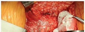
Deep Dissection and Identification of Structures
A Charnley or self-retaining retractor is placed beneath the fascial flaps to maintain exposure. The deep layer of the hip is now visible, obscured by a layer of areolar tissue. Internal rotation of the leg places the short external rotators under tension, facilitating their identification.
The piriformis tendon is identified superiorly. The sciatic nerve must be routinely palpated and visually identified as it emerges inferior to the piriformis and courses superficially over the obturator internus and gemelli. While formal neurolysis is rarely indicated in primary arthroplasty, identifying the nerve prevents inadvertent injury during retractor placement.

Release of the Short External Rotators
Stay sutures are placed in the conjoined tendon of the short external rotators (piriformis, superior gemellus, obturator internus, and inferior gemellus) close to their insertion on the greater trochanter. These muscles are then transected adjacent to their insertion. The tagged muscles are reflected posteriorly over the sciatic nerve; this maneuver provides a protective soft-tissue cushion for the nerve during the remainder of the procedure.
The quadratus femoris is typically left intact unless extensive inferior exposure is required. If release is necessary, it should be done cautiously, leaving a cuff of tissue at the trochanteric insertion, and avoiding deep dissection to protect the ascending branch of the medial femoral circumflex artery.
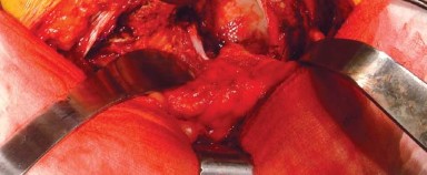
Capsulotomy and Dislocation
With the short external rotators reflected, the posterior joint capsule is exposed. A capsulotomy (T-shaped, H-shaped, or L-shaped) or a capsulectomy is performed based on surgeon preference and the underlying pathology. For arthroplasty, preserving the capsule for later repair is strongly advocated to reduce dislocation rates.
The hip is then dislocated posteriorly through a combination of flexion, adduction, and progressive internal rotation. Care must be taken during this maneuver; excessive torsional force in osteoporotic bone can result in an iatrogenic spiral fracture of the femoral shaft.
Closure and Enhanced Soft Tissue Repair
Following the definitive procedure (e.g., component implantation or fracture fixation), meticulous closure is critical. The "enhanced posterior soft tissue repair" involves the robust reattachment of the posterior capsule and the short external rotators to the greater trochanter, often via transosseous drill holes. This step significantly restores posterior soft-tissue tension and has drastically reduced the incidence of postoperative posterior dislocation. The fascia lata and gluteal aponeurosis are then closed with heavy, interrupted or continuous sutures, followed by standard subcutaneous and skin closure.
Complications and Management
While the posterior approach is safe and reproducible, it carries specific risks inherent to the regional anatomy. Anticipation, meticulous technique, and prompt recognition are required to manage these complications effectively.
Common Complications
The most feared complication specific to this exposure is iatrogenic injury to the sciatic nerve. The peroneal division is particularly susceptible to traction injury due to its lateral position and relative tethering at the fibular head. Retractor misplacement (e.g., placing a Hohmann retractor directly on the nerve instead of the bone) or excessive tension during anterior retraction of the femur can cause neuropraxia or axonotmesis.
Posterior dislocation is another historically significant complication. Because the posterior capsular and muscular restraints are compromised during the approach, the hip is inherently unstable in flexion, adduction, and internal rotation.
Complications Incidence and Salvage Strategies
| Complication | Estimated Incidence | Etiology / Risk Factors | Salvage / Management Strategy |
|---|---|---|---|
| Sciatic Nerve Palsy | 1% - 3% (Primary THA); up to 10% in complex trauma/revision. | Traction from retractors, direct laceration, hematoma compression, limb lengthening >4cm. | Immediate: Remove retractors, ensure no impingement. Postop: AFO for foot drop, gabapentinoids, EMG at 6 weeks if no recovery. Evacuate expanding hematomas emergently. |
| Posterior Dislocation | 1% - 4% (Historically higher; reduced with enhanced repair). | Failure of soft tissue repair, component malposition (retroversion), patient non-compliance. | Closed reduction under sedation. Revision surgery for recurrent instability (addressing component version, using dual mobility or constrained liners). |
| Heterotopic Ossification (HO) | 15% - 50% (Radiographic); 2% - 5% (Clinically significant). | Muscle trauma (gluteus maximus splitting), bone debris, male gender, ankylosing spondylitis. | Prophylaxis: Indomethacin or single-dose localized radiation. Treatment: Surgical excision once bone is mature (typically >6-12 months) and margins are sharp on CT. |
| Vascular Injury (Bleeding) | < 1% (Major vessel); Common (Minor vessels). | Laceration of inferior gluteal artery branches or medial femoral circumflex artery. | Meticulous intraoperative hemostasis. Ligation of bleeding vessels. Avoid blind clamping deep in the sciatic notch to prevent nerve injury. |
| Deep Surgical Site Infection | 0.5% - 2% | Prolonged operative time, obesity, diabetes, immunosuppression. | Acute (<4 weeks): DAIR (Debridement, Antibiotics, Implant Retention). Chronic: Two-stage revision arthroplasty. |
Post Operative Rehabilitation Protocols
Rehabilitation following a posterior approach is tailored to protect the healing posterior soft tissues while maximizing early mobilization to prevent thromboembolic and pulmonary complications.
Acute Postoperative Phase (0-6 Weeks)
The hallmark of the early rehabilitation phase is the strict adherence to "posterior hip precautions." Patients are instructed to avoid the combined positions of:
1. Hip flexion greater than 90 degrees.
2. Adduction of the operative leg past the midline of the body.
3. Internal rotation of the operative leg.
These precautions are typically maintained for 6 weeks to allow the transosseous repair of the capsule and short external rotators to heal. Weight-bearing status is procedure-dependent. Uncemented THAs and most cemented THAs are allowed weight-bearing as tolerated (WBAT) immediately. Conversely, patients who have undergone ORIF for an acetabular fracture are typically restricted to touch-down weight-bearing (TDWB) or flat-foot weight-bearing (FFWB) for 6 to 12 weeks to prevent fracture displacement.
Intermediate and Advanced Phases
Phase II (6-12 weeks) focuses on restoring normal gait mechanics, weaning off assistive devices, and initiating abductor strengthening. Isometric gluteal sets and progressive resistance exercises are introduced. Phase III (3-6 months) involves a return to higher-level functional activities. While low-impact activities (swimming, cycling, golf) are encouraged, high-impact activities (running, jumping) are generally discouraged following arthroplasty to minimize bearing surface wear and aseptic loosening.
Summary of Key Literature and Guidelines
The academic foundation of the posterior approach is built upon several seminal papers and evolving modern literature.
- Anatomical Foundations: The classic descriptions by Moore (1957) and Osborne (1930) established the utility of the gluteus maximus splitting approach for arthroplasty. Marcy and Fletcher (1954) contributed significantly by delineating the internervous plane between the gluteus maximus and medius, though the muscle-splitting approach remains more prevalent due to its superior exposure of the distal posterior column.
- Soft Tissue Repair: Pellicci et al. (1998) published a landmark study demonstrating that an enhanced posterior soft tissue repair (repairing the capsule and short external rotators to the greater trochanter) drastically reduced the dislocation rate following posterior THA from approximately 4% to less than 1%. This has become the standard of care globally.
- Approach Comparisons: Modern orthopedic literature frequently compares the posterior approach to the direct anterior approach (DAA). Meta-analyses (e.g., Higgins et al., 2015) generally demonstrate that while the DAA may offer slightly faster early functional recovery in the first 2 to 6 weeks, functional outcomes, pain scores, and complication rates equalize by 6 months to 1 year. The posterior approach consistently demonstrates shorter operative times and lower rates of intraoperative femoral fractures compared to the DAA.
- Trauma Guidelines: For acetabular fractures, the guidelines established by Letournel and Judet remain the definitive reference. They dictate that the Kocher-Langenbeck approach is the mandatory exposure for posterior wall, posterior column, and certain transverse or T-type fractures where the posterior displacement is dominant.
Mastery of the posterior approach requires a synthesis of this three-dimensional anatomy, respect for the biomechanical envelope of the hip, and strict adherence to evidence-based surgical techniques and postoperative protocols.
You Might Also Like