Orthopedic Ob Shoulder And Elb Review | Dr Hutaif Shoul -...
20 Jun 2026
57 min read
173 Views
Key Takeaway
This interactive board review contains 100 randomly selected orthopedic surgery questions with clinical images, immediate feedback, and detailed references.
Orthopedic Ob Shoulder And Elb Review | Dr Hutaif Shoul -...
Comprehensive 100-Question Exam
00:00
Start Quiz
Question 1
A 22-year-old male rugby player presents with his fourth anterior shoulder dislocation. A pre-operative CT scan demonstrates 25% anterior glenoid bone loss. Which of the following surgical interventions provides the most biomechanically sound and durable stabilization for this patient?
Explanation
The Latarjet procedure involves transferring the coracoid process with the attached conjoined tendon to the anterior glenoid neck. It is indicated in patients with recurrent anterior shoulder instability who have critical glenoid bone loss (typically >20-25%). It provides a triple blocking effect: bone block, sling effect of the conjoined tendon, and capsular repair.
Question 2
A 45-year-old female falls from a ladder and sustains an injury to her right elbow. Radiographs similar to the typical appearance of this injury pattern
demonstrate a posterior elbow dislocation, a radial head fracture, and a coronoid fracture. According to the standard lateral-to-medial surgical protocol (e.g., Pugh et al.), what is the correct sequence of structural fixation?
demonstrate a posterior elbow dislocation, a radial head fracture, and a coronoid fracture. According to the standard lateral-to-medial surgical protocol (e.g., Pugh et al.), what is the correct sequence of structural fixation?
Explanation
In the surgical management of a terrible triad injury of the elbow, a deep-to-superficial, anterior-to-posterior, or medial-to-lateral progression is generally utilized. The widely accepted standard sequence from a lateral or global approach is: 1) fixation of the coronoid fracture and anterior capsule, 2) fixation or replacement of the radial head, and 3) repair of the lateral ulnar collateral ligament (LUCL). If the elbow remains unstable after this sequence, the MCL may be explored and repaired.
Question 3
A 74-year-old male complains of right shoulder pain and an inability to elevate his arm actively beyond 30 degrees (pseudoparalysis). Radiographs demonstrate superior migration of the humeral head with severe acromiohumeral narrowing (Hamada grade 4). MRI confirms a massive, retracted, unrepairable tear of the supraspinatus and infraspinatus with fatty infiltration. The deltoid muscle is fully intact, and axillary nerve function is normal. What is the most appropriate surgical treatment?
Explanation
Reverse total shoulder arthroplasty (RTSA) is the gold standard for elderly patients with rotator cuff arthropathy and pseudoparalysis, provided the deltoid and axillary nerve are intact. By medializing the center of rotation and placing the humerus distally, it increases the deltoid moment arm, allowing for functional arm elevation despite a deficient rotator cuff.
Question 4
A 6-year-old boy falls off monkey bars and sustains an extension-type Gartland III supracondylar humerus fracture. On evaluation in the ER, his hand is pink but the radial pulse is absent. The patient is taken emergently to the OR for closed reduction and percutaneous pinning. Post-operatively, the limb remains well-perfused (pink) with brisk capillary refill, but the radial pulse remains non-palpable. What is the most appropriate next step in management?
Explanation
A 'pink, pulseless' hand following reduction of a supracondylar humerus fracture is a well-recognized clinical scenario. Provided that the hand is definitively warm, pink, and has capillary refill < 2 seconds, collateral circulation is deemed adequate. The recommended management is observation and close monitoring for 24-48 hours. Open exploration is indicated only for a 'white, pulseless' hand or if signs of ischemia develop.
Question 5
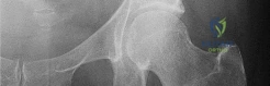
A 35-year-old male presents to the ER following a first-time generalized tonic-clonic seizure. He complains of right shoulder pain and his arm is locked in adduction and internal rotation. Passive external rotation is severely restricted. An AP radiograph shows a 'lightbulb' sign
. What is the most common associated bony defect seen with this specific injury pattern?
. What is the most common associated bony defect seen with this specific injury pattern?
Explanation
The patient has a posterior shoulder dislocation, classically associated with seizures, electrocution, or high-energy trauma. The 'lightbulb' sign is typical on AP radiographs due to internal rotation of the humerus. The most common associated bony defect is a reverse Hill-Sachs lesion, which is an impaction fracture of the anteromedial aspect of the humeral head against the posterior glenoid rim.
Question 6
A Monteggia fracture-dislocation consists of a fracture of the ulnar diaphysis and dislocation of the radial head. According to the Bado classification, which type is characterized by posterior or posterolateral dislocation of the radial head with an apex-posterior angulated diaphyseal ulnar fracture, and is most commonly seen in adult populations?
Explanation
The Bado classification categorizes Monteggia fractures based on the direction of radial head dislocation. Type I (anterior dislocation, most common in children), Type II (posterior/posterolateral dislocation, most common in adults), Type III (lateral dislocation), and Type IV (associated with both radius and ulna fractures). Therefore, Type II matches the description.
Question 7
A 12-year-old gymnast falls during a tumbling routine and sustains an acute right elbow dislocation. In the emergency department, a closed reduction is performed successfully. Post-reduction radiographs reveal a medial epicondyle fracture. Which of the following is considered an absolute indication for open reduction and internal fixation (ORIF) of the medial epicondyle?
Explanation
Absolute indications for ORIF of a medial epicondyle fracture include an incarcerated fragment within the elbow joint that cannot be extracted closed, and an open fracture. Relative indications include displacement > 5 mm, ulnar nerve dysfunction, and high-demand athletes requiring valgus stability. Incarceration within the joint is the only absolute indication among the choices.
Question 8
A 28-year-old competitive weightlifter feels a sudden 'pop' and sharp pain in his anterior chest wall while performing a heavy bench press. On examination, he has extensive ecchymosis over the anterior axillary fold and weakness in internal rotation and adduction. The vast majority of pectoralis major muscle ruptures occur at which specific anatomical location?
Explanation
The vast majority of pectoralis major ruptures occur at the tendinous insertion onto the proximal humerus, specifically involving the sternocostal head. This injury typically occurs during eccentric loading (e.g., the descent phase of a bench press). Acute surgical repair yields the best functional outcomes for athletes.
Question 9
A 30-year-old cyclist falls directly onto the point of his shoulder. Radiographs demonstrate an acromioclavicular (AC) joint separation where the clavicle is elevated by 150% relative to the acromion. He is diagnosed with a Rockwood Type V injury. Which of the following structures are disrupted in this specific injury grade?
Explanation
Rockwood Type V AC separations involve disruption of both the AC ligaments and the CC ligaments, along with significant stripping and disruption of the deltotrapezial fascia from the distal clavicle. This extreme displacement (>100% to 300% superiorly) requires surgical reconstruction for optimal functional recovery.
Question 10
A 45-year-old male undergoes a single-incision anterior approach for repair of a complete distal biceps tendon rupture. During the post-operative follow-up, he complains of numbness and tingling over the lateral aspect of his proximal forearm. Which nerve was most likely injured or stretched by retraction during this procedure?
Explanation
The lateral antebrachial cutaneous nerve (LABCN) is the most frequently injured nerve during a single-incision anterior approach for distal biceps tendon repair due to its superficial course lateral to the biceps tendon. While the posterior interosseous nerve (PIN) is at risk, it is more classically injured during the second (lateral) incision of a two-incision approach if dissection splits the supinator incorrectly.
Question 11
A 30-year-old female falls onto an outstretched hand. A radiograph similar to the one shown
reveals a coronal shear fracture. According to the current classification systems, a coronal shear fracture of the capitellum that extends medially to include a large contiguous portion of the trochlea is classified as:
reveals a coronal shear fracture. According to the current classification systems, a coronal shear fracture of the capitellum that extends medially to include a large contiguous portion of the trochlea is classified as:
Explanation
The McKee modification describes a Type IV capitellar fracture, which is a coronal shear fracture that involves not only the capitellum but extends medially to include the lateral ridge and a significant portion of the trochlea. Type I (Hahn-Steinthal) involves a large fragment of the capitellum with subchondral bone. Type II (Kocher-Lorenz) is an articular cartilage fragment with minimal bone. Type III is a comminuted fracture.
Question 12
A 65-year-old osteoporotic female sustains a proximal humerus fracture.
According to the Hertel criteria, which combination of radiographic findings provides the most accurate and sensitive prediction of subsequent humeral head avascular necrosis (ischemia)?
According to the Hertel criteria, which combination of radiographic findings provides the most accurate and sensitive prediction of subsequent humeral head avascular necrosis (ischemia)?
Explanation
Hertel et al. (2004) identified specific criteria that strongly predict humeral head ischemia following proximal humerus fractures. The combination most predictive of avascular necrosis (positive predictive value of 97%) consists of: a short calcar segment attached to the articular surface (metaphyseal extension < 8 mm), disruption of the medial hinge (> 2 mm displacement), and an anatomic neck fracture pattern (separation of the articular segment from the tuberosities).
Question 13
A 70-year-old female with advanced, medication-refractory rheumatoid arthritis undergoes a primary semi-constrained (linked) total elbow arthroplasty (TEA). To prevent mechanical failure and aseptic loosening, she must be counseled to strictly observe which of the following lifetime weight-lifting restrictions for the operative arm?
Explanation
Patients undergoing total elbow arthroplasty must adhere to strict lifelong weight-lifting restrictions due to the risk of implant failure, bushing wear, and aseptic loosening at the cement-bone interface. The standard recommendation (such as the Mayo Clinic protocol) restricts the patient to a maximum of 5 to 10 pounds for a single event with the operative arm, and 1 to 2 pounds for repetitive activities.
Question 14
An 18-year-old football player is tackled forcefully on his shoulder while lying on the ground. He presents to the ER with severe pain at the base of his anterior neck, dysphagia, and difficulty breathing. His right arm is supported across his chest. What is the most appropriate initial imaging modality to definitively diagnose the specific displacement of this injury?
Explanation
The patient's clinical presentation (dysphagia, shortness of breath, mechanism of injury) is highly suspicious for a posterior sternoclavicular (SC) joint dislocation, which is a surgical emergency if there is mediastinal impingement. While a serendipity view can demonstrate asymmetry, a CT scan of the chest is the gold standard imaging modality to evaluate the exact position of the medial clavicle and its proximity to vital mediastinal structures (trachea, esophagus, great vessels).
Question 15
A 25-year-old motorcyclist is involved in a high-speed collision. He presents to the trauma bay with massive swelling over the left shoulder, an absent left radial pulse, and complete flaccidity and anesthesia of the left upper extremity. Radiographs demonstrate an intact clavicle but massive lateral displacement of the scapula relative to the thoracic spine. What is the most appropriate next step in the definitive management of this patient?
Explanation
This patient has scapulothoracic dissociation, characterized by massive lateral displacement of the scapula with highly associated catastrophic vascular (subclavian or axillary artery) and neurologic (complete brachial plexus avulsion) injuries. Given the absent pulse, the immediate next step must be vascular evaluation via CT angiography or emergent vascular surgery consultation to restore perfusion. Fixing the bone or exploring the plexus immediately takes lower priority to saving the limb from ischemia.
Question 16
A 32-year-old male bodybuilder complains of medial elbow pain and an audible, palpable snapping sensation when performing triceps extensions. Examination reveals a 'double snap' over the medial epicondyle as the elbow is moved from extension into flexion. Which two anatomical structures are most likely subluxating over the medial epicondyle?
Explanation
Snapping triceps syndrome typically involves the dynamic subluxation of the ulnar nerve followed by the medial head of the triceps over the medial epicondyle during elbow flexion. This creates a distinct 'double snap' on examination. Treatment often requires ulnar nerve transposition and concurrent resection or management of the prominent medial head of the triceps.
Question 17
A 40-year-old male presents with weakness in his right shoulder that began 3 weeks ago. He initially experienced excruciating, acute shoulder pain that woke him from sleep and lasted for several days before subsiding. He denies any trauma but notes a viral respiratory infection a month prior. On exam, he has prominent scapular winging and profound weakness in forward elevation. EMG shows denervation in the supraspinatus, infraspinatus, and serratus anterior. What is the most appropriate management?
Explanation
This clinical presentation is classic for Parsonage-Turner Syndrome (idiopathic brachial neuritis). It typically begins with acute, severe shoulder pain that gradually subsides, followed by profound weakness/paralysis of shoulder girdle muscles (commonly involving the long thoracic or suprascapular nerves). The condition is self-limiting in the vast majority of cases. The treatment is primarily supportive with observation, pain management, and physical therapy to maintain ROM.
Question 18
A 35-year-old laborer with an irreparable brachial plexus injury (affecting the C5-C6 roots) and a flail shoulder but functional hand and elbow undergoes a glenohumeral arthrodesis. To maximize postoperative functional outcome, allowing the patient to reach his mouth and perineum, the arthrodesis should be secured in which of the following positions?
Explanation
The optimal position for a shoulder arthrodesis is generally considered to be 20-30 degrees of abduction, 20-30 degrees of forward flexion, and 20-30 degrees of internal rotation. This position allows the hand to reach the mouth (via elbow flexion) and the back pocket/perineum, maximizing the functional workspace for activities of daily living.
Question 19
A 35-year-old male presents with a rigidly stiff elbow 5 months after suffering a traumatic brain injury and prolonged ICU stay.
Radiographs confirm extensive bridging heterotopic ossification (HO) anteriorly. He is neurologically intact. Regarding the surgical excision of this HO, which of the following statements reflects the most currently accepted treatment principle?
Radiographs confirm extensive bridging heterotopic ossification (HO) anteriorly. He is neurologically intact. Regarding the surgical excision of this HO, which of the following statements reflects the most currently accepted treatment principle?
Explanation
Current evidence suggests that waiting 12-18 months or for normal alkaline phosphatase levels is unnecessary and prolongs disability. Early excision (around 4-6 months) is safe and effective as long as the HO demonstrates radiographic maturity (sharp margins and trabecular pattern). Early mobilization, not prolonged immobilization, is crucial postoperatively. Ulnar neurolysis is frequently required due to the global capsular release often needed.
Question 20
A 45-year-old recreational tennis player presents with chronic anterior shoulder pain. The pain is strongly reproduced by passive forward flexion, internal rotation, and adduction. MRI of the shoulder reveals subcoracoid stenosis and localized edema in the subscapularis tendon insertion. Based on these findings, he is diagnosed with coracoid impingement syndrome. On an axial MRI slice, what anatomical distance between the tip of the coracoid and the lesser tuberosity is classically considered diagnostic for this condition?
Explanation
Coracoid impingement occurs when the subscapularis tendon is compressed between the coracoid process and the lesser tuberosity. While the normal coracohumeral distance on an axial MRI or CT scan is typically 8 to 11 mm, a narrowed distance of less than 6 mm (in internal rotation) is considered highly suggestive and diagnostic of coracoid impingement.
Question 21
A 35-year-old male falls from a ladder and sustains an elbow dislocation. Radiographs demonstrate a posterior elbow dislocation associated with a radial head fracture and a coronoid process fracture. What is the standard algorithmic sequence of surgical reconstruction for this injury pattern to restore elbow stability?
Explanation
This patient has a 'terrible triad' injury of the elbow (elbow dislocation, radial head fracture, and coronoid fracture). The standard surgical sequence proposed by Pugh and McKee is 1) Fixation or replacement of the coronoid (to restore the anterior buttress), 2) Repair or replacement of the radial head (to restore the anterior and lateral column), and 3) Repair of the lateral collateral ligament (LCL) complex to its isometric origin on the lateral epicondyle. MCL repair is rarely necessary unless the elbow remains unstable in extension after the first three steps are completed.
Question 22
A 40-year-old female presents to the emergency department after a generalized tonic-clonic seizure. She has a locked, internally rotated shoulder.
CT imaging reveals a locked posterior shoulder dislocation with a reverse Hill-Sachs lesion involving 35% of the humeral head articular surface. Which surgical procedure is most appropriate?
CT imaging reveals a locked posterior shoulder dislocation with a reverse Hill-Sachs lesion involving 35% of the humeral head articular surface. Which surgical procedure is most appropriate?
Explanation
The patient has a posterior shoulder dislocation with a medium-sized reverse Hill-Sachs (anteromedial humeral head) defect. The management algorithm depends on the defect size: <20% can often be managed non-operatively or with simple subscapularis transfer (McLaughlin procedure); 20% to 40% defects are best treated with a modified McLaughlin procedure (transfer of the lesser tuberosity with the attached subscapularis tendon into the defect) to provide structural bone fill and dynamic stabilization; >40-50% defects typically require structural allograft or arthroplasty (hemiarthroplasty or TSA depending on glenoid condition and patient age).
Question 23
A 72-year-old male is 2 years post-operative from a reverse total shoulder arthroplasty (RTSA). Routine radiographs reveal prominent inferior scapular notching.
Which of the following surgical techniques or implant choices during the index procedure would have best minimized the risk of developing this complication?
Which of the following surgical techniques or implant choices during the index procedure would have best minimized the risk of developing this complication?
Explanation
Scapular notching is a common complication following Grammont-style reverse total shoulder arthroplasty, caused by mechanical impingement of the humeral component against the inferior scapular neck during adduction. Techniques to minimize scapular notching include inferior translation of the glenosphere (overhanging the inferior rim by 2-4 mm), inferior tilt of the glenosphere, and using lateralized components (either a lateralized glenosphere or bony-increased offset [BIO] RTSA) to increase clearance between the humeral metaphysis and the scapular neck.
Question 24
A 25-year-old female presents with lateral elbow pain and a sensation of the elbow 'clunking' out of place when pushing herself up from a chair with her forearms supinated. Physical examination demonstrates apprehension and subluxation during a lateral pivot shift test. This condition is primarily caused by insufficiency of which ligamentous structure?
Explanation
The patient's history and positive lateral pivot shift test are classic for posterolateral rotatory instability (PLRI) of the elbow. PLRI is caused by insufficiency or rupture of the lateral ulnar collateral ligament (LUCL). The LUCL originates on the lateral epicondyle and inserts on the supinator crest of the ulna, acting as the primary restraint to varus and external rotation stress of the elbow.
Question 25
A 28-year-old elite volleyball player presents with painless weakness in external rotation of his right shoulder. His abduction strength and forward elevation are symmetric to the contralateral side. Physical examination reveals isolated atrophy of the infraspinatus muscle belly.
Where is the most likely location of nerve compression?
Where is the most likely location of nerve compression?
Explanation
The patient presents with isolated infraspinatus atrophy and weakness in external rotation, with preserved supraspinatus function (normal abduction). This presentation is characteristic of suprascapular nerve compression at the spinoglenoid notch. Compression at the more proximal suprascapular notch (beneath the transverse scapular ligament) would typically affect both the supraspinatus and infraspinatus, leading to weakness in both abduction and external rotation. A paralabral cyst (often associated with posterior SLAP tears in overhead athletes) is a common cause of compression at the spinoglenoid notch.
Question 26
A 22-year-old rugby player sustains a traumatic anterior shoulder dislocation. After reduction, he experiences recurrent instability.
An MR arthrogram demonstrates a 'J-sign' on the coronal oblique view with extravasation of contrast inferiorly. Which of the following is the most likely diagnosis?
An MR arthrogram demonstrates a 'J-sign' on the coronal oblique view with extravasation of contrast inferiorly. Which of the following is the most likely diagnosis?
Explanation
A 'J-sign' on an MR arthrogram coronal oblique view is pathognomonic for a Humeral Avulsion of the Glenohumeral Ligament (HAGL) lesion. Normally, the inferior glenohumeral ligament (IGHL) forms a U-shaped dependent pouch. When it avulses from its humeral attachment, the contrast extends inferiorly down the humeral shaft, converting the 'U' shape into a 'J' shape. Arthroscopically, this exposes the bare muscle belly of the subscapularis.
Question 27
A 45-year-old male undergoes a single-incision anterior approach repair for an acute distal biceps tendon rupture. Postoperatively, he has an excellent return of flexion and supination strength but complains of numbness and tingling radiating down the radial aspect of his volar forearm. Which nerve is most likely injured?
Explanation
The lateral antebrachial cutaneous nerve (LABCN), which is the terminal sensory branch of the musculocutaneous nerve, is the most commonly injured nerve during a single-incision anterior approach for distal biceps tendon repair. It exits laterally from beneath the biceps muscle belly and courses distally in the subcutaneous tissue, making it highly susceptible to injury or retraction neuropraxia. In contrast, the posterior interosseous nerve (PIN) is more at risk during a two-incision approach or if dissection proceeds too far radially or if retractors are placed aggressively against the radius.
Question 28
A 32-year-old male sustains a fall onto his extended arm and presents with an elbow injury. CT imaging reveals a fracture of the anteromedial facet of the coronoid process. This specific fracture pattern is highly associated with which of the following injury mechanisms and instability patterns?
Explanation
Fractures of the anteromedial facet of the coronoid (O'Driscoll Type 2) are pathognomonic for a varus posteromedial rotatory instability mechanism. As the elbow undergoes varus stress, the anteromedial facet of the coronoid impacts the medial trochlea, causing the fracture. This mechanism inevitably causes rupture of the lateral collateral ligament (LCL) complex from the lateral epicondyle. Treatment often requires fixation of the anteromedial facet (to restore the medial buttress) and repair of the LCL.
Question 29
A 42-year-old male is involved in a high-speed motor vehicle collision and sustains an isolated extra-articular fracture of the scapular neck. Which of the following radiographic parameters is the most widely accepted absolute indication for operative internal fixation?
Explanation
The normal glenopolar angle (GPA) is between 30 to 45 degrees. A severely decreased GPA (< 22 degrees) indicates significant rotational malalignment of the glenoid fragment relative to the scapular body, which alters rotator cuff biomechanics and glenohumeral kinematics, leading to poor clinical outcomes. Therefore, a GPA < 22 degrees is a primary indication for open reduction and internal fixation of a scapular neck fracture. Other relative indications include medial translation > 20 mm, or angular displacement > 40 degrees.
Question 30
A 12-year-old right-hand-dominant baseball pitcher presents with chronic medial elbow pain that worsens during the late cocking and early acceleration phases of throwing.
Radiographs demonstrate widening and irregularity of the medial epicondyle apophysis. What is the primary pathophysiologic biomechanical mechanism responsible for this condition (Little Leaguer's elbow)?
Radiographs demonstrate widening and irregularity of the medial epicondyle apophysis. What is the primary pathophysiologic biomechanical mechanism responsible for this condition (Little Leaguer's elbow)?
Explanation
Little Leaguer's elbow encompasses a spectrum of injuries in the skeletally immature overhead athlete, most classically medial epicondyle apophysitis. The biomechanical mechanism is repetitive valgus overload during the throwing motion, which creates excessive tensile stress across the medial elbow structures (causing traction apophysitis of the medial epicondyle) and simultaneous compressive stress across the lateral radiocapitellar joint (which can lead to osteochondritis dissecans of the capitellum).
Question 31
A 75-year-old female undergoes a reverse total shoulder arthroplasty (RTSA) for a highly comminuted, osteoporotic 4-part proximal humerus fracture. Despite surgical efforts, the tuberosities fail to heal to the shaft postoperatively. Compared to patients with healed tuberosities, what functional deficit is most prominent in this patient?
Explanation
In the setting of a reverse total shoulder arthroplasty for a proximal humerus fracture, healing of the greater tuberosity is highly correlated with improved clinical outcomes. Because the deltoid is highly tensioned and mechanically advantaged by the RTSA construct, active forward elevation is generally preserved even if the tuberosities resorb. However, failure of the greater tuberosity (and attached infraspinatus/teres minor) to heal results in a significant deficit in active external rotation and lower patient satisfaction scores.
Question 32
A 32-year-old cyclist falls directly onto his shoulder and sustains a displaced distal clavicle fracture. Radiographs show the fracture line is medial to the coracoclavicular (CC) ligaments. The CC ligaments remain intact and attached to the distal fracture fragment, while the proximal fragment is displaced significantly superiorly by the trapezius. Under the Neer classification, what type of fracture is this, and what is its hallmark characteristic?
Explanation
This describes a Neer Type IIA distal clavicle fracture. The fracture occurs medial to the coracoclavicular (CC) ligaments. The ligaments remain intact and attached to the distal fragment, pulling it inferiorly, while the proximal fragment is displaced superiorly by the pull of the trapezius and SCM muscles. This significant displacement and mechanical instability lead to a very high rate of non-union (up to 30-40%) if treated non-operatively, making surgery the standard of care for displaced Type II distal clavicle fractures.
Question 33
A 19-year-old male presents to the emergency department after being tackled in a rugby match. He complains of severe medial chest pain, shortness of breath, and difficulty swallowing. Physical examination reveals a palpable depression at the medial end of the right clavicle. An urgent chest CT confirms a posterior sternoclavicular dislocation. What is the most appropriate next step in management?
Explanation
Posterior sternoclavicular dislocations are orthopedic emergencies because of the proximity to critical mediastinal structures (trachea, esophagus, great vessels). Up to 30% are associated with complications such as pneumothorax or vascular injury. Because attempts at reduction can exacerbate a pre-existing occult vascular tear or cause a new one, closed reduction must be performed in the operating room under general anesthesia with a cardiothoracic surgeon readily available. K-wire fixation across the SC joint is strictly contraindicated due to the high risk of fatal wire migration.
Question 34
A 30-year-old male powerlifter feels a 'tearing' sensation in his anterior axilla while performing a one-rep max bench press. He presents with extensive ecchymosis, loss of the anterior axillary fold contour, and weakness in internal rotation and adduction. In a complete rupture of the pectoralis major during this specific athletic activity, which anatomic segment typically fails first?
Explanation
Pectoralis major ruptures almost exclusively occur during eccentric loading, most commonly the bench press. Due to the 180-degree twist of the pectoralis major tendon at its insertion, the inferior fibers of the sternocostal head insert most superiorly on the humerus. During the eccentric phase of a bench press (shoulder extended, abducted, externally rotated), the inferior (sternal) fibers are placed under maximal stretch and tension, causing them to rupture first, followed by the superior (clavicular) head in a complete tear.
Question 35
A 50-year-old female with poorly controlled Type 1 Diabetes Mellitus presents with insidious onset of profound, painful restriction of both active and passive shoulder motion. She is diagnosed with the 'freezing' phase of adhesive capsulitis. Which of the following cytokine profiles and histologic findings are most characteristic of the primary pathology in this condition?
Explanation
Adhesive capsulitis (frozen shoulder) is a fibrotic condition characterized by global restriction of glenohumeral motion. The core pathology lies in the rotator interval and the coracohumeral ligament (CHL), where there is a dense proliferation of fibroblasts and myofibroblasts. This process is driven by fibrogenic cytokines, most notably transforming growth factor-beta (TGF-beta) and platelet-derived growth factor (PDGF). Unlike rheumatoid arthritis, it is primarily a fibrotic, rather than highly inflammatory (IL-1, TNF-alpha), disease.
Question 36
A 40-year-old female sustains a coronal shear fracture of the distal humerus following a fall. CT imaging demonstrates a fracture line that separates the entire capitellum and the lateral aspect of the trochlea in a single piece from the posterior humeral column. According to the Bryan and Morrey classification as modified by McKee, what type of fracture is this?
Explanation
Coronal shear fractures of the distal humerus are classified by Bryan and Morrey. Type I (Hahn-Steinthal) is a large osseous fragment containing the capitellum. Type II (Kocher-Lorenz) is a purely articular cartilage sleeve avulsion with very little bone. Type III (Broberg-Morrey) is a severely comminuted capitellum. McKee modified the classification by adding Type IV, which describes a coronal shear fracture that involves the capitellum AND the lateral aspect of the trochlea (often termed a 'capitellotrochlear' fracture). Recognition is critical because Type IV fractures require fixation of the trochlear component to prevent late instability or arthrosis.
Question 37
A 24-year-old collegiate swimmer presents with a chronic, painful 'clunking' and grinding sensation at the superomedial border of her right scapula during active shoulder elevation. A fluoroscopic guided injection provides temporary relief, but conservative management with periscapular strengthening has ultimately failed. If surgical intervention is pursued, which procedure is most commonly indicated?
Explanation
The patient has 'snapping scapula syndrome' (scapulothoracic crepitus). It is often caused by an inflamed scapulothoracic bursa or bony abnormalities (like an osteochondroma or prominent Luschka's tubercle) at the superomedial angle of the scapula, where it articulates with the ribs. When non-operative treatment fails, the standard surgical intervention is an open or arthroscopic resection of the superomedial angle of the scapula along with excision of the inflamed interposing bursa.
Question 38
A 45-year-old male presents with acute posterior elbow pain and an inability to actively extend his elbow against gravity after decelerating a heavy barbell. A lateral elbow radiograph demonstrates a small osseous avulsion fragment ('fleck sign') located 2 cm proximal to the olecranon fossa. What is the most appropriate surgical treatment consideration to avoid a common postoperative complication?
Explanation
This patient has an acute triceps tendon rupture, characterized by the 'fleck sign' (avulsion of a piece of the olecranon). Surgical repair is indicated for complete tears to restore active extension power. The triceps tendon should be reattached to its anatomic footprint on the olecranon via transosseous tunnels or suture anchors. A critical technical pearl is to tie the sutures with the elbow in approximately 90 degrees of flexion; tying them in full extension can overtension the repair, resulting in a profound permanent loss of elbow flexion.
Question 39
A 35-year-old male with a severe traumatic brain injury develops massive heterotopic ossification (HO) around his right elbow following a supracondylar humerus fracture, resulting in complete ankylosis. What is the optimal criteria and timing for surgical excision of the heterotopic bone to maximize range of motion and minimize the risk of recurrence?
Explanation
Surgical excision of heterotopic ossification (HO) is technically demanding and carries a risk of recurrence. The classic criteria for safe excision include 1) neurologic recovery or stability, 2) normal serum alkaline phosphatase levels, and 3) radiographic evidence of mature bone with a clear trabecular pattern and sharp cortical margins. Historically, waiting 12-18 months was recommended, but modern literature suggests earlier excision (typically around 6 months) is safe as long as the bone appears radiographically mature and the patient is neurologically stable. Postoperative prophylaxis with radiation or indomethacin is critical.
Question 40
A 28-year-old male sustains a high-energy fall, resulting in a highly comminuted, unsalvageable radial head fracture, acute wrist pain with dorsal prominence of the distal ulna, and tearing of the interosseous membrane. Which of the following treatments for the radial head is strictly contraindicated in this specific injury pattern?
Explanation
The patient has an Essex-Lopresti lesion (radial head fracture, disruption of the distal radioulnar joint [DRUJ], and tearing of the longitudinal interosseous membrane). In this longitudinal instability pattern, the radial head acts as the critical secondary stabilizer preventing proximal migration of the radius. Therefore, simple radial head excision (or the use of non-structural silicone implants that cannot resist axial loads) is absolutely contraindicated, as it will lead to proximal radial migration, ulnocarpal impaction, and severe wrist and elbow dysfunction. A rigid metallic radial head replacement is required.
Question 41
A 35-year-old male sustains a terrible triad elbow injury. Intraoperatively, after rigid fixation of the coronoid and radial head, and repair of the lateral ulnar collateral ligament (LUCL), the elbow remains unstable in extension. According to the standard surgical protocol, what is the next best step in management?
Explanation
In a terrible triad injury, if the elbow remains unstable after restoring the anterior and lateral columns (coronoid, radial head, and LUCL), the next step in the treatment algorithm is repair of the medial collateral ligament (MCL). A hinged external fixator is reserved for persistent instability after MCL repair.
Question 42
A 68-year-old female is 3 years post-operative from a reverse total shoulder arthroplasty. Radiographs reveal scapular notching extending beyond the inferior screw of the glenoid baseplate, but not reaching the central peg. What grade is this according to the Sirveaux classification?
Explanation
According to the Sirveaux classification for scapular notching, Grade 1 involves the scapular pillar, Grade 2 reaches the inferior screw, Grade 3 extends beyond the inferior screw, and Grade 4 extends to the central peg. Therefore, this is Grade 3.
Question 43
A 60-year-old female sustains a two-part surgical neck fracture of the proximal humerus. Examination reveals decreased sensation over the lateral aspect of the deltoid. Which of the following muscles is also most likely to exhibit weakness due to this specific neurologic injury?
Explanation
The patient has an axillary nerve injury, which is a common complication of proximal humerus fractures. The axillary nerve innervates both the deltoid and the teres minor muscles.
Question 44
A 40-year-old male undergoes operative fixation of a complex, comminuted distal humerus fracture. To effectively prevent the formation of heterotopic ossification, which of the following prophylactic regimens is most appropriate?
Explanation
Single-dose radiation therapy (typically 700 cGy) administered within 48-72 hours post-operatively is highly effective for preventing heterotopic ossification. Alternatively, a 3- to 6-week course of Indomethacin (e.g., 75mg sustained release daily) can be used.
Question 45
A 30-year-old male suffers a seizure and sustains a locked posterior shoulder dislocation.
CT scan shows an anteromedial humeral head defect (reverse Hill-Sachs lesion) involving 30% of the articular surface. What is the most appropriate surgical management?
CT scan shows an anteromedial humeral head defect (reverse Hill-Sachs lesion) involving 30% of the articular surface. What is the most appropriate surgical management?
Explanation
For reverse Hill-Sachs defects between 20% and 40%, transfer of the lesser tuberosity and subscapularis tendon into the defect (Neer modification of the McLaughlin procedure) is indicated. Defects >40% typically require allograft reconstruction or arthroplasty.
Question 46
A 42-year-old bodybuilder undergoes a single-incision anterior repair for a distal biceps tendon rupture. Postoperatively, he complains of numbness along the radial aspect of his forearm. Which nerve was most likely injured during the surgical approach?
Explanation
The lateral antebrachial cutaneous nerve (LABCN) exits between the biceps and brachialis and is the most commonly injured nerve during a single-incision anterior distal biceps repair due to lateral retraction.
Question 47
A 55-year-old manual laborer presents with a massive, irreparable posterosuperior rotator cuff tear. His subscapularis is intact, and he has no glenohumeral arthritis. He undergoes an arthroscopic superior capsular reconstruction (SCR). The primary biomechanical purpose of the dermal allograft is to prevent which of the following?
Explanation
Superior capsular reconstruction (SCR) is designed to restore the static restraint to superior translation of the humeral head, stabilizing the fulcrum for the remaining intact muscles (deltoid) to elevate the arm.
Question 48
A 28-year-old male weightlifter feels a 'pop' in his anterior chest wall while performing a bench press. Examination reveals ecchymosis and loss of the anterior axillary fold contour. MRI confirms a complete rupture at the sternal head insertion. Which specific motion will exhibit the most significant weakness?
Explanation
The pectoralis major functions primarily in internal rotation, adduction, and forward flexion of the arm. Ruptures typically cause marked weakness in resisted internal rotation and adduction.
Question 49
A 32-year-old female presents with shoulder pain and a deformity of her scapula following a posterior triangle lymph node biopsy. On examination, the scapula is translated laterally and superiorly, with prominent winging that worsens with active shoulder abduction. Injury to which nerve is the most likely cause?
Explanation
Injury to the spinal accessory nerve paralyzes the trapezius, causing lateral winging of the scapula that worsens with abduction. Medial winging is typically caused by long thoracic nerve injury (serratus anterior).
Question 50
A 14-year-old elite baseball pitcher complains of lateral shoulder pain during the late cocking phase of throwing. Radiographs demonstrate widening and irregularity of the proximal humeral physis. What is the most appropriate initial management for this condition?
Explanation
Little Leaguer's shoulder represents an epiphysiolysis of the proximal humerus due to repetitive rotational stress. The primary treatment is absolute rest from throwing for typically 3 months until radiographic resolution.
Question 51
A 28-year-old female presents with recurrent elbow clicking and a sense of instability when pushing herself up from a chair. A pivot-shift test of the elbow produces subluxation and apprehension. This condition is primarily caused by insufficiency of which anatomic structure?
Explanation
Posterolateral rotatory instability (PLRI) of the elbow presents with a positive pivot-shift test and is caused by deficiency or attenuation of the lateral ulnar collateral ligament (LUCL).
Question 52
A 29-year-old elite volleyball player presents with insidious onset of right shoulder weakness. Clinical examination reveals isolated profound atrophy of the infraspinatus with completely preserved supraspinatus bulk. An MRI is most likely to reveal a paralabral cyst compressing the nerve at which anatomical location?
Explanation
Compression of the suprascapular nerve at the spinoglenoid notch affects only the infraspinatus muscle. Compression more proximally at the suprascapular notch would affect both the supraspinatus and infraspinatus.
Question 53
A 24-year-old active male presents with recurrent anterior shoulder instability. A 3D CT scan demonstrates 12% anterior glenoid bone loss. An MRI reveals an off-track Hill-Sachs lesion. What is the most appropriate surgical management?
Explanation
In patients with subcritical glenoid bone loss (<15-20%) and an off-track (engaging) Hill-Sachs lesion, arthroscopic Bankart repair combined with remplissage is indicated. This fills the humeral defect with the infraspinatus tendon, preventing engagement during abduction and external rotation.
Question 54
A 45-year-old male presents with a locked posterior shoulder dislocation following a seizure. CT imaging shows an anteromedial humeral head impression fracture (reverse Hill-Sachs lesion) involving 30% of the articular surface. The glenoid is intact. What is the most appropriate surgical treatment?
Explanation
For a reverse Hill-Sachs lesion involving 20% to 40% of the articular surface, the modified McLaughlin procedure is indicated. It involves transferring the lesser tuberosity and attached subscapularis into the anteromedial humeral defect to restore stability.
Question 55
A 38-year-old female sustains a fall on an outstretched hand and presents with elbow pain. A lateral elbow radiograph demonstrates the "double-arc sign." What specific injury pattern does this radiographic finding indicate?
Explanation
The double-arc sign on a lateral elbow radiograph indicates a McKee Type IV coronal shear fracture. The two arcs represent the subchondral bone of the capitellum and the lateral ridge of the trochlea, necessitating operative fixation.
Question 56
A 40-year-old bodybuilder undergoes a single-incision anterior approach for the repair of a distal biceps tendon rupture. Postoperatively, he complains of numbness and tingling along the radial border of his forearm. Which of the following nerves is most likely injured?
Explanation
The lateral antebrachial cutaneous nerve (LABC) is the most commonly injured nerve during a single-incision anterior approach for distal biceps repair. It exits between the biceps and brachialis and supplies sensation to the lateral forearm.
Question 57
A 72-year-old male with long-standing pseudoparalysis of the right shoulder is evaluated for a massive, irreparable rotator cuff tear. MRI reveals Goutallier grade 4 fatty infiltration of the supraspinatus, infraspinatus, and teres minor. He has a positive Hornblower's sign. Which surgical option is most appropriate?
Explanation
A positive Hornblower's sign and teres minor fatty atrophy indicate profound loss of active external rotation. A standard RTSA alone will not restore external rotation, so a combined RTSA with a latissimus dorsi transfer (L'Episcopo) is required.
Question 58
A 30-year-old female presents with shoulder pain and an inability to abduct her arm beyond 90 degrees after undergoing a lymph node biopsy in the posterior triangle of the neck. On exam, she has lateral winging of the scapula. Which nerve was most likely injured?
Explanation
Injury to the spinal accessory nerve, often iatrogenic from posterior cervical triangle surgery, denervates the trapezius. This results in lateral scapular winging and weakness in shoulder abduction/elevation.
Question 59
A 55-year-old male undergoes a radial head excision for an unrepairable radial head fracture. Three months later, he develops severe wrist pain and proximal migration of the radius. Injury to which of the following structures is the primary cause of this complication?
Explanation
This is an Essex-Lopresti lesion, characterized by longitudinal radioulnar dissociation due to disruption of the interosseous membrane. Radial head excision is contraindicated in these injuries, as the radial head is a vital secondary stabilizer to proximal migration.
Question 60
A 28-year-old professional volleyball player presents with insidious onset of posterior shoulder pain and weakness. MRI reveals a paralabral cyst located strictly at the spinoglenoid notch. Which of the following clinical deficits is most likely present?
Explanation
A cyst at the spinoglenoid notch compresses the suprascapular nerve after it has already innervated the supraspinatus. This leads to isolated denervation of the infraspinatus, resulting in weakness in external rotation only.
Question 61
A 32-year-old male felt a pop in his anterior axilla while performing a heavy bench press. Exam reveals a loss of the anterior axillary fold. If this is a complete rupture, what is the most common pathoanatomy of this injury?
Explanation
Pectoralis major ruptures typically occur during eccentric loading. The sternal head is most commonly injured, usually tearing near or at its tendinous insertion on the humerus because it is under maximal tension when the arm is extended and abducted.
Question 62
Following a traumatic posterolateral elbow dislocation, a 40-year-old patient complains of a clicking sensation and instability when pushing off from a chair. The primary anatomical structure responsible for preventing this specific instability is the:
Explanation
The patient is describing posterolateral rotatory instability (PLRI) of the elbow. The lateral ulnar collateral ligament (LUCL) is the primary static restraint to PLRI.
Question 63
A 21-year-old collegiate baseball pitcher presents with posterior elbow pain during the deceleration phase of throwing. He reports a loss of terminal extension. Radiographs are most likely to show osteophyte formation in which location?
Explanation
Valgus extension overload in throwers causes impingement of the olecranon in the olecranon fossa. This repetitive microtrauma characteristically leads to osteophyte formation at the posteromedial tip of the olecranon.
Question 64
A 52-year-old diabetic female presents with an insidious onset of shoulder pain and stiffness over 6 months. She is diagnosed with adhesive capsulitis. Which plane of motion typically demonstrates the earliest and most severe loss in this condition?
Explanation
Adhesive capsulitis (frozen shoulder) classically presents with a severe loss of passive and active external rotation with the arm at the side. This is primarily due to contracture of the coracohumeral ligament and the rotator interval.
Question 65
A 78-year-old female with osteoporosis sustains a severely comminuted 4-part proximal humerus fracture with head-splitting. Which of the following is the primary biomechanical advantage of treating this with a reverse total shoulder arthroplasty (RTSA) rather than a hemiarthroplasty?
Explanation
In elderly patients with poor bone quality, tuberosity healing is unreliable. RTSA is favored because it medializes and distalizes the center of rotation, maximizing the deltoid moment arm to provide forward elevation even if the tuberosities fail to heal.
Question 66
A 25-year-old cyclist crashes and sustains an acromioclavicular (AC) joint injury. Radiographs show the clavicle is displaced superiorly with a coracoclavicular (CC) distance that is 150% of the contralateral side. According to the Rockwood classification, what type of injury is this?
Explanation
A Rockwood Type V AC joint separation is defined by >100% (to 300%) superior displacement of the distal clavicle compared to the contralateral side. This involves disruption of the AC and CC ligaments with significant deltotrapezial fascial stripping.
Question 67
A 35-year-old male sustains a high-energy anterior transolecranon fracture-dislocation. What is the most critical surgical step to restore elbow stability in this specific injury pattern?
Explanation
Transolecranon fracture-dislocations involve the ulna breaking and the forearm displacing anteriorly, often sparing the collateral ligaments. Restoring the exact dimensions of the greater sigmoid notch with a dorsal contour plate is critical to restore joint congruence and stability.
Question 68
A 10-year-old gymnast falls and sustains a displaced medial epicondyle fracture. Which of the following is an absolute indication for open reduction and internal fixation?
Explanation
Incarceration of the medial epicondyle fragment within the elbow joint is an absolute indication for surgery. Other relative indications include open fracture, significant displacement (>5-15 mm depending on criteria), or gross valgus instability.
Question 69
A 45-year-old male is involved in an MVA and sustains a "floating shoulder" (ipsilateral displaced midshaft clavicle fracture and a displaced extra-articular scapular neck fracture). What is a primary radiographic indication for surgical fixation of this injury complex?
Explanation
A floating shoulder may be treated nonoperatively if minimally displaced. Surgical fixation is indicated for significant deformity, generally defined as >1 cm of medial translation or >40 degrees of angular displacement of the glenoid/scapular neck.
Question 70
A 65-year-old female with advanced rheumatoid arthritis undergoes an uncomplicated linked total elbow arthroplasty (TEA). Postoperatively, she must be counseled regarding permanent lifting restrictions to prevent aseptic loosening. What is the generally accepted maximum lifetime weight-lifting limit for a single arm after TEA?
Explanation
Linked total elbow arthroplasties are highly susceptible to early wear and aseptic loosening. Patients are typically restricted to a lifetime lifting limit of 1 pound repetitively and 5 to 10 pounds for a single event.
Question 71
A 55-year-old manual laborer presents with persistent anterior shoulder pain. MRI confirms an isolated Type II SLAP tear. Given the patient's age and occupation, what is the most reliable surgical intervention to provide pain relief and functional recovery?
Explanation
In patients over 40-50 years of age, especially laborers, biceps tenodesis is highly preferred over SLAP repair. SLAP repairs in older patients have a high failure rate and frequently lead to postoperative stiffness.
Question 72
During surgical release for recalcitrant lateral epicondylitis, the pathologic tissue is identified deep to the extensor aponeurosis. Histologic evaluation of this tissue typically reveals angiofibroblastic hyperplasia. Which specific tendinous structure is primarily involved?
Explanation
Lateral epicondylitis primarily involves microtearing and degenerative changes (angiofibroblastic hyperplasia) of the origin of the extensor carpi radialis brevis (ECRB) tendon.
Question 73
A 35-year-old male presents with elbow instability following a sprain. On examination, he has a positive lateral pivot-shift test. Which structure is primarily responsible for preventing posterolateral rotatory instability (PLRI) of the elbow?
Explanation
The lateral ulnar collateral ligament (LUCL) is the primary restraint to varus and external rotation stress, preventing PLRI. Disruption typically occurs from a fall on an outstretched hand with the arm in supination.
Question 74
In planning a reverse total shoulder arthroplasty (rTSA) for a 70-year-old female with advanced rotator cuff tear arthropathy, how does the prosthesis alter the biomechanical center of rotation compared to the native glenohumeral joint?
Explanation
Reverse TSA medializes and distalizes (inferiorly moves) the center of rotation. This increases the deltoid moment arm and restores adequate tension to the muscle, compensating for the deficient rotator cuff.
Question 75
A 40-year-old laborer undergoes operative repair of a distal biceps tendon rupture utilizing a classic two-incision technique. Which complication is historically more associated with the two-incision technique compared to the single anterior incision technique?
Explanation
Radioulnar synostosis (or heterotopic ossification) is historically more common with the two-incision technique due to muscle-splitting near the interosseous membrane. The single-incision technique carries a higher risk of lateral antebrachial cutaneous and PIN injuries.
Question 76
A 25-year-old athlete undergoes arthroscopic stabilization for recurrent anterior shoulder instability. MRI reveals an engaging Hill-Sachs lesion and 10% anterior glenoid bone loss. What is the primary biomechanical rationale for performing an arthroscopic remplissage alongside a Bankart repair?
Explanation
Remplissage involves tenodesis of the infraspinatus into the Hill-Sachs defect. This prevents the defect from engaging the anterior glenoid rim by effectively converting it into an extra-articular lesion.
Question 77
A 38-year-old male sustains a fall resulting in elbow trauma. CT imaging reveals an isolated anteromedial facet fracture of the coronoid process. This specific fracture pattern is most strongly associated with which of the following mechanisms and instability patterns?
Explanation
Varus posteromedial rotatory instability (VPMRI) typically involves an anteromedial facet fracture of the coronoid accompanied by disruption of the lateral collateral ligament complex. It results from a varus force applied to the elbow combined with axial loading.
Question 78
During surgical approach and fixation of a displaced 4-part proximal humerus fracture, preservation of the primary blood supply to the humeral head is critical. Recent anatomical studies indicate that the predominant blood supply to the humeral head is derived from which vessel?
Explanation
Recent microvascular studies demonstrate that the posterior humeral circumflex artery provides the main blood supply to the humeral head (posterior, superior, and inferior aspects). Historically, the anterior circumflex (arcuate artery) was incorrectly thought to be the primary supply.
Question 79
A 28-year-old elite volleyball player presents with painless weakness in external rotation. Physical examination reveals isolated atrophy of the infraspinatus with a normal supraspinatus. MRI demonstrates a paralabral cyst. Where is the cyst most likely located to cause these specific findings?
Explanation
Compression of the suprascapular nerve at the spinoglenoid notch affects only the motor branch to the infraspinatus, causing isolated external rotation weakness. Proximal compression at the suprascapular notch would affect both the supraspinatus and infraspinatus.
Question 80
Following operative fixation of a terrible triad elbow injury involving LCL repair, coronoid fixation, and radial head arthroplasty, what is the most appropriate early postoperative rehabilitation protocol to permit motion while minimizing the risk of subluxation?
Explanation
Early active range of motion is crucial. Performing extension in pronation tightens the medial structures and relies on the intact medial soft tissue envelope to protect the repaired lateral structures, preventing posterolateral subluxation.
Question 81
A 25-year-old cyclist falls directly onto his shoulder. Radiographs demonstrate a 150% superior displacement of the clavicle relative to the acromion, consistent with a Rockwood Type V injury. Which ligaments must be surgically reconstructed to properly restore superior-inferior stability?
Explanation
Rockwood Type V injuries involve severe disruption of both the AC and coracoclavicular (CC) ligaments with >100% superior displacement. Surgical reconstruction primarily targets the conoid and trapezoid (CC) ligaments to restore vertical stability.
Question 82
In planning an anatomic total shoulder arthroplasty for primary osteoarthritis, a CT scan reveals a Walch B2 glenoid morphology. What specifically characterizes this type of glenoid wear?
Explanation
A Walch B2 glenoid is defined by biconcavity due to asymmetric posterior wear, often accompanied by posterior subluxation of the humeral head. This presents a high risk of glenoid component loosening if not addressed with eccentric reaming or augmented components.
Question 83
A 45-year-old male presents to the emergency department with an acute anterior dislocation of the glenohumeral joint. What is the most common neurological injury associated with this dislocation pattern in an adult patient, and what muscle function is consequently impaired?
Explanation
The axillary nerve is most frequently injured in anterior shoulder dislocations due to its anatomical course near the inferior capsule. It innervates the deltoid and teres minor, leading to weakness in shoulder abduction and external rotation.
Question 84
A 32-year-old female sustains a fracture involving the capitellum with extension medially into the lateral trochlear ridge.
According to the Bryan and Morrey classification, which type best describes this fracture?
According to the Bryan and Morrey classification, which type best describes this fracture?
Explanation
A coronal shear fracture of the capitellum that extends medially to involve the lateral trochlear ridge is classified as a Type IV fracture according to the McKee modification. Recognizing this medial extension is crucial for surgical planning and ensuring adequate fixation.
Question 85
During arthroscopic evaluation of a patient with anterior shoulder pain and a positive belly-press test, an isolated full-thickness tear of the subscapularis tendon is identified. Which anatomical structure is consistently utilized as a landmark identifying the superior border of the subscapularis?
Explanation
The long head of the biceps tendon, located within the bicipital groove, serves as a crucial anatomical landmark that separates the superior border of the subscapularis (lesser tuberosity) from the supraspinatus (greater tuberosity).
Question 86
A 28-year-old powerlifter feels a sudden "pop" in his anterior axillary fold during a heavy bench press. Examination reveals loss of the normal anterior axillary contour and marked weakness in internal rotation and adduction. What is the most common anatomical site of rupture for the pectoralis major?
Explanation
Pectoralis major ruptures almost exclusively occur in males doing heavy resistance training, most commonly failing at the tendinous insertion onto the lateral lip of the bicipital groove. Surgical repair to the humeral footprint is recommended to restore peak torque.
Question 87
A 12-year-old baseball pitcher presents with chronic medial elbow pain. Radiographs demonstrate widening and fragmentation of the medial epicondyle apophysis. Which specific biomechanical force is the primary driver of this condition, commonly known as "Little League Elbow"?
Explanation
"Little League Elbow" represents a medial epicondyle apophysitis caused by repetitive valgus stress during the late cocking and early acceleration phases of pitching. This creates immense tension on the medial structures, causing microtrauma to the open apophysis.
None
Previous ChapterOrthopedic With Answer Upper L Review | Dr Hutaif Gener -...
Next Chapter Ortho Shoulder And Elbow Review | Dr Hutaif Shoulder & - ...

Medically Verified Content by
Prof. Dr. Mohammed Hutaif Clinic
Consultant Orthopedic & Spine Surgeon