Ortho Shoulder And Elbow Review | Dr Hutaif Shoulder & - ...
Key Takeaway
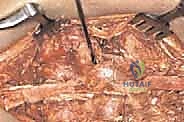
This topic focuses on ORTHO MCQS Shoulder and Elbow 0192, Following surgical repair of a radial head fracture complicated by lateral collateral ligament (LCL) avulsion, initial postoperative rehabilitation should include elbow extension exercises performed with the forearm in pronation. This specific positioning helps protect the compromised LCL. A clear explanation of this approach forms a crucial response a discussion point for therapists to ensure optimal recovery and stability.
Ortho Shoulder And Elbow Review | Dr Hutaif Shoulder & - ...
Comprehensive 100-Question Exam
00:00
Start Quiz
Question 1
A 24-year-old competitive rugby player presents with recurrent anterior shoulder instability. A 3D CT scan reveals a 24% anterior glenoid bone defect. He has failed an extensive course of non-operative management. What is the most appropriate surgical intervention?
Explanation
Question 2
A 72-year-old female presents with chronic shoulder pain and an inability to actively elevate her arm above 40 degrees. On examination, she has a positive hornblower's sign but her deltoid function is intact. Radiographs demonstrate superior migration of the humeral head and acetabularization of the coracoacromial arch (Hamada grade 3). Which of the following is the most reliable surgical option?
Explanation
Question 3
In the surgical management of a 'terrible triad' injury of the elbow (elbow dislocation, radial head fracture, and coronoid fracture), what is the most accepted sequential order of structure repair to restore stability?
Explanation
Question 4
Recent quantitative anatomic and perfusion studies have re-evaluated the principal vascular supply to the humeral head. Which of the following arteries is now recognized as providing the majority of the blood supply to the humeral head?
Explanation
Question 5
A 28-year-old professional volleyball player complains of vague posterior shoulder pain and selective weakness in external rotation. Exam reveals isolated atrophy of the infraspinatus fossa. MRI is most likely to show a paralabral cyst causing nerve entrapment in which of the following locations?
Explanation
Question 6
Posterolateral rotatory instability (PLRI) of the elbow is a clinical entity most frequently caused by insufficiency of which of the following capsuloligamentous structures?
Explanation
Question 7
A 45-year-old male undergoes a single-incision anterior approach for the repair of a distal biceps tendon rupture. Post-operatively, he complains of numbness over the lateral aspect of his forearm. Which nerve is most likely injured during this surgical exposure?
Explanation
Question 8
During the repair of an acute pectoralis major tendon rupture, the surgeon isolates the distinct heads of the tendon. Which of the following accurately describes the anatomic insertion of the sternal head of the pectoralis major onto the humerus relative to the clavicular head?
Explanation
Question 9
A 5-year-old boy falls on an outstretched hand. Elbow radiographs reveal an ossific density within the medial aspect of the elbow joint space. The normal medial epicondyle ossification center is absent. To avoid misdiagnosing this incarcerated fragment as a normal developing ossification center, which ossification center's chronological appearance must be verified absent according to normal pediatric development?
Explanation
Question 10
A 40-year-old male presents to the ED after a seizure. He holds his left arm in internal rotation. Radiographs confirm a posterior shoulder dislocation. CT scan reveals an anteromedial humeral head impaction fracture (reverse Hill-Sachs lesion) involving 30% of the articular surface. Which of the following is the most appropriate surgical intervention?
Explanation
Question 11
A 25-year-old patient presents with right shoulder pain and weakness when lifting objects above his head. Examination reveals medial scapular winging that worsens when the patient pushes against a wall. Injury to which of the following nerves is the most likely cause?
Explanation
Question 12
In the surgical reconstruction of the medial ulnar collateral ligament (UCL) of the elbow in throwing athletes, the graft is primarily positioned to replicate the biomechanical function of which specific structural component?
Explanation
Question 13
When performing an olecranon osteotomy to gain exposure for fixing a complex intra-articular distal humerus fracture (AO/OTA 13-C3), what is the optimal shape of the osteotomy to maximize postoperative stability, healing surface area, and rotational control?
Explanation
Question 14
Which of the following factors has been shown in the literature to be the most significant independent risk factor for nonunion in the nonoperative management of midshaft clavicle fractures?
Explanation
Question 15
A 55-year-old female with Type I diabetes mellitus presents with progressive, severe restriction of both active and passive shoulder motion. She is diagnosed with primary adhesive capsulitis. Which cytokine is most strongly implicated in driving the excessive capsular fibroblastic proliferation seen in this condition?
Explanation
Question 16
Glenohumeral Internal Rotation Deficit (GIRD) in the overhead throwing athlete is biomechanically linked to the development of a Type II SLAP tear. Which capsular abnormality is considered the primary driver of this internal rotation deficit and the resultant peel-back mechanism?
Explanation
Question 17
During a Reverse Total Shoulder Arthroplasty (RTSA), superior or posterior retractor placement and aberrant screw placement in the superior or posterior glenoid vault can most likely cause iatrogenic injury to which of the following nerves?
Explanation
Question 18
A 12-year-old Little League pitcher presents with medial elbow pain. Radiographs demonstrate widening of the medial epicondyle apophysis without displacement. What is the most common underlying pathomechanical force responsible for this condition (Little Leaguer's elbow)?
Explanation
Question 19
In the surgical treatment of high-grade acromioclavicular (AC) joint separations, reconstruction often targets the coracoclavicular (CC) ligaments. Which of the following accurately describes the anatomy of the native CC ligaments?
Explanation
Question 20
A 35-year-old female sustains a highly comminuted radial head fracture from a fall. Intraoperatively, the radial head is deemed unreconstructable. Prior to deciding on a radial head arthroplasty versus excision, what intraoperative finding strongly dictates the absolute necessity for a radial head prosthesis?
Explanation
Question 21
Which of the following best describes the biomechanical advantage of shifting the center of rotation medially and distally in a reverse total shoulder arthroplasty (rTSA)?
Explanation
Question 22
During a Latarjet procedure, retractors are often placed deep to the conjoint tendon. To avoid neuropraxia or permanent injury to the musculocutaneous nerve, retractor placement must be carefully monitored. What is the generally accepted 'safe zone' for retractor placement in relation to the coracoid process?
Explanation
Question 23
A 35-year-old male sustains a fracture involving the anteromedial facet of the coronoid process after a fall on an outstretched hand. If this fracture is not properly stabilized, the elbow is at highest risk for developing which specific pattern of instability?
Explanation
Question 24
What is the most widely accepted surgical sequence for repairing a 'terrible triad' injury of the elbow (elbow dislocation, radial head fracture, and coronoid fracture)?
Explanation
Question 25
A lower trapezius tendon transfer is performed for a patient with a massive, irreparable posterosuperior rotator cuff tear to restore active external rotation. What is the primary motor innervation to the transferred muscle?
Explanation
Question 26
A 24-year-old trauma patient presents with scapulothoracic dissociation characterized by massive lateral displacement of the scapula on the AP chest radiograph. Due to the high mortality and morbidity associated with this injury, which of the following must be evaluated emergently?
Explanation
Question 27
A 22-year-old rugby player sustains a posterior sternoclavicular (SC) joint dislocation. Which anatomical structure lies directly posterior to the medial clavicle and is at highest risk of catastrophic injury during reduction or from the injury itself?
Explanation
Question 28
A 45-year-old female presents with a coronal shear fracture of the distal humerus. The radiograph demonstrates a 'double arc' sign on the lateral view. This radiographic finding is pathognomonic for which specific injury pattern?
Explanation
Question 29
A 28-year-old elite overhead athlete presents with painless weakness in the dominant shoulder. Physical examination reveals isolated severe atrophy of the infraspinatus muscle, but completely preserved muscle bulk and strength of the supraspinatus. Where is the most likely anatomical site of nerve compression?
Explanation
Question 30
In the design of a linked, semiconstrained total elbow arthroplasty (TEA), what is the primary biomechanical function of incorporating a 'sloppy hinge' (allowing 5-10 degrees of varus-valgus laxity)?
Explanation
Question 31
In a complete rupture of the pectoralis major tendon sustained during weightlifting (e.g., bench press), which specific portion of the muscle-tendon unit is anatomically most prone to failure, and what is its normal insertion pattern?
Explanation
Question 32
During an anatomic coracoclavicular (CC) ligament reconstruction for a chronic Type V acromioclavicular joint separation, bone tunnels are drilled in the clavicle to recreate the native anatomy. What is the approximate anatomical distance from the distal end of the clavicle to the center of the conoid and trapezoid insertions, respectively?
Explanation
Question 33
Which specific portion of the ulnar collateral ligament (UCL) complex of the elbow serves as the primary restraint to valgus stress from 30 to 120 degrees of flexion, and what is its anatomic ulnar insertion?
Explanation
Question 34
When performing an open subpectoral biceps tenodesis, deep retraction is required to expose the humerus. If retractors are placed too far medially, which nerve is at greatest risk of iatrogenic injury?
Explanation
Question 35
A 24-year-old male with recurrent anterior shoulder instability is evaluated preoperatively with 3D CT. The concept of the 'glenoid track' is used to evaluate the interplay between glenoid bone loss and a Hill-Sachs lesion. Which of the following correctly defines an 'off-track' Hill-Sachs lesion?
Explanation
Question 36
In the surgical management of a displaced 3-part proximal humerus fracture using a locking plate, failure to restore adequate medial cortical support (the medial hinge) most frequently leads to which of the following mechanical failures?
Explanation
Question 37
A superior capsular reconstruction (SCR) using a thick dermal allograft is performed for a patient with a massive, irreparable posterosuperior rotator cuff tear. Biomechanically, what is the primary structural role of the graft in this procedure?
Explanation
Question 38
A 40-year-old male presents with a locked posterior shoulder dislocation sustained during a seizure. Imaging reveals a reverse Hill-Sachs (impaction) defect involving 35% of the anterior articular surface of the humeral head. Assuming an intact glenohumeral joint otherwise, which of the following is the most appropriate surgical management?
Explanation
Question 39
The distal biceps tendon normally inserts onto the radial tuberosity. To maximize the biomechanical supination moment arm (the 'cam effect') during an anatomical surgical repair of a distal biceps rupture, where should the tendon be optimally positioned on the tuberosity?
Explanation
Question 40
In a patient presenting with primary adhesive capsulitis (frozen shoulder), the classic physical examination finding is marked restriction of passive external rotation with the arm adducted at the side. This specific restriction is most anatomically attributed to contracture of which of the following structures?
Explanation
Question 41
Varus posteromedial rotatory instability (VPMRI) of the elbow is characterized by a fracture of the anteromedial facet of the coronoid. What is the typical mechanism of injury that produces this specific instability pattern?
Explanation
Question 42
During a Latarjet procedure for recurrent anterior shoulder instability, careful retraction of the conjoint tendon is essential. Over-retraction medially places which of the following nerves at the greatest risk of iatrogenic injury?
Explanation
Question 43
A 70-year-old female undergoes a reverse total shoulder arthroplasty (RTSA) for rotator cuff tear arthropathy. To minimize the risk of inferior scapular notching, which is a common complication, how should the glenosphere baseplate optimally be positioned?
Explanation
Question 44
In posterolateral rotatory instability (PLRI) of the elbow, the primary deficient structure is the lateral ulnar collateral ligament (LUCL). What are the precise anatomic origin and insertion of the LUCL?
Explanation
Question 45
A 58-year-old male weightlifter presents with advanced glenohumeral osteoarthritis. A CT scan reveals a biconcave glenoid with severe retroversion and posterior subluxation of the humeral head. According to the Walch classification, what type of glenoid morphology does this represent?
Explanation
Question 46
A 28-year-old overhead athlete presents with posterior shoulder pain and weakness in external rotation. Forward elevation and internal rotation strength are normal. MRI reveals a paralabral cyst in the spinoglenoid notch. Which physical examination finding is most likely to be exclusively present?
Explanation
Question 47
A patient undergoes surgical repair of an acute distal biceps tendon rupture using a single-incision anterior approach. Postoperatively, he complains of burning pain and numbness along the lateral aspect of his forearm. Which nerve was most likely injured during the surgical exposure?
Explanation
Question 48
A 35-year-old male presents with a locked posterior shoulder dislocation following a seizure. CT imaging shows a reverse Hill-Sachs lesion (anteromedial humeral head defect) involving approximately 35% of the articular surface. The joint is unstable after closed reduction. What is the most appropriate surgical management?
Explanation
Question 49
The anterior bundle of the medial collateral ligament (AMCL) is the primary restraint to valgus instability of the elbow. What is the precise distal insertion site of the AMCL?
Explanation
Question 50
When evaluating a proximal humerus fracture to determine the risk of developing avascular necrosis (AVN), which of the following radiographic findings (Hertel's criteria) is the most reliable predictor of ischemia to the humeral head?
Explanation
Question 51
A 45-year-old male presents with severe cubital tunnel syndrome that has failed conservative management. During an in situ ulnar nerve decompression, which of the following structures forms the anatomic roof of the cubital tunnel and must be released?
Explanation
Question 52
A 14-year-old male baseball pitcher presents with lateral elbow pain and catching. Radiographs and MRI demonstrate a detached osteochondral fragment in the capitellum with an underlying fluid signal. What is the most appropriate diagnosis?
Explanation
Question 53
A 30-year-old male weightlifter complains of poorly localized posterior shoulder pain and numbness over the lateral deltoid. Physical examination reveals focal point tenderness in the quadrilateral space. Which vascular structure is at risk of compression in this space alongside the affected nerve?
Explanation
Question 54
A 22-year-old rugby player sustains a direct blow to the medial clavicle, resulting in a posterior sternoclavicular (SC) joint dislocation. Which ligamentous structure is considered the strongest and primary stabilizer preventing anterior-posterior translation of the SC joint?
Explanation
Question 55
During a radial head arthroplasty for a comminuted radial head fracture, the surgeon inadvertently overstuffs the radiocapitellar joint by 4 mm. Which of the following biomechanical consequences is most likely to occur as a direct result of this technical error?
Explanation
Question 56
A 35-year-old female presents with shoulder weakness 3 months after a posterior triangle cervical lymph node biopsy. On physical examination, the shoulder exhibits lateral winging of the scapula when she abducts her arm. Injury to which nerve is responsible for this physical finding?
Explanation
Question 57
In the biomechanics of the acromioclavicular (AC) joint, the coracoclavicular (CC) ligaments provide primary vertical and axial stability. Which of the following statements accurately describes the specific anatomy and function of the CC ligaments?
Explanation
Question 58
A 12-year-old elite baseball pitcher presents with poorly localized shoulder pain that worsens strictly with throwing. Radiographs reveal widening and lateral fragmentation of the proximal humeral physis compared to the contralateral side. What is the most appropriate initial management for this condition?
Explanation
Question 59
A 25-year-old male bodybuilder feels a 'pop' in his anterior axilla while performing heavy bench presses. MRI confirms a complete rupture of the pectoralis major tendon. Regarding the anatomy of the pectoralis major insertion on the humerus, which of the following is true?
Explanation
Question 60
A 42-year-old male sustains a complete distal triceps tendon rupture after a fall on an outstretched hand. During surgical repair using a transosseous cruciate technique, it is crucial to reattach the tendon to its anatomic footprint. Where is the normal anatomic insertion footprint of the triceps tendon?
Explanation
Question 61
A 45-year-old male laborer presents with acute anterior elbow pain and ecchymosis after attempting to lift a heavy box. Examination reveals a positive hook test. During a single-incision anterior surgical repair using suture anchors, which nerve is at the highest risk of injury?
Explanation
Question 62
A 62-year-old male presents with advanced glenohumeral osteoarthritis. CT scan reveals a retroverted biconcave glenoid (Walch B2) with 20 degrees of retroversion. He has an intact rotator cuff. What is the most appropriate surgical management?
Explanation
Question 63
A 35-year-old female presents with shoulder pain and weakness 4 weeks after a posterior cervical triangle lymph node biopsy. On examination, the affected shoulder droops, and the scapula rests lateral to the midline and wings when she abducts her arm. Injury to which of the following nerves is the most likely cause?
Explanation
Question 64
A 32-year-old female complains of a recurrent clicking and giving way of her elbow when pushing up from a chair. Physical examination demonstrates apprehension with the elbow supinated, axially loaded, and moving from extension into flexion. Which ligamentous structure is primarily deficient?
Explanation
Question 65
A 22-year-old elite collegiate baseball pitcher presents with deep shoulder pain and decreased throwing velocity. MRI arthrogram shows a peel-back of the superior labrum with detachment of the biceps anchor (Type II SLAP tear). Conservative treatment has failed. What is the most appropriate surgical approach?
Explanation
Question 66
A 75-year-old female with osteoporosis sustains a 4-part proximal humerus fracture. Radiographs show severe comminution of the calcar and the humeral head split into two fragments. What is the most reliable surgical option to restore active elevation?
Explanation
Question 67
A 40-year-old man falls onto an outstretched hand and presents with anterior shoulder pain. He has a positive bear hug test and belly press test. The external rotation lag sign is negative. MRI confirms an isolated tear of the subscapularis tendon. Which of the following associated findings is most likely present?
Explanation
Question 68
A 28-year-old competitive weightlifter feels a pop in his anterior axillary fold while performing a heavy bench press. Examination reveals loss of the anterior axillary contour and weakness in internal rotation and adduction. MRI shows a rupture of the sternocostal head of the pectoralis major. Where does the sternocostal head normally insert?
Explanation
Question 69
A 34-year-old female sustains a coronal shear fracture of the distal humerus involving the capitellum and the lateral aspect of the trochlea. The fracture extends into the lateral epicondyle. Which surgical approach provides the most optimal visualization for open reduction and internal fixation?
Explanation
Question 70
A 55-year-old male presents with numbness in his small and ring fingers and intrinsic muscle weakness. Electrodiagnostic studies confirm severe ulnar neuropathy at the elbow. During an open surgical release, which structure forms the primary roof of the cubital tunnel?
Explanation
Question 71
A 42-year-old male suffers a seizure and subsequently complains of shoulder pain and inability to externally rotate his arm. An axillary lateral radiograph reveals a posterior shoulder dislocation with an anteromedial humeral head defect (reverse Hill-Sachs lesion) involving 35% of the articular surface. What is the most appropriate management?
Explanation
Question 72
A 19-year-old male is tackled during a football game and sustains a posterior sternoclavicular joint dislocation. He reports mild difficulty swallowing but has normal vital signs. Attempted closed reduction in the operating room under general anesthesia fails. What is the next most appropriate step in management?
Explanation
Question 73
A 68-year-old female with severe rheumatoid arthritis undergoes a primary linked semi-constrained total elbow arthroplasty (TEA). Post-operatively, what is the generally recommended permanent lifetime lifting restriction for the operated arm?
Explanation
Question 74
A 24-year-old manual worker presents with painful crepitus and snapping at the superomedial border of his scapula. Non-operative management has failed after 6 months. Imaging reveals a skeletal prominence at the superomedial angle. What is the anatomical name of this structure commonly responsible for this syndrome?
Explanation
Question 75
A 30-year-old elite volleyball player complains of vague posterior shoulder pain and weakness in external rotation. Examination shows isolated atrophy of the infraspinatus fossa. MRI shows a paralabral cyst at the spinoglenoid notch. Which finding is most likely to be present on physical examination?
Explanation
Question 76
A 45-year-old male hears a pop in his anterior elbow while lifting a heavy box. On examination, he has a positive hook test. He undergoes a single-incision distal biceps tendon repair. Postoperatively, he complains of numbness along the lateral aspect of his forearm. Which structure was most likely injured during the surgical exposure?
Explanation
Question 77
A 30-year-old male presents with lateral elbow pain and a clicking sensation when extending and supinating the elbow. He is diagnosed with posterolateral rotatory instability (PLRI) and is scheduled for ligament reconstruction. What are the correct isometric points for reconstructing the primary ligament deficient in this condition?
Explanation
Question 78
Reverse total shoulder arthroplasty (rTSA) relies heavily on the deltoid muscle to compensate for a deficient rotator cuff. How does the standard Grammont-style rTSA alter glenohumeral biomechanics to achieve this?
Explanation
Question 79
A 65-year-old female sustains a complex proximal humerus fracture. According to the Hertel criteria, which of the following radiographic features is the most reliable predictor of humeral head ischemia?
Explanation
Question 80
A 25-year-old cyclist falls directly onto his shoulder, sustaining a Type V acromioclavicular (AC) joint separation. The surgeon plans a coracoclavicular (CC) ligament reconstruction. Which of the following accurately describes the anatomic insertions of the native CC ligaments on the clavicle?
Explanation
Question 81
A 22-year-old overhead athlete presents with persistent anterior shoulder apprehension. Magnetic resonance arthrography (MRA) demonstrates a "J sign" with extravasation of contrast into the axillary pouch due to an avulsion of the inferior glenohumeral ligament from the humeral neck. What is the eponymous term for this lesion?
Explanation
Question 82
A 30-year-old competitive weightlifter feels a sudden "pop" while performing a heavy bench press. Examination reveals loss of the anterior axillary fold and weakness in internal rotation. MRI confirms a rupture of the sternal head of the pectoralis major. Where does the sternal head normally insert relative to the clavicular head on the humerus?
Explanation
Question 83
A 40-year-old male sustains a locked posterior shoulder dislocation during a seizure. A CT scan reveals an anterior articular impaction fracture (reverse Hill-Sachs lesion) involving 30% of the humeral head articular surface. What is the most appropriate surgical management?
Explanation
Question 84
A 35-year-old female falls onto an outstretched hand. A lateral radiograph of the elbow demonstrates a "double arc sign." What specific injury does this radiographic finding indicate?
Explanation
Question 85
A 45-year-old tennis player fails 12 months of conservative management for lateral epicondylitis. An open debridement of the extensor carpi radialis brevis (ECRB) is planned. Topographically, where is the ECRB origin located relative to the extensor carpi radialis longus (ECRL)?
Explanation
Question 86
A 68-year-old male with severe glenohumeral osteoarthritis undergoes preoperative planning for an anatomic total shoulder arthroplasty. A 3D CT scan reveals a Walch B2 glenoid with 22 degrees of retroversion and significant posterior subluxation. What is the most appropriate intraoperative management of the glenoid?
Explanation
Question 87
A 28-year-old elite volleyball player presents with insidious onset of shoulder pain and isolated weakness in external rotation. An MRI demonstrates a paralabral cyst in the spinoglenoid notch compressing a nerve. This cyst is most commonly associated with which of the following intra-articular pathologies?
Explanation
Question 88
A 35-year-old male develops medial scapular winging following a prolonged viral illness. Electromyography confirms neuropathy of the affected muscle's primary nerve. Which nerve roots predominantly supply this injured nerve?
Explanation
None
