Tibial Eminence Fracture Case Study: Comprehensive Diagnostic & Imaging Insights
Key Takeaway
Tibial eminence fractures are diagnosed via detailed clinical examination, noting acute knee pain, swelling, limited ROM, and positive Lachman/Anterior Drawer tests, especially after sports injury. Confirmatory imaging includes X-rays showing avulsion, CT for fragment morphology and displacement (e.g., Type III), and MRI to assess associated soft tissue injuries like meniscal entrapment.
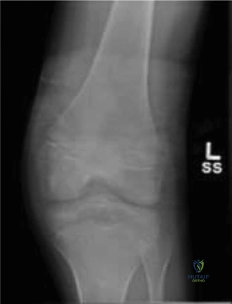
A 24-year-old collegiate athlete sustains a valgus injury with axial load. He presents with a tense hemarthrosis and a fixed flexion deformity of 15 degrees. Radiographs are obtained as shown below. What is your diagnosis, and how does the radiographic appearance correlate with the clinical finding of a fixed flexion deformity?
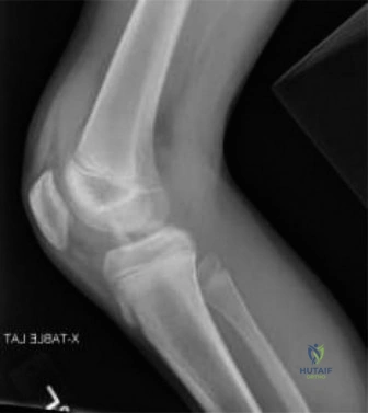
Candidate: The radiograph shows a displaced tibial eminence avulsion fracture, consistent with a Meyers and McKeever Type III injury. The fixed flexion deformity occurs because the avulsed fragment and the attached ACL are displaced superiorly into the intercondylar notch, acting as a physical block to terminal extension.
Failure to mention the specific mechanism causing the impingement or ignoring the possibility of soft-tissue interposition. Simply stating "it's a fracture" lacks the biomechanical reasoning expected at the FRCS level.
Identify the Meyers and McKeever Type III injury. Explain that the clinical mechanical block is due to the superior migration of the fragment combined with interposition of the anterior horn of the medial meniscus or the transverse intermeniscal ligament within the fracture crater, which prevents the reduction of the fragment and inhibits full extension.
Given the patient's age and desire for high-level return to sport, you decide on surgery. The MRI findings are presented below. What do you see, and how does this influence your operative planning regarding potential concomitant injuries?
Candidate: The MRI confirms the displaced eminence avulsion and demonstrates that the ACL fibers are attached to the fragment. It also shows the anterior horn of the medial meniscus is trapped within the fracture site. I need to perform an arthroscopic-assisted reduction, ensuring I clear the meniscal entrapment before attempting fixation.
Focusing only on the bone and ignoring the ACL substance. High-energy injuries in adults can lead to interstitial ACL fiber stretching or partial tears; failing to check this means you might fix the bone but leave the patient with a functionally lax ligament.
Systematically address: 1) Confirmation of fragment displacement. 2) Identification of soft-tissue entrapment (meniscus/intermeniscal ligament). 3) Assessment of the ACL substance integrity. 4) Preoperative planning to ensure anatomic reduction of the "footprint" to restore native kinematics, noting that suture fixation is preferred over rigid screw fixation in adults to avoid hardware prominence and future impingement.
