Operative Management of Scoliosis and Kyphosis: A Comprehensive Surgical Guide
Key Takeaway
This comprehensive guide details the operative management of scoliosis and kyphosis, tailored for orthopedic surgeons and fellows. It explores the biomechanics, surgical indications, and advanced instrumentation techniques for adolescent idiopathic, neuromuscular, and congenital spinal deformities. Key surgical approaches, including posterior pedicle screw fixation, anterior thoracoscopic instrumentation, and pelvic fixation strategies, are critically analyzed to optimize sagittal and coronal balance while minimizing postoperative complications.
Introduction to Spinal Deformity
The operative management of scoliosis and kyphosis represents one of the most complex and biomechanically demanding disciplines within orthopedic surgery. Spinal deformity is a three-dimensional derangement involving coronal translation, sagittal plane alteration, and axial rotation. Successful surgical intervention requires a profound understanding of spinal biomechanics, meticulous preoperative planning, and precise execution of instrumentation and fusion techniques. This guide synthesizes the foundational principles and advanced surgical strategies for managing idiopathic, neuromuscular, congenital, and syndromic spinal deformities.
Adolescent Idiopathic Scoliosis (AIS)
Adolescent Idiopathic Scoliosis (AIS) is the most common pediatric spinal deformity, presenting in patients aged 10 to skeletal maturity. Operative intervention is generally indicated for curves exceeding 45 to 50 degrees, curves progressing despite orthotic management, or those causing significant coronal or sagittal imbalance.
Radiographic Evaluation and Curve Patterns
Comprehensive radiographic evaluation is the cornerstone of surgical planning. Standard imaging includes standing posteroanterior (PA) and lateral full-spine radiographs, supplemented by supine side-bending or traction films to assess curve flexibility.
- Cobb Angle Measurement: Determines the magnitude of the coronal deformity.
- Vertebral Rotation: Assessed via the Nash-Moe method or pedicle offset.
- Sagittal Balance: Critical for preventing postoperative flatback syndrome; involves assessing thoracic kyphosis, lumbar lordosis, and pelvic parameters (Pelvic Incidence, Pelvic Tilt, Sacral Slope).
- Curve Classification: The Lenke Classification system guides fusion levels by categorizing curves into six types based on the structural nature of the proximal thoracic, main thoracic, and thoracolumbar/lumbar curves, combined with lumbar spine modifiers and sagittal thoracic modifiers.
Posterior Surgeries for Idiopathic Scoliosis
Posterior spinal fusion (PSF) with instrumentation remains the gold standard for the majority of AIS curve patterns. The primary surgical goals are to halt curve progression, achieve maximum safe three-dimensional correction, and obtain a solid arthrodesis.
Posterior Spinal Instrumentation
The evolution of posterior instrumentation has transitioned from Harrington rods to multihook segmental systems (e.g., Cotrel-Dubousset), and currently to all-pedicle screw constructs.
- Multihook Segmental Instrumentation: While largely supplanted by pedicle screws, hook constructs remain relevant in specific scenarios, such as severe osteoporosis or anomalous pedicle anatomy. Hook site preparation requires meticulous ligamentum flavum excision and precise seating of pedicle, transverse process, or sublaminar hooks to prevent spinal canal encroachment.
- Pedicle Fixation: All-pedicle screw constructs offer superior three-dimensional correction, enhanced pull-out strength, and the ability to save fusion levels.
- Lumbar Pedicle Screws: Inserted via the intersection of the pars interarticularis, the transverse process, and the superior articular facet.
- Thoracic Pedicle Screws: Require a highly nuanced freehand technique or navigation. The starting point varies from the lateral aspect of the facet in the upper thoracic spine to a more medial trajectory in the lower thoracic spine.
Surgical Warning: Breaches of the medial pedicle wall in the thoracic spine risk catastrophic spinal cord injury. Continuous intraoperative neuromonitoring (IONM), including Somatosensory Evoked Potentials (SSEPs) and Motor Evoked Potentials (MEPs), is mandatory during pedicle preparation and screw insertion.
Posterior Thoracoplasty and Concave Rib Osteotomies
For patients with severe rib prominences that do not resolve with spinal derotation, a posterior thoracoplasty may be indicated. This involves the subperiosteal resection of the prominent convex ribs. Conversely, concave rib osteotomies can be utilized to mobilize rigid curves and improve pulmonary mechanics in severe deformities.
Anterior Instrumentation for Idiopathic Scoliosis
Anterior spinal fusion (ASF) and instrumentation are primarily indicated for single thoracolumbar or lumbar curves (Lenke Type 5). The anterior approach allows for excellent coronal correction and derotation while saving distal fusion levels compared to posterior approaches.
Video-Assisted Thoracoscopy (VATS)
Endoscopic anterior instrumentation via VATS minimizes the morbidity associated with an open thoracotomy. It allows for anterior release, diskectomy, and instrumentation.
Clinical Pearl: VATS requires single-lung ventilation and specialized endoscopic instrumentation. Pitfalls include inadequate diskectomy leading to pseudarthrosis and the risk of vascular injury to the segmental vessels or aorta.
Neuromuscular Scoliosis
Neuromuscular scoliosis arises from myopathic or neuropathic conditions, including Cerebral Palsy (CP), Duchenne Muscular Dystrophy (DMD), Spinal Muscular Atrophy (SMA), and Myelomeningocele. These curves are typically long, sweeping, "C-shaped" deformities that progress rapidly and are associated with pelvic obliquity.
Preoperative Considerations
Patients with neuromuscular scoliosis often present with significant medical comorbidities. Preoperative optimization must address pulmonary function, cardiac status, nutritional deficiencies, and seizure control.
Operative Treatment and Sacropelvic Fixation
The primary goal of surgery in neuromuscular scoliosis is to achieve a balanced spine over a level pelvis, thereby optimizing sitting balance and preventing pressure sores.
- Luque Rod Instrumentation with Sublaminar Wiring: Historically utilized for its load-sharing capabilities, though largely replaced due to the neurologic risks of sublaminar wire passage.
- Unit Rod Segmental Spinal Instrumentation: A pre-bent, U-shaped rod that provides excellent pelvic fixation and coronal correction. It is particularly effective in non-ambulatory patients with cerebral palsy.
- Iliac Fixation with Iliac Screws: Modern constructs utilize bilateral iliac or S2-Alar-Iliac (S2AI) screws connected to the main longitudinal rods. S2AI screws offer the advantage of being in line with the cephalad construct, eliminating the need for offset connectors and reducing implant prominence.
Congenital Scoliosis
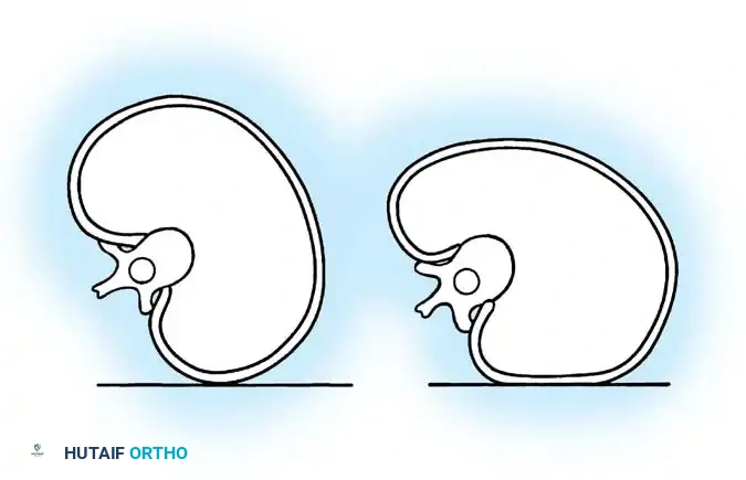
Congenital scoliosis results from anomalous vertebral development during the first trimester, categorized into failures of formation (e.g., hemivertebra), failures of segmentation (e.g., unilateral unsegmented bar), or mixed anomalies.
Natural History and Patient Evaluation
The risk of progression depends heavily on the anomaly type. A unilateral unsegmented bar with a contralateral fully segmented hemivertebra carries the highest risk of rapid, relentless progression. Evaluation must include an MRI to rule out intraspinal anomalies (e.g., tethered cord, syringomyelia, diastematomyelia), which are present in up to 40% of patients. Renal and cardiac ultrasounds are also mandatory due to the high incidence of VACTERL associations.
Operative Treatment
Early surgical intervention is often required to prevent severe, rigid deformities.
- Hemivertebra Excision: The procedure of choice for a fully segmented, progressive hemivertebra. It can be performed via a combined anterior-posterior approach or an all-posterior transpedicular approach. The hemivertebra and adjacent discs are resected, and the defect is closed using pedicle screw compression.
- Combined Anterior and Posterior Convex Hemiepiphysiodesis: Indicated for young children with progressive curves to arrest growth on the convex side while allowing continued growth on the concave side.
- Thoracic Insufficiency Syndrome (TIS): Severe congenital scoliosis or fused ribs can restrict lung growth, leading to TIS. Expansion thoracoplasty using the Vertical Expandable Prosthetic Titanium Rib (VEPTR) or magnetically controlled growing rods (MCGR) expands the constricted hemithorax, indirectly correcting the scoliosis while permitting pulmonary development.
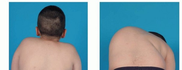
Kyphosis
Scheuermann Disease
Scheuermann kyphosis is a rigid structural hyperkyphosis of the thoracic or thoracolumbar spine. Diagnostic radiographic criteria include anterior wedging of greater than 5 degrees in at least three consecutive vertebrae, endplate irregularities, and Schmorl nodes.
Operative Treatment
Surgery is indicated for curves exceeding 75 degrees, curves associated with intractable pain, or significant cosmetic deformity.
* Surgical Approach: Modern treatment predominantly utilizes an all-posterior approach with multilevel Ponte osteotomies (resection of the facet joints and ligamentum flavum) to restore flexibility, followed by pedicle screw instrumentation and compression across the convexities.
* Biomechanical Goal: Restoration of sagittal balance without inducing junctional kyphosis. The fusion must extend from the proximal measured Cobb vertebra to the first lordotic disc space distally.
Congenital Kyphosis
Congenital kyphosis is highly prone to progression and carries a significant risk of spinal cord compression and paraplegia, particularly in Type I (failure of formation) anomalies.
Surgical Warning: In situ posterior fusion is only effective in very young children (under age 3) before significant deformity develops. Once a severe angular kyphosis is present, an anterior decompression (corpectomy) and strut grafting, combined with posterior stabilization, is mandatory to decompress the spinal cord and stabilize the spine.
Spondylolysis and Spondylolisthesis
Spondylolysis is a defect in the pars interarticularis, most commonly at L5. Spondylolisthesis is the anterior translation of one vertebra over another.
Treatment of Severe (High-Dysplastic) Spondylolisthesis
High-grade spondylolisthesis (Meyerding Grade III, IV, and V/Spondyloptosis) presents a formidable surgical challenge. Patients often exhibit a characteristic "pelvic waddle" gait, hamstring tightness, and severe lumbosacral kyphosis.
- In Situ Fusion vs. Reduction: While in situ posterolateral fusion from L4 to S1 is safe, it does not correct the slip angle or sagittal profile. Partial reduction using pedicle screw instrumentation improves the biomechanical environment for fusion and restores sagittal balance but carries a higher risk of L5 nerve root stretch injury.
- L5 Vertebrectomy (Gaines Procedure): For severe spondyloptosis, a two-stage L5 vertebrectomy may be indicated. The first stage involves an anterior L5 corpectomy. The second stage involves posterior resection of the L5 posterior elements and reduction of L4 directly onto S1.
Unusual Causes of Spinal Deformity
Neurofibromatosis (NF1)
Spinal deformity is the most common orthopedic manifestation of NF1. Curves are classified as non-dystrophic (resembling AIS) or dystrophic.
* Dystrophic Scoliosis: Characterized by short, sharp, angular curves, vertebral scalloping, penciling of the ribs, and enlarged neural foramina. Dystrophic curves have a notoriously high rate of progression and pseudarthrosis.
* Operative Strategy: Circumferential (anterior and posterior) fusion is strongly recommended for dystrophic curves to prevent pseudarthrosis and the "adding-on" phenomenon.
Marfan Syndrome
Patients with Marfan syndrome develop scoliosis that is often rigid and associated with dural ectasia. Surgical intervention requires robust fixation due to poor bone quality and a high risk of curve progression even after skeletal maturity.
Complications of Spinal Deformity Surgery
The operative management of scoliosis and kyphosis carries significant inherent risks. Meticulous technique and vigilance are required to mitigate these complications.
- Neurologic Deficit: The most devastating complication. Risk factors include severe angular kyphosis, congenital anomalies, and excessive distraction. Continuous IONM is critical. If signal loss occurs, the surgeon must immediately assess blood pressure, reverse any corrective forces, and consider a wake-up test.
- Infection: More common in neuromuscular patients due to poor nutrition and incontinence. Treatment requires aggressive surgical debridement, implant retention (if stable), and targeted intravenous antibiotics.
- Pseudarthrosis: Failure of fusion can lead to implant failure and loss of correction. It is mitigated by meticulous decortication, generous use of autograft/allograft, and rigid segmental fixation.
- Adding-On Phenomenon: Progression of the deformity beyond the instrumented levels. This is avoided by strictly adhering to the principles of selecting the stable vertebra and the lowest instrumented vertebra (LIV) during preoperative planning.
📚 Medical References
- scoliosis and kyphosis radiographs: intraobserver and interobserver variation, J Bone Joint Surg 72A:328, 1990.
- Cobb JR: Outline for the study of scoliosis in instructional course lectures. In The American Academy of Orthopaedic Surgeons: Instructional course lectures, vol 5, Ann Arbor, Mich, 1948, JW Edwards. Deacon P, Flood BM, Dickson RA: Idiopathic scoliosis in three dimension: a radiographic and morphometric analysis, J Bone Joint Surg 66B:509, 1984.
- DeSmet A, Fritz SL, Asher MA: A method for minimizing the radiation exposure from scoliosis radiographs, J Bone Joint Surg 63A:156, 1981.
- DeSmet A, Goin JE, Asher MA, et al: A clinical study of the differences between the scoliotic angles measured on the PA versus the AP radiographs, J Bone Joint Surg 64A:489, 1982.
- Drummond D, Ranallo F, Lonstein J, et al: Radiation hazards and scoliosis management, Spine 8:741, 1983.
- Edwards CC 2nd, Lenke LG, Peelte M, et al: Selective thoracic fusion for adolescent idiopathic scoliosis with C modifi er lumbar curves: 2to 16-year radiographic and clinical results, Spine 29:536, 2004.
- Farren J: Routine radiographic assessment of the scoliotic spine, Radiography 47:92, 1981.
- Ferguson AB: Roentgen interpretations and decisions in scoliosis. In The American Academy of Orthopaedic Surgeons: Instructional course lectures, vol 7, Ann Arbor, Mich, 1950, JW Edwards. Gelb DE, Lenke LG, Bridwell KH, et al: An analysis of sagittal spinal alignment in 100 asymptomatic middle and older age volunteers, Spine 20:1351, 1995.
- Gray JE, Hoffman AE, Peterson HA: Reduction of radiation exposure during radiography for scoliosis, J Bone Joint Surg 65A:5, 1983.
- Gunzburg R, Gunzburg J, Wagner J, et al: Radiologic interpretation of lumbar vertebral rotation, Spine 16:660, 1991.
- Hopkins R, Grundy M, Sherr-Mehl M: X-ray fi lters in scoliosis x-rays, Orthop Trans 8:148, 1984.
- King HA, Moe JH, Bradford DS, et al: The selection of the fusion levels in thoracic idiopathic scoliosis, J Bone Joint Surg 65A:1302, 1983.
- Kleinman RE, Csongradi JJ, Rinsky LA, et al: A radiographic assessment of spinal fl exibility in scoliosis, Clin Orthop Relat Res 162:47, 1982.
- Klepps SJ, Lenke LG, Bridwell KH, et al: Prospective comparison of fl exibility radiographs in adolescent idiopathic scoliosis, Spine 26:E74, 2001.
- Kuklo TR, Potter BK, Lenke LG: Vertebral rotation and thoracic torsion in adolescent idiopathic scoliosis: what is the best radiographic correlate? J Spinal Disord Tech 18:139, 2005.
- Lenke LG, Edwards CC 2nd, Bridwell KH: The Lenke classifi cation of adolescent idiopathic scoliosis: how it organizes curve patterns as a template to perform selective fusions of the spine, Spine 28:S199, 2003.
- [Lenke LG, Linville DA, Bridwell KH: Sagittal balance considerations in adults. In Margulies JY, Aebi M, Farcy JPC, eds: Revision spine surgery, St Louis, 1999, Mosby.
Leuonowaski K, King JD, Nelson MD: Routine use of magnetic resonance imaging in idiopathic scoliosis patients less than 11 years of age, Spine 17:S109, 1992.](https://pubmed.ncbi.nlm.nih.gov/?term=Lenke%20LG%2C%20Linville%20DA%2C%20Bridwell%20KH%3A%20Sagittal%20balance%20considerations%20in%20adults.%20In%20Margulies%20JY%2C%20Aebi%20M%2C%20Farcy%20JPC%2C%20eds%3A%20Revision%20spine%20surgery%2C%20St%20Louis%2C%201999%2C%20Mosby.%0A%0ALeuonowaski%20K%2C%20King%20JD%2C%20Nelson%20MD%3A%20Routine%20use%20of%20magnetic%20resonance%20imaging%20in%20idiopathic%20scoliosis%20patients%20less%20than%2011%20years%20of%20age%2C%20Spine%2017%3AS109%2C%201992.)
- Lonstein JE, Carlson JM: The prediction of curve progression in untreated idiopathic scoliosis during growth, J Bone Joint Surg 66A:1061, 1984.
- Maenza RA: Juvenile and adolescent idiopathic scoliosis: magnetic resonance imaging evaluation and clinical indications, J Pediatr Orthop 12B:295, 2003.
- Mehta MH: Radiographic estimation of vertebral rotation in scoliosis, J Bone Joint Surg 55B:513, 1973.
- Morrissy RT, Goldsmith GS, Hall EC, et al: Measurement of the Cobb angle on radiographs of patients who have scoliosis: evaluation of intrinsic error, J Bone Joint Surg 72A:320, 1990.
- Nash C, Moe J: A study of vertebral rotation, J Bone Joint Surg 51A:223, 1969.
- Perdriolle R, Vidal J: Morphology of scoliosis: three-dimension evolution, Orthopedics 10:909, 1987.
- Ponseti IV, Friedman B: Prognosis in idiopathic scoliosis, J Bone Joint Surg 32A:381, 1950.
- Probst-Proctor SL, Bleck EE: Radiographic determination of lordosis and kyphosis in normal and scoliotic children, J Pediatr Orthop 3:344, 1983.
- Pun WK, Lak KDK, Lee W, et al: A simple method to estimate the rib hump in scoliosis, Spine 12:342, 1987.
- Richards BS: Measurement error in assessment of vertebral rotation using the Perdriolle torsiometer, Spine 17:513, 1992.
- Risser JC: Important practical facts in the treatment of scoliosis. In The American Academy of Orthopaedic Surgeons: Instructional course lectures, vol 5, Ann Arbor, Mich, 1948, JW Edwards. Risser JC: The iliac apophysis: an invaluable sign in the management of scoliosis, Clin Orthop 11:111, 1958.
- Russell GG, Raso VJ, Hill D, et al: A comparison of four computerized methods of measuring vertebral rotation, Spine 15:24, 1990.
- Sahlstrand T: The clinical value of Moiré topography in the management of scoliosis, Spine 11:409, 1986.
- Sanders JO, Herring JA, Brown RH: Behavior of the immature Risser 0 (spine) in idiopathic scoliosis following posterior spinal instrumentation and fusion. Paper presented at the 28th annual meeting of the Scoliosis Research Society, Dublin, Sept 1993.
- Stagnara P: Examen du scoliotique. In Deviations laterales du rachis: scolioses, encyclopedie mediocochirurgicale, vol 7, Paris, 1974, Appareil Locomoteur. Stokes IAF, Moreland MS: Measurement of the shape of the surface of the back in patients with scoliosis: the standing and forward-bending positions, J Bone Joint Surg 69A:203, 1987.
- Stokes IAF, Shuma-Hartswick D, Moreland MS: Spine and backshape changes in scoliosis, Acta Orthop Scand 59:128, 1988.
- Vedantam R, Lenke LG, Keeney JA, et al: Comparison of standing sagittal alignment in asymptomatic adolescents versus adults, Spine 23:211, 1998.
- Wamboldt A, Spencer DL: A segmental analysis of the distribution of lumbar lordosis in the normal spine, Orthop Trans 11:92, 1987.
- Weisz I, Jefferson RJ, Turner-Smith AR, et al: ISIS scanning: a useful assessment to technique in the management of scoliosis, Spine 13:405, 1988.
- Nonoperative Management of Idiopathic Scoliosis Aaro S, Burstrom R, Dahlborn M: The derotating effect of the Boston brace: a comparison between computer tomography and a conventional method, Spine 6:477, 1981.
- Andrews G, MacEwen GD: Idiopathic scoliosis: an 11-year followup study of the role of the Milwaukee brace in curve control and trunco-pelvic alignment, Orthopedics 12:809, 1989.
- Apter A, Morein G, Munitz H, et al: The psychological sequelae of the Milwaukee brace in adolescent girls, Clin Orthop 131:156, 1978.
- Axelgaard J, Brown JC: Lateral electrode surface stimulation for the treatment of progressive idiopathic scoliosis, Spine 8:242, 1983.
- Bancel P, Kaelin A, Hall J, et al: The Boston brace: results of a clinical and radiologic study of 401 patients, Orthop Trans 8:33, 1984.
- Bassett GS, Bunnell WP: Effect of a thoracolumbar orthosis on lateral trunk shift in idiopathic scoliosis, J Pediatr Orthop 6:182, 1986.
- Benson DR, Wolf AW, Shoji H: Can the Milwaukee patient participate in competitive athletics? Am J Sports Med 5:7, 1977.
- Bjerkreim I, Carlsen B, Korsell E: Preoperative Cotrel traction in idiopathic scoliosis, Acta Orthop Scand 53:901, 1982.
- Bradford DS, Tanguy A, Vanselow J: Surface electrical stimulation in the treatment of idiopathic scoliosis: preliminary results in 30 patients, Spine 8:757, 1983.
- Bunnell WP: Treatment of idiopathic scoliosis, Orthop Clin North Am 10:813, 1979.
- Bunnell WP: Nonoperative treatment of spinal deformity: the case for observation, Instr Course Lect 34:106, 1985.
- Bylund P, Aaro S, Gottfries B: Is lateral electric surface stimulation an effective treatment for scoliosis? J Pediatr Orthop 7:298, 1987.
- Carr WA, Moe JH, Winter RB, et al: Treatment of idiopathic scoliosis in the Milwaukee brace, J Bone Joint Surg 62A:599, 1980.
- Clayson D, Luz-Alterman S, Cataletto MM, et al: Long-term psychological sequelae of surgically versus nonsurgically treated scoliosis, Spine 12:983, 1987.
- Cochran T, Nachemson A: Long-term anatomic and functional changes in patients with adolescent idiopathic scoliosis treated with the Milwaukee brace, Spine 10:27, 1985.
- Danielsson AJ, Nachemson AL: Back pain and function 22 years after brace treatment for adolescent idiopathic scoliosis: a casecontrol study—part I, Spine 28:2078, 2003.
- Dickson RA: Spinal deformity—adolescent idiopathic scoliosis: nonoperative treatment, Spine 24:2601, 1999.
- DiRaimondo CV, Green NE: Brace-wear compliance in patients with adolescent idiopathic scoliosis, J Pediatr Orthop 8:143, 1988.
- Dove J, Hsu LC, Yau AC: The cervical spine after halo-pelvic traction: an analysis of the complications of 83 patients, J Bone Joint Surg 62B:158, 1980.
- Durham JW, Moskowitz A, Whitney J: Surface electrical stimulation versus brace in treatment of idiopathic scoliosis, Spine 15:888, 1990.
- Edgar MA, Chapman RH, Glasgow MM: Preoperative correction in adolescent idiopathic scoliosis, J Bone Joint Surg 64A:530, 1982.
- Edmonson AS, Morris JT: Follow-up study of Milwaukee brace treatment in patients with idiopathic scoliosis, Clin Orthop 126:58, 1977.
- Edmonson AS, Smith GR: Long-term follow-up study of Milwaukee brace treatment in patients with idiopathic scoliosis. Proceedings of the Scoliosis Research Society, Denver, Sept 22, 1982.
- Emans JB, Kaelin A, Bancel P, et al: The Boston bracing system for idiopathic scoliosis: follow-up results of 295 patients, Spine 11:792, 1986.
- Fallstrom K, Cochran T, Nachemson A: Long-term effects on personality development in patients with adolescent idiopathic scoliosis: infl uence of type of treatment, Spine 11:756, 1986.
- Federico DJ, Renshaw TS: Results of treatment of idiopathic scoliosis with the Charleston bending orthosis, Spine 15:886, 1990.
- Focarile FA, Bonaldi A, Giarolo M, et al: Effectiveness of nonsurgical treatment for idiopathic scoliosis: overview of available evidence, Spine 16:395, 1991.
- Goldberg C, Poitras B, Mayo NE: Electro-spinal stimulation in children with adolescent and juvenile scoliosis, Spine 13:482, 1988.
- Goldberg CJ, Dowling FE, Hall JE, et al: A statistical comparison between natural history of idiopathic scoliosis and brace treatment in skeletally immature adolescent girls, Spine 18:902, 1993.
- Green NE: Part-time bracing of idiopathic scoliosis, J Bone Joint Surg 68A:738, 1986.
- Hanks GA, Zimmer B, Nogi J: TLSO treatment of idiopathic scoliosis: an analysis of the Wilmington jacket, Spine 13:626, 1988.
- Hassan J, Bjerkreim I: Progression in idiopathic scoliosis after conservative treatment, Acta Orthop Scand 54:88, 1983.
- Humbyrd DE, Latimer FR, Lonstein JE, et al: Brain abscess as a complication of halo traction, Spine 6:364, 1981.
- Jonassen-Rajala E, Josefsson E, Lundberg B, et al: Boston thoracic brace in the treatment of idiopathic scoliosis, Clin Orthop Relat Res 183:37, 1984.
- Kahanovitz N, Levine DB, Lardone J: The part-time Milwaukee brace treatment of juvenile idiopathic scoliosis: long-term follow-up, Clin Orthop Relat Res 167:145, 1982.
- Kahanovitz N, Weiser S: LESS compliance in adolescent female scoliosis patients. Proceedings of the Scoliosis Research Society, Coronado, Calif, Sept 1985.
- Kahanovitz N, Weiser S: The psychological impact of idiopathic scoliosis on the adolescent female, Spine 14:483, 1989.
- Katz DE, Richards S, Browne RH, et al: A comparison between the Boston brace and the Charleston bending brace in adolescent idiopathic scoliosis, Spine 22:1302, 1997.
- Kehl OK, Morrissy RT: Brace treatment in adolescent idiopathic scoliosis: an update on concepts and technique, Clin Orthop Relat Res 229:34, 1988.
- Keller RB: Nonoperative treatment of adolescent idiopathic scoliosis, Instr Course Lect 38:129, 1989.
- Laurnen EL, Tupper JW, Mullen MP: The Boston brace in thoracic scoliosis: a preliminary report, Spine 8:388, 1983.
- Leslie IJ, Dorgan JC, Bentley G, et al: A prospective study of deep vein thrombosis of the leg in children on halo-femoral traction, J Bone Joint Surg 63B:168, 1981.
- Lindh M: The effect of sagittal curve changes on brace correction of idiopathic scoliosis, Spine 5:26, 1980.
- Lonstein JE: Cast techniques. In Bradford DS, et al, eds: Moe’s textbook of scoliosis and other spinal deformities, 2nd ed, Philadelphia, 1987, WB Sanders. Lonstein JE, Winter RB: Adolescent idiopathic scoliosis: nonoperative treatment, Orthop Clin North Am 19:239, 1988.
- Lonstein JE, Winter RB: The Milwaukee brace for the treatment of adolescent idiopathic scoliosis: review of 1020 patients, J Bone Joint Surg 76A:1207, 1994.
- MacLean WE, Green NE, Pierre CB, et al: Stress and coping with scoliosis: psychological effects on adolescents and their families, J Pediatr Orthop 9:257, 1989.
- McCollough NC III: Nonoperative treatment of idiopathic scoliosis using surface electrical stimulation, Spine 11:802, 1986.
- McCollough NC, Schultz M, Javech N, et al: Miami TLSO in the management of scoliosis: preliminary results from 100 cases, J Pediatr Orthop 1:141, 1981.
- Meade KP, Bunch WH, Vanderby R Jr, et al: Progression of unsupported curves in adolescent idiopathic scoliosis, Spine 12:520, 1987.
- Miller JAA, Nachemson AL, Schultz AB: Effectiveness of braces in mild idiopathic scoliosis, Spine 9:632, 1984.
- Moe JH: Methods of correction and surgical techniques in scoliosis, Orthop Clin North Am 3:17, 1972.
- Montgomery F, Willner S: Prognosis of brace-treated scoliosis: comparison of the Boston and Milwaukee methods in 244 girls, Acta Orthop Scand 60:383, 1989.
- Montgomery F, Willner S, Appelgren G: Long-term follow-up of patients with adolescent idiopathic scoliosis treated conservatively: an analysis of the clinical value of progression, J Pediatr Orthop 10:48, 1990.
- Mubarak SJ, Camp JF, Valetich W, et al: Halo application in the infant, J Pediatr Orthop 9:612, 1989.
- Nachemson AL, Cochran TP, Fällström K, et al: Somatic, social, and psychologic effects of treatment for idiopathic scoliosis, Orthop Trans 7:508, 1983.
- Nachemson AL, Nordwall A: Effectiveness of preoperative Cotrel traction for idiopathic scoliosis, J Bone Joint Surg 59A:504, 1977.
- Nachemson AL, Peterson L: Scoliosis Research Society brace study report. Part 1. Effectiveness of brace treatment in moderate adolescent idiopathic scoliosis. Paper presented at the 28th annual meeting of the Scoliosis Research Society, Dublin, Sept 1993.
- Nash CL: Current concepts review: scoliosis bracing, J Bone Joint Surg 62A:848, 1980.
- Nickel VL, Perry J, Garrett A, et al: The halo: a spinal skeletal traction fi xation device, J Bone Joint Surg 50A:1400, 1968.
- O’Brien JP, Yau AC, Hodgson AR: Halo-pelvic traction: a technic for severe spinal deformities, Clin Orthop Relat Res 93:179, 1973.
- O’Brien JP, Yau ACMC, Smith TK, et al: Halo-pelvic traction: a preliminary report on a method of external skeletal fi xation for correcting deformities and maintaining fi xation of the spine, J Bone Joint Surg 53B:217, 1971.
- Perry J: The halo in spinal abnormalities: practical factors and avoidance of complications, Orthop Clin North Am 3:69, 1972.
- Piazza MR, Bassett GS: Curve progression after treatment with the Wilmington brace for idiopathic scoliosis, J Pediatr Orthop 10:39, 1990.
- Price CT, Scott DS, Reed FE Jr, et al: Nighttime bracing for idiopathic scoliosis with the Charleston bending brace: preliminary report, Spine 16:1294, 1990.
- Refsum HE, Naess-Andreson CF, Lange JE: Pulmonary function and gas exchange at rest and exercise in adolescent girls with mild idiopathic scoliosis during treatment with Boston thoracic brace, Spine 15:420, 1990.
- Renshaw TS: Orthotic treatment of idiopathic scoliosis and kyphosis, Instr Course Lect 34:110, 1985.
- Risser JC: Plaster body-jackets, Am J Orthop 3:19, 1961.
- Risser JC, Norquist DM, Lauder CH Jr, et al: Three types of body casts. In The American Academy of Orthopaedic Surgeons: Instructional course lectures, vol 10, Ann Arbor, Mich, 1953, JW Edwards. Rowe DE, Berstein SM, Riddick MF, et al: A meta-analysis of the effi cacy of nonoperative treatments for idiopathic scoliosis, J Bone Joint Surg 79A:664, 1997.
- Rudicel S, Renshaw TS: The effect of the Milwaukee brace on spinal decompensation in idiopathic scoliosis, Spine 8:385, 1983.
- Schultz A, Haderspeck K, Takashima S: Correction of scoliosis by muscle stimulation: biomechanical analyses, Spine 6:468, 1981.
- Shuffl ebarger HL, Kaiser RP: Nonoperative treatment of idiopathic scoliosis: a 10-year study, Orthop Trans 7:11, 1983.
- Sullivan JA, Davidson R, Renshaw TS, et al: Further evaluation of the Scolitron treatment of idiopathic adolescent scoliosis, Spine 11:903, 1986.
- Swank SM, Brown JC, Jennings MV, et al: Lateral electrical surface stimulation in idiopathic scoliosis: experience in two private practices, Spine 14:1293, 1989.
- Swank SM, Winter RB, Moe JH: Scoliosis and cor pulmonale, Spine 7:343, 1982.
- Toledo LC, Toledo CH, MacEwen GD: Halo traction with the Circolectric bed in the treatment of severe spinal deformities: a preliminary report, J Pediatr Orthop 2:554, 1982.
- Tolo VT: Treatment follow-up or discharge, Spine 13:1189, 1988.
- Uden A, Willner S: The effect of lumbar fl exion and Boston thoracic brace on the curves in idiopathic scoliosis, Spine 8:846, 1983.
- Wiley JW, Thomson JD, Mitchell TM, et al: The effectiveness of the Boston brace in treatment of large curves in adolescent idiopathic scoliosis, Spine 25:2326, 2000.
- Willers U, Mormelli H, Aaro S, et al: Long-term results of Boston brace treatment on vertebral rotation in idiopathic scoliosis, Spine 18:432, 1993.
- Willner S: Effect of the Boston thoracic brace on the frontal and sagittal curves of the spine, Acta Orthop Scand 55:457, 1984.
- Winter RB, Lonstein JE: Brace or not to brace: the true value of school screening, Spine 22:1283, 1997 (editorial). Winter RB, Lonstein JE, Drogt J, et al: The effectiveness of bracing in the nonoperative treatment of idiopathic scoliosis, Spine 11:790, 1986.
- Wynarsky GT, Schultz AB: Trunk muscle activities in braced scoliosis patients, Spine 14:1283, 1989.
- Ylikoski M, Peltonen J, Poussa M: Biological factors and predictability of bracing in adolescent idiopathic scoliosis, J Pediatr Orthop 9:680, 1989.
- Operative Treatment of Idiopathic Scoliosis Aaro S, Ohlen G: The effect of Harrington instrumentation on the sagittal confi guration and mobility of the spine in scoliosis, Spine 8:570, 1983.
- Akbarnia BA: Selection of methodology in surgical treatment of adolescent idiopathic scoliosis, Orthop Clin North Am 19:319, 1988.
- Akbarnia BA, Marks DS, Boachie-Adjei O, et al: Dual growing rod technique for the treatment of progressive early-onset scoliosis: a multicenter study, Spine 30:S46, 2005.
- Akbarnia BA, McCarthy RE: Pediatric Isola instrumentation without fusion for the treatment of progressive early-onset scoliosis. In McCarthy R, ed: Spinal instrumentation techniques, Chicago, 1998, Scoliosis Research Society. Albanese SA, Bobechko WP: Spine deformity in familial dysautonomia (Riley-Day syndrome), J Pediatr Orthop 7:179, 1987.
- Apel DM, Marrero G, King J, et al: Avoiding paraplegia during anterior spinal surgery: the role of somatosensory-evoked potential monitoring with temporary occlusion of segmental spinal arteries, Spine 16:365, 1991.
- Aprin H, Bowen JR, MacEwen GD, et al: Spine fusion in patients with spinal muscle atrophy, J Bone Joint Surg 64A:1179, 1982.
- Asher M, Heinig C, Carson W, et al: ISOLA spinal implant system: principles, design, and applications. In An HS, Cotler JM, eds: Spinal instrumentation, Baltimore, 1992, Williams & Wilkins. Asher MA, Strippgen WE, Heinig CF, et al: ISOLA spine implant system: principles and practice, Cleveland, 1991, AcroMed. Ashman RB, Herring JA, Johnston CE II: Texas Scottish Rite Hospital (TSRH) instrumentation system. In Bridwell KH, DeWald RL, eds: The textbook of spinal surgery, Philadelphia, 1991, JB Lippincott. Aurori BF, Weierman RJ, Lowell HA, et al: Pseudarthrosis after spinal fusion for scoliosis: a comparison of autogenic and allogenic bone grafts, Clin Orthop Relat Res 199:153, 1985.
- Bailey TE, Mahoney OM: The use of banked autologous blood in patients undergoing surgery for spinal deformity, J Bone Joint Surg 69A:329, 1987.
- Balderston RA: Cotrel-Dubousset instrumentation. In An HS, Cotler JM, eds: Spinal instrumentation, Baltimore, 1992, Williams & Wilkins. Banta CJ, King AG, Dabezies EG, et al: Measurement of effective pedicle diameter in the human spine, Orthopedics 12:939, 1989.
- Barr SJ, Scutte AM, Emans JB: Screws versus hooks: results in double-major curves in adolescent idiopathic scoliosis, Spine 22:1369, 1997.
- Bell GR, Gurd AR, Orlowski JP, et al: The syndrome of inappropriate antidiuretic-hormone secretion following spinal fusion, J Bone Joint Surg 68A:720, 1986.
- Ben-David B: Spinal cord monitoring, Orthop Clin North Am 19:427, 1988.
- Benson L, Ibrahim K, Goldberg B: Coronal balance in CotrelDubousset instrumentation: compensation versus decompensation. Paper presented at the annual meeting of the Scoliosis Research Society, Honolulu, Sept 1990.
- Bergoin M, Bollini G, Hornung H: Is the Cotrel-Dubousset really universal in the surgical treatment of idiopathic scoliosis? J Pediatr Orthop 8:45, 1988.
- Bernhardt M, Bridwell KH: Segmental analysis of the sagittal plane alignment of the normal thoracic and lumbar spines and thoracolumbar junction, Spine 14:717, 1989.
- Berry JL, Moran JM, Berg WS, et al: A morphometric study of human lumbar and selected thoracic vertebrae, Spine 12:362, 1987.
- Beschetti GD, Moore JS, Smith JG, et al: Techniques for exposure of the anterior thoracic and lumbar spine, Spine: State of the Art Reviews 12:599, 1998.
- Betz RR, Harms J, Clements DH, et al: Comparison of anterior and posterior instrumentation for correction of adolescent thoracic idiopathic scoliosis, Spine 24:225, 1999.
- Betz RR, Kim J, D’Andrea LP, et al: An innovative technique of vertebral body stapling for the treatment of patients with adolescent idiopathic scoliosis: a feasibility, safety, and utility study, Spine 28:S255, 2003.
- Bieber E, Tolo V, Uematsu S: Spinal cord monitoring during posterior spinal instrumentation and fusion, Clin Orthop Relat Res 229:121, 1988.
- Birch JG, Herring JA, Roach JW, et al: Cotrel-Dubousset instrumentation in idiopathic scoliosis: a preliminary report, Clin Orthop Relat Res 227:24, 1988.
- Blackman R: Multiple level anterior thoracic diskectomy using an endoscopic exposure. Paper presented at the 28th annual meeting of the Scoliosis Research Society, Dublin, Sept 18-23, 1993.
- Blackman RG, Picetti G, O’Neal K: Endoscopic thoracic spine surgery. In White AH, Schofferman JA, eds: Spine care: operative treatment, vol 2, St Louis, 1995, Mosby. Bradford DS: Techniques of surgery. In Bradford DS, et al, eds: Moe’s textbook of scoliosis and other spinal deformities, 2nd ed, Philadelphia, 1987, WB Saunders. Bradshaw K, Webb JK, Fraser AM: Clinical evaluation of spinal cord monitoring in scoliosis surgery, Spine 9:636, 1984.
- Bridwell KH: Idiopathic scoliosis. In Bridwell KH, DeWald RL, eds: The textbook of spinal surgery, Philadelphia, 1991, JB Lippincott. Bridwell KH, Betz RR, Capelli AM, et al: Sagittal plane analysis in idiopathic scoliosis patients treated with Cotrel-Dubousset instrumentation, Spine 15:921, 1990.
- Bridwell KH, McCallister JW, Betz RR, et al: Coronal decompensation produced by Cotrel-Dubousset “derotation” maneuver for idiopathic right thoracic scoliosis, Spine 16:769, 1991.
- Brinker MR, Willis JK, Cook SD, et al: Neurologic testing with somatosensory-evoked potentials in idiopathic scoliosis, Spine 17:277, 1992.
- Brodsky JW, Dickson JH, Erwin WD, et al: Hypotensive anesthesia for scoliosis surgery in Jehovah’s Witnesses, Spine 16:304, 1991.
- Broome G, Simpson AHRW, Catalan J, et al: The modifi ed Schollner costoplasty, J Bone Joint Surg 72B:894, 1990.
- Brown JC, Zelter JS, Swank SM, et al: Surgical and functional results of spine fusion in spinal muscle atrophy, Spine 14:763, 1989.
- Brown RH, Nash CL Jr: Current status of spinal cord monitoring, Spine 4:466, 1979.
- Bullmann V, Fallenberg EM, Meier N. et al: Anterior dual rod instrumentation in idiopathic thoracic scoliosis: a computed tomography analysis of screw placement relative to the aorta and the spinal canal, Spine 30:2078, 2005.
- Bullmann V, Halm HF, Niemeyer T, et al: Dual-rod correction and instrumentation of idiopathic scoliosis with the HalmZielke instrumentation, Spine 15:1306, 2003.
- Casey MP, Asher MA, Jacobs RR, et al: The effect of Harrington rod contouring on lumbar lordosis, Spine 12:750, 1987.
- Chen SH, Huang TJ, Lee YY, et al: Pulmonary function after thoracoplasty in adolescent idiopathic scoliosis, Clin Orthop Relat Res 399:152, 2002.
- Cheng I, Kim Y, Gupta MC, et al: Apical sublaminar wires versus pedicle screws—which provides better results for surgical correction of adolescent idiopathic scoliosis? Spine 30:2104, 2005.
- Chopin D, Morin C: Cotrel-Dubousset instrumentation (CDI) for adolescent and pediatric scoliosis. In Bridwell KH, DeWald RL, eds: The textbook of spinal surgery, Philadelphia, 1991, JB Lippincott. Clark CE, Shuffl ebarger HL: Cotrel-Dubousset instrumentation for Scheuermann’s kyphosis. Paper presented at the annual meeting of the American Academy of Orthopaedie Surgeons, New Orleans, Feb, 1990.
- Cobb JR: The treatment of scoliosis, Conn Med J 7:467, 1943.
- Cobb JR: Technique, after-treatment, and results of spine fusion for scoliosis. In The American Academy of Orthopaedic Surgeons: Instructional course lectures, vol 9, Ann Arbor, Mich, 1952, JW Edwards. Cobb JR: The problem of the primary curve, J Bone Joint Surg 42A:1413, 1960.
- Cochran T, Irstam L, Nachemson A: Long-term anatomic and function changes in patients with adolescent idiopathic scoliosis treated by Harrington rod fusions, Spine 8:576, 1983.
- Corovessis PG, Zielke K: Does the combined ventral derotation system (VDS) followed by Harrington instrumentation improve the vital capacity in patients with idiopathic double major curve pattern scoliosis? Clin Orthop Relat Res 283:130, 1992.
- Cotrel Y, Dubousset J: New segmental posterior instrumentation of the spine, Orthop Trans 9:118, 1985.
- Cotrel Y, Dubousset J, Guillaumat M: New universal instrumentation and spinal surgery, Clin Orthop Relat Res 227:10, 1988.
- Coventry MB, Tapper EM: Pelvic instability: a consequence of removing iliac bone for grafting, J Bone Joint Surg 54A:83, 1972.
- Crawford AH: Video-assisted thoracoscopy. Minimally invasive spine surgery, Spine: State of the Art Reviews 11:341, 1997.
- Crawford AH, Wall EJ, Wolf R: Video-assisted thoracoscopy, Orthop Clin North Am 30:367, 1999.
- Crawford AH, Wolf RK, Wall EJ, et al: Pediatric spinal deformity. In Regan JJ, McAfee PC, Mack MF, eds: Atlas of endoscopic spine surgery, St Louis, 1995, Quality Medical Publishing. Cundy PJ, Paterson DC, Hillier TM, et al: Cotrel-Dubousset instrumentation and vertebral rotation in adolescent idiopathic scoliosis, J Bone Joint Surg 72B:670, 1990.
- Cunningham BW, Kotani Y, McNulty PS, et al: Video-assisted thoracoscopic surgery versus open thoracotomy for anterior thoracic spinal fusion: a comparative radiographic, biomechanical, and histologic analysis in a sheep model, Spine 23:1333, 1998.
- D’Andrea LP, Betz RR, Lenke LG, et al: The effect of continued posterior spinal growth on sagittal contour in patients treated by anterior instrumentation for idiopathic scoliosis, Spine 25:813, 2000.
- Daher YH, Lonstein JE, Winter RB, et al: Spinal surgery in spinal muscle atrophy, J Pediatr Orthop 5:391, 1985.
- Daher YH, Winter RB, Lonstein JE, et al: Spinal deformities in patients with Friedrich’s ataxia: a review of 19 patients, J Pediatr Orthop 5:553, 1985.
- Daher YH, Winter RB, Lonstein JE, et al: Spinal deformities in patients with Charcot-Marie-Tooth: a review of 12 patients, Clin Orthop Relat Res 202:219, 1986.
- Danielsson AJ, Nachemson AL: Back pain and function 23 years after fusion for adolescent idiopathic scoliosis: a case-control study—part II, Spine 28:E373, 2003.
- Dawson EG, Sherman JE, Kanim LEA, et al: Spinal cord monitoring: results of the Scoliosis Research Society and the European Spinal Society survey, Spine 16:S361, 1991.
- Denis F: Cotrel-Dubousset instrumentation in the treatment of idiopathic scoliosis, Orthop Clin North Am 19:291, 1988.
- Dickman CA, Mican C: Thoracoscopic approaches for the treatment of anterior thoracic spinal pathology, Barrow Neurological Institute Quarterly 12:4, 1996.
- Dickman CA, Rosenthal D, Karahalios DG, et al: Thoracic vertebrectomy and reconstruction using a microsurgical thoracoscopic approach, Neurosurgery 38:279, 1996.
- Dickson JH, Erwin WD, Rossi D: Harrington instrumentation and arthrodesis for idiopathic scoliosis: a 21-year follow-up, J Bone Joint Surg 72A:678, 1990.
- Dodd CAF, Fergusson CM, Freedman L, et al: Allograft versus autograft bone in scoliosis surgery, J Bone Joint Surg 70B:431, 1988.
- Dove J, Lin YT, Shen YS, et al: Aortic aneurysm complicating spinal fi xation with Dwyer’s apparatus: report of a case, Spine 6:524, 1981.
- Dowell JK, Powell JM, Webb PJ, et al: Factors infl uencing the result of posterior spinal fusion in the treatment of adolescent idiopathic scoliosis, Spine 15:803, 1990.
- Dubousset J, Katti E, Seringe R: Epiphysiodesis of the spine in young children for congenital spinal deformations, J Pediatr Orthop 1:123, 1993.
- Dunn HK: Spinal instrumentation. I. Principles of posterior and anterior instrumentation, Instr Course Lect 32:192, 1983.
- Dwyer AF, Newton NC, Sherwood AA: An anterior approach to scoliosis: a preliminary report, Clin Orthop Relat Res 62:192, 1969.
- Dwyer AP, O’Brien JP, Seal PP, et al: The late complications after the Dwyer anterior spinal instrumentation for scoliosis, J Bone Joint Surg 59B:117, 1977.
- Edgar MA, Mehta MH: Long-term follow-up of fused and unfused idiopathic scoliosis, J Bone Joint Surg 70B:712, 1988.
- Edmonds HL, Paloheimo MP, Backman MH, et al: Transcranial magnetic motor evoked potentials (tcMMEP) for functional monitoring of motor pathways during scoliosis surgery, Spine 14:683, 1989.
- Edwards CC 2nd, Lenke LG, Peelle M, et al: Selective thoracic fusion for adolescent idiopathic scoliosis with C modifi er lumbar curves: 2to 16-year radiographic and clinical results, Spine 29:536, 2004.
- Eker ML, Betz RR, Trent PS, et al: Computer tomography evaluation of Cotrel-Dubousset instrumentation in idiopathic scoliosis, Spine 13:1141, 1988.
- Emans JB, Caubet JF, Ordonez CL, et al: The treatment of spine and chest wall deformities with fused ribs by expansion thoracostomy and insertion of vertical expandable prosthetic titanium rib: growth of thoracic spine and improvement of lung volumes, Spine 1:S58, 2005.
- Engler G: Preoperative and intraoperative considerations in adolescent idiopathic scoliosis, Instr Course Lect 38:137, 1989.
- Engler GL, Spielholz NI, Bernhard WN, et al: Somatosensoryevoked potentials during Harrington instrumentation for scoliosis, J Bone Joint Surg 60A:528, 1978.
- Fabry G, Melkebeek JV, Bockx E: Back pain after Harrington rod instrumentation for idiopathic scoliosis, Spine 14:620, 1989.
- Farcy J, Weidenbaum M, Roye DP Jr: Correction of thoracic scoliosis using the Cotrel-Dubousset technique, Surg Rounds Orthop 1:11, 1987.
- Fernyhough JC, Schimandle JJ, Weigel MC, et al: Chronic donor site pain complicating bone graft harvesting from the posterior iliac crest for spinal fusion, Spine 17:1474, 1992.
- Fitch RD, Turi M, Bowman BE, et al: Comparison of CotrelDubousset and Harrington rod instrumentations in idiopathic scoliosis, J Pediatr Orthop 10:44, 1990.
- Flinchum D: Rib resection in the treatment of scoliosis, South Med J 56:1378, 1963.
- Flinchum D: Scoliosis trouble, J Med Assoc Ga 52:67, 1963.
- Fraser RD: A wide muscle-splitting approach to the lumbosacral spine, J Bone Joint Surg 64B:44, 1982.
- Fraser RD, Gogan WJ: A modifi ed muscle-splitting approach to the lumbosacral spine, Spine 17:943, 1992.
- Freeman BL, Betz RR: The pediatric spine. In Canale ST, Beaty JH, eds: Operative pediatric orthopaedics, St Louis, 1995, Mosby. Gaines RW Jr, York DH, Watts C: Identifi cation of spinal cord pathways responsible for the peroneal-evoked response in the dog, Spine 9:810, 1984.
- Gertzbein SD, Robbins SE: Accuracy of pedicular screw placement in vivo, Spine 15:11, 1990.
- [Giehl JP, Zielke K: Zielke procedures in scoliosis correction. In Bridwell KH, DeWald RL, eds: The textbook of spinal surgery, Philadelphia, 1991, JB Lippincott. Goldstein LA: Concave rib resection and ligament release for correction of idiopathic scoliosis. In American Academy of
Orthopaedic Surgeons: Symposium on the spine, St Louis, 1969, Mosby. Gollehon D, Kahanovitz N, Happel LT: Temperature effects on the feline cortical and spinal evoked potentials, Spine 8:443, 1983.](https://pubmed.ncbi.nlm.nih.gov/?term=Giehl%20JP%2C%20Zielke%20K%3A%20Zielke%20procedures%20in%20scoliosis%20correction.%20In%20Bridwell%20KH%2C%20DeWald%20RL%2C%20eds%3A%20The%20textbook%20of%20spinal%20surgery%2C%20Philadelphia%2C%201991%2C%20JB%20Lippincott.%20Goldstein%20LA%3A%20Concave%20rib%20resection%20and%20ligament%20release%20for%20correction%20of%20idiopathic%20scoliosis.%20In%20American%20Academy%20of%0A%0AOrthopaedic%20Surgeons%3A%20Symposium%20on%20the%20spine%2C%20St%20Louis%2C%201969%2C%20Mosby.%20Gollehon%20D%2C%20Kahanovitz%20N%2C%20Happel%20LT%3A%20Temperature%20effects%20on%20the%20feline%20cortical%20and%20spinal%20evoked%20potentials%2C%20Spine%208%3A443%2C%201983.)
- Goodnough LT, Marcus RE: Effect of autologous blood donation in patients undergoing elective spine surgery, Spine 17:172, 1992.
- Graham EJ, Lenke LG, Lowe TG, et al: Prospective pulmonary function evaluation following open thoracotomy for anterior spinal fusion in adolescent idiopathic scoliosis, Spine 25:2319, 2000.
- Granata C, Merlini L, Magni E, et al: Spinal muscle atrophy: natural history and orthopaedic treatment of scoliosis, Spine 14:760, 1989.
- Gray JM, Smith BW, Ashley RK, et al: Derotational analysis of Cotrel-Dubousset instrumentation in idiopathic scoliosis, Spine 16:S391, 1991.
- Guidera KJ, Hoote J, Weatherly W, et al: Cotrel-Dubousset instrumentation: results in 52 patients, Spine 18:427, 1993.
- Hales DD, Dawson EG, Delamarter R: Late neurological complications of Harrington-rod instrumentation, J Bone Joint Surg 71A:1053, 1989.
- Hall JE: The anterior approach to spinal deformities, Orthop Clin North Am 3:81, 1972.
- Hall JE: Preoperative assessment of the patient with a spinal deformity, Instr Course Lect 34:127, 1985.
- Hall JE, Millis MB, Snyder BD: Short segment anterior instrumentation for thoracolumbar scoliosis. In Bridwell KH, DeWald RL, eds: The textbook of spinal surgery, 2nd ed, Philadelphia, 1997, Lippincott-Raven. Halsall AP, James DF, Kostuik JP, et al: An experimental evaluation of spinal fl exibility with respect to scoliosis surgery, Spine 8:482, 1983.
- Hamill CL, Lenke LG, Bridwell KH: Use of pedicle screw fi xation to improve correction in the lumbar spine in patients with idiopathic scoliosis. Is it warranted? Spine 21:1241, 1996.
- Hammerberg KW, Rodts MF, DeWald RL: Zielke instrumentation, Orthopedics 11:1365, 1988.
- Harms J: Surgical treatment of
You Might Also Like