
Comprehensive Introduction and Patho-Epidemiology
The foundation of orthopaedic trauma surgery rests upon a thorough understanding of biomechanics, soft tissue management, and the physiological status of the patient. The decision to operate is rarely binary; rather, it is a complex calculus that weighs the benefits of anatomical reduction and early mobilization against the inherent risks of surgical trauma, infection, and implant failure. Orthopaedic trauma encompasses a vast spectrum of pathology, ranging from low-energy fragility fractures in the geriatric population to catastrophic, high-energy polytrauma in young adults. This bimodal epidemiological distribution requires the modern orthopaedic surgeon to possess an expansive armamentarium of clinical knowledge, surgical skills, and physiological understanding to tailor interventions precisely to the individual patient.
The Evolution of Orthopaedic Consensus
Historically, orthopaedic schools of thought were rigidly divided into two distinct camps. The first group championed nonoperative modalities—such as closed reduction, casting, splinting, and skeletal traction—and were broadly labeled as proponents of "conservative treatment." The second school, heavily influenced by the early teachings of the Arbeitsgemeinschaft für Osteosynthesefragen (AO), advocated for the aggressive surgical treatment of nearly all fractures, prioritizing absolute anatomical reduction and rigid internal fixation to achieve primary bone healing.
Today, these historical distinctions have become largely obsolete, giving way to a more nuanced, biologically respectful approach. The modern orthopaedic community operates under a unified "conservative orthopaedic consensus." In this contemporary paradigm, the term "conservative" does not mean "nonoperative"; rather, it signifies the overarching goal to conserve as much functional potential of the injured extremity as possible while minimizing iatrogenic harm. True conservative management is defined by the preservation of function and biology, not merely the avoidance of the operating room. In many complex injuries, aggressive surgical intervention is the most conservative approach to saving the limb and restoring patient mobility.
For example, a complex, comminuted intra-articular fracture of the distal femur or tibial plateau inherently destroys the congruity of the joint. In such circumstances, a meticulously planned open reduction and internal fixation (ORIF) is the patient’s only viable chance for regaining a functional, pain-free extremity and mitigating the rapid onset of post-traumatic osteoarthritis. Here, surgery is the conservative choice. Conversely, an isolated, simple, closed, and stable midshaft tibial or fibular fracture presents multiple treatment avenues. While it can be treated with plate osteosynthesis or intramedullary nailing, nonoperative management with a long-leg walking cast followed by functional cast bracing remains a highly effective, conservative option that avoids the risks of anesthesia, surgical site infection, and disruption of the periosteal blood supply.
Pathophysiology of Polytrauma and the Systemic Response
The epidemiological burden of orthopaedic trauma is profound, with high-energy mechanisms (motor vehicle collisions, falls from height, industrial accidents) frequently resulting in polytrauma. In these scenarios, the orthopaedic injury cannot be viewed in isolation. The initial trauma inflicts the "first hit," triggering a massive Systemic Inflammatory Response Syndrome (SIRS). This systemic cascade is characterized by the release of pro-inflammatory cytokines (IL-1, IL-6, TNF-alpha), which can rapidly lead to endothelial damage, increased capillary permeability, and acute respiratory distress syndrome (ARDS).
Surgical intervention represents a necessary "second hit" to the patient's physiology. If this second hit is delivered while the patient is at the peak of their SIRS response, it can precipitate multi-organ failure (MOF) and death. Conversely, the body eventually enters a Compensatory Anti-inflammatory Response Syndrome (CARS), leading to a state of relative immunosuppression. Understanding this delicate immunological timeline is paramount. The modern trauma surgeon must continuously assess the patient's physiological state—evaluating parameters such as lactate clearance, base deficit, coagulation profiles, and core temperature—to determine whether the patient can withstand definitive surgical fixation or if they require a staged, damage-control approach.
Detailed Surgical Anatomy and Biomechanics
The successful surgical treatment of orthopaedic trauma requires an intimate, three-dimensional understanding of musculoskeletal anatomy, neurovascular relationships, and the fundamental principles of bone biomechanics. Bone is an anisotropic, viscoelastic material; its mechanical properties change depending on the direction and rate of the applied load. Cortical bone is highly dense and withstands compressive forces well but is vulnerable to tension and shear, whereas cancellous bone is highly porous, absorbing energy and distributing loads across articular surfaces.
The Soft Tissue Envelope and Vascular Supply
The skeletal system does not exist in a vacuum; it is entirely dependent on the surrounding soft tissue envelope for its vascular supply and biological healing potential. The blood supply to a long bone is dual in nature, comprising the high-pressure endosteal nutrient artery system (supplying the inner two-thirds of the cortex) and the low-pressure periosteal capillary network (supplying the outer one-third). High-energy fractures severely disrupt the endosteal supply, rendering the fracture fragments acutely dependent on the periosteal circulation.
Surgical approaches must therefore be meticulously designed to respect these vascular territories. Extensive periosteal stripping during open reduction devascularizes the bone, increasing the risk of necrosis, infection, and nonunion. The modern transition toward Minimally Invasive Plate Osteosynthesis (MIPO) and intramedullary nailing reflects a profound respect for the soft tissue envelope. Furthermore, the surgeon must acutely evaluate the integument for signs of severe trauma, such as fracture blisters, degloving injuries (Morel-Lavallée lesions), and impending compartment syndrome. Operating through compromised, edematous skin with altered dermal perfusion is a leading cause of catastrophic wound dehiscence and deep surgical site infections.
Biomechanics of Fracture Fixation and Perren's Strain Theory
The mechanical environment dictated by the chosen surgical implant directly determines the biological pathway of bone healing. This relationship is elegantly described by Perren’s Strain Theory. Strain is defined as the change in gap length divided by the original gap length ($/Delta L / L$). The type of tissue that forms within a fracture gap is dictated by the amount of strain it can tolerate before rupturing: granulation tissue tolerates 100% strain, fibrous tissue 17%, fibrocartilage 2-10%, and solid woven bone only 2%.
When a surgeon utilizes absolute stability techniques—such as lag screws and compression plating—the interfragmentary strain is reduced to less than 2%. This environment suppresses callus formation and allows for primary (direct) bone healing, where cutting cones of osteoclasts cross the fracture line, followed closely by osteoblasts laying down new osteons. Absolute stability is mandatory for intra-articular fractures to restore perfect joint congruity and prevent step-offs.
Conversely, when a surgeon employs relative stability techniques—such as intramedullary nailing, bridge plating, or external fixation—the construct permits micromotion at the fracture site (strain between 2% and 10%). This micromotion stimulates the formation of a robust cartilaginous callus, leading to secondary (indirect) bone healing via endochondral ossification. Relative stability is the treatment of choice for complex, comminuted diaphyseal and metaphyseal fractures, where attempting absolute reduction of every fragment would necessitate unacceptable devascularization of the bone.
Exhaustive Indications and Contraindications
Rather than relying on a rigid list of absolute indications, the modern orthopaedic surgeon must evaluate the "personality" of the fracture. This involves assessing the skeletal injury, the soft tissue envelope, and the patient's systemic physiological state. The decision to proceed with surgical reduction and stabilization is a dynamic process, heavily influenced by the presence of concomitant injuries, the patient's metabolic reserves, and the functional demands of the individual.
Polytrauma, the ISS, and Systemic Health
The context of the injury drastically alters the indication for surgery. Consider the aforementioned simple midshaft tibial fracture. If this exact same fracture occurs in a polytraumatized patient with an ipsilateral femoral fracture (a "floating knee"), an ipsilateral tibial plateau fracture, or severe systemic injuries, the treatment paradigm shifts immediately. In the polytrauma setting, surgical repair of the tibia with an intramedullary nail, external fixation, or plate and screws becomes mandatory. High Injury Severity Score (ISS) patients require early stabilization of long bones to mitigate SIRS, prevent ARDS, and facilitate nursing care. Furthermore, upper extremity injuries that necessitate the use of crutches or walkers require the lower extremities to be structurally stable to allow for weight-bearing and mobilization.
The physiological and immunological status of the patient plays a critical role in surgical decision-making. Patients with advanced systemic diseases, malnutrition, poorly controlled diabetes, or compromised immune systems present unique challenges. Historically, and still relevant today, the HIV status of a patient requires careful consideration. If patients have advanced HIV infection (e.g., a low CD4 count or high viral load), their immune status may be compromised to such a degree that they are at a significantly increased risk of nosocomial and surgical site infections. In such severely immunocompromised states, the threshold for extensive open surgical approaches may be raised, favoring minimally invasive techniques or external fixation to minimize soft tissue stripping and infection risk.
Indications and Contraindications Matrix
The following table delineates the absolute and relative indications and contraindications for the surgical management of orthopaedic trauma.
| Parameter | Absolute | Relative |
|---|---|---|
| Indications | • Open fractures (require emergent I&D) • Polytrauma with long bone fractures • Irreducible joint dislocations • Fractures with vascular compromise • Compartment syndrome • Displaced intra-articular fractures |
• Displaced diaphyseal fractures • Fractures failing conservative management • Pathological fractures • Bilateral upper/lower extremity fractures • Nonunions and malunions |
| Contraindications | • Active, untreated systemic sepsis • Severe, unresuscitated hemorrhagic shock • Medically unfit for anesthesia (ASA V) • Active infection at the planned surgical site • Terminal illness with minimal life expectancy |
• Severe soft tissue compromise (fracture blisters) • Extreme osteopenia/osteoporosis • Non-ambulatory baseline status • Severe peripheral vascular disease • Uncontrolled diabetes mellitus (HbA1c > 8.5%) |
Pre-Operative Planning, Templating, and Patient Positioning
The success of any orthopaedic trauma procedure is largely determined before the initial incision is made. Meticulous preoperative planning is the hallmark of a master surgeon. This phase requires a comprehensive review of all imaging modalities, a strategic formulation of the surgical approach, the anticipation of potential intraoperative difficulties, and the preparation of backup plans. The axiom "failing to plan is planning to fail" holds absolute truth in the trauma operating theater.
The "Span, Scan, and Plan" Protocol
For complex periarticular fractures, particularly those involving the tibial plateau, tibial pilon, and distal femur, the soft tissue envelope is often profoundly compromised by high-energy trauma. Immediate open surgery through swollen, blistered skin carries an unacceptably high risk of wound necrosis and deep infection. In these scenarios, the "Span, Scan, and Plan" protocol is the gold standard.
First, the joint is spanned emergently with a temporary external fixator to restore length, alignment, and rotation, which simultaneously relieves tension on the compromised soft tissues. Second, once the limb is stabilized out to length, a high-resolution, fine-cut Computed Tomography (CT) scan with 3D reconstructions is obtained to map the articular comminution accurately. Finally, the definitive surgery is meticulously planned. The surgeon waits for the soft tissues to recover—indicated by the resolution of edema and the appearance of the "wrinkle sign"—which typically takes 10 to 21 days. During this waiting period, the surgeon utilizes digital templating software to select the appropriate implants, determine screw trajectories, and formulate a step-by-step reduction tactic.
Templating, Positioning, and Fluoroscopy
Digital templating is a mandatory step that requires calibrated radiographs (using a magnification marker, typically a 25mm sphere) to accurately predict implant size, length, and the required position of lag screws. The surgeon should template not only the injured extremity but also the contralateral uninjured limb to establish the patient's native anatomical length, mechanical axis, and joint orientation angles.
Patient positioning and operating room setup are equally critical. The patient must be positioned on a radiolucent table that permits unhindered fluoroscopic access in both the anteroposterior (AP) and lateral planes. The C-arm must be positioned such that it can arc freely without compromising the sterile field or colliding with the surgical team. Depending on the fracture, the patient may be positioned supine, prone, lateral decubitus, or on a specialized fracture table. A tourniquet should be applied when operating on the extremities to provide a bloodless field, though tourniquet time must be strictly monitored (ideally kept under 120 minutes) to prevent ischemic muscle damage and neuropraxia.
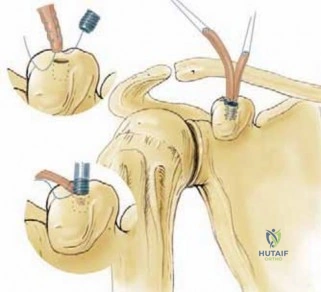
Step-by-Step Surgical Approach and Fixation Technique
The execution of the surgical plan demands a deep respect for anatomical internervous and intermuscular planes. By utilizing these natural anatomical corridors, the surgeon minimizes iatrogenic denervation and devascularization of the musculature. The choice of fixation technique—whether it be lag screws, neutralization plates, bridge plating, intramedullary nailing, or external fixation—is dictated by the biomechanical requirements of the specific fracture pattern as established during preoperative planning.
Timing of Surgical Treatment
The optimal timing for surgical intervention is one of the most critical variables in orthopaedic trauma. Surgical procedures are broadly stratified into three distinct categories based on physiological urgency:
- Emergency Procedures (Immediate to < 6 Hours): These include open fractures requiring emergent administration of intravenous antibiotics and immediate surgical débridement; compartment syndrome requiring immediate fasciotomy; fractures with vascular compromise requiring reduction and shunting; and irreducible dislocations of major joints to prevent avascular necrosis (AVN).
- Urgent Procedures (24 to 72 Hours): These allow for a brief window to optimize the patient medically. They include polytrauma long bone stabilization in physiologically stable patients (Early Total Care) to reduce the risk of fat embolism syndrome; hip fractures in the elderly (where surgery within 48 hours significantly reduces mortality); and "second look" procedures for severe open fractures.
- Elective and Delayed Procedures (3 Days to 4 Weeks): These are operations that can be safely delayed to allow for soft tissue recovery, detailed preoperative planning, or the arrival of custom implants. This includes the definitive fixation of complex intra-articular fractures following the "Span, Scan, and Plan" protocol.
The Pathophysiology of Delayed Surgery (> 4 TO 6 WEEKS)
While delaying surgery is often necessary for soft tissue optimization, excessive delays introduce a formidable set of surgical challenges. The biological and mechanical environment of the fracture site alters significantly as the body attempts to heal the unreduced fracture. If open reductions are delayed for longer than 4 to 6 weeks, the surgeon will encounter severe obstacles.
First, musculotendinous units contract and adapt to the shortened, deformed position of the fracture, making the restoration of anatomical length exceedingly difficult and often requiring femoral distractors or skeletal traction. Second, the initial hematoma organizes into dense, fibrous scar tissue, obliterating the clearly defined internervous tissue planes and increasing the risk of iatrogenic neurovascular injury. Finally, the fracture ends undergo osteoclastic resorption, rounding off the sharp interdigitating cortical edges that normally aid in anatomical reduction, while early woven bone (callus) forms, which must be meticulously taken down. Operating on a fracture at 6 weeks is fundamentally different from operating at 6 days; the procedure transitions from a standard ORIF to a complex nonunion or malunion takedown, often requiring autogenous bone grafting to stimulate osteogenesis.
Complications, Incidence Rates, and Salvage Management
Despite meticulous planning and flawless surgical execution, complications in orthopaedic trauma are inevitable. The trauma surgeon must be adept not only at preventing these adverse events but also at recognizing them early and executing definitive salvage procedures. Complications can be broadly categorized into biological failures (infection, nonunion), mechanical failures (implant breakage, loss of fixation), and systemic complications (venous thromboembolism, fat embolism syndrome).
Implant Retention, Removal, and Refracture Risks
Surgical intervention inherently involves the introduction of foreign materials. The lifecycle of these implants must be factored into the initial surgical indication. Implants frequently require removal due to prominent hardware, deep infection, or patient discomfort. Hardware removal carries the attendant risks of a second surgical procedure, including neurovascular injury and anesthetic complications. Refractures have been well-documented following the removal of both internal implants and external fixation frames due to stress shielding (where the rigid implant bypasses physiological load, leading to cortical osteopenia) or the creation of stress risers at empty screw holes. Never underestimate the complexity of hardware removal; stripped screw heads, cold-welded locking mechanisms, and bony overgrowth can turn a "simple" removal into a grueling procedure.
Nonunion and Infection
Nonunion is a catastrophic biological failure defined as a fracture that has not healed within the expected timeframe and shows no radiographic progression of healing over three consecutive months. Nonunions are classified biologically: hypertrophic nonunions (characterized by abundant callus, indicating adequate biology but insufficient mechanical stability) and atrophic nonunions (characterized by absent callus and resorbed bone ends, indicating a failure of biology). Hypertrophic nonunions are salvaged by improving mechanical stability (e.g., exchanging to a larger intramedullary nail), whereas atrophic nonunions require biological augmentation (e.g., autologous iliac crest bone grafting) in addition to stable fixation.
Surgical Site Infection (SSI) and post-traumatic osteomyelitis represent the most devastating complications. The presence of a biofilm on orthopaedic implants renders systemic antibiotics largely ineffective. Deep infections require aggressive surgical débridement, removal of loose hardware, copious irrigation, and the placement of local antibiotic delivery systems (e.g., PMMA beads), followed by a prolonged course of culture-directed intravenous antibiotics.
Complications and Salvage Matrix
| Complication | Estimated Incidence | Primary Salvage Management Strategy |
|---|---|---|
| Surgical Site Infection (Deep) | 2% - 15% (varies by open fracture grade) | Radical I&D, hardware removal (if loose), antibiotic spacers, IV antibiotics, soft tissue flap coverage. |
| Hypertrophic Nonunion | 5% - 10% | Increase mechanical stability (e.g., exchange nailing, compression plating). No bone graft typically needed. |
| Atrophic Nonunion | 3% - 8% | Biological augmentation (autogenous bone graft, RIA, BMPs) combined with absolute rigid internal fixation. |
| Hardware Failure / Breakage | 1% - 5% | Revision ORIF with longer/thicker implants, addressing the underlying cause of delayed union or nonunion. |
| Venous Thromboembolism (DVT/PE) | 1% - 10% (despite prophylaxis) | Systemic anticoagulation (LMWH, DOACs, or Warfarin); consider IVC filter if anticoagulation is contraindicated. |
Phased Post-Operative Rehabilitation Protocols
The surgical procedure is merely the first phase of the patient's recovery journey. The ultimate success of orthopaedic trauma surgery is heavily dependent on a structured, phased, and meticulously supervised post-operative rehabilitation protocol. The surgeon must communicate clearly with the physical therapy team regarding the mechanical stability of the construct, the quality of the patient's bone, and the specific limitations required to protect the soft tissue envelope and the healing fracture.
Phase I: Immediate Post-Operative Period (Weeks 0-2)
The primary goals of the immediate post-operative phase are pain control, edema management, wound healing, and the prevention of systemic complications such as deep vein thrombosis (DVT) and pneumonia. Patients are typically mobilized out of bed on post-operative day one. Weight-bearing status is strictly dictated by the fracture pattern and fixation method. Diaphyseal fractures treated with intramedullary nails may allow for Weight-Bearing As Tolerated (WBAT), whereas complex periarticular fractures treated with plates require strict Non-Weight-Bearing (NWB) or Toe-Touch Weight-Bearing (TTWB) to prevent implant failure. Early, passive Range of Motion (ROM) of adjacent joints is initiated to prevent arthrofibrosis and capsular contracture, provided it does not place undue stress on the surgical repair.
Phase II: Intermediate Healing Phase (Weeks 2-8)
During this phase, the soft tissues have generally healed, and sutures or staples are removed. The focus shifts to restoring active range of motion, improving muscular endurance, and gradually increasing weight-bearing forces as dictated by radiographic evidence of callus formation (in cases of secondary healing) or the obliteration of fracture lines (in primary healing). Proprioceptive training is initiated, and patients are transitioned from walkers to crutches or canes. The surgeon closely monitors serial radiographs to ensure there is no loss of reduction, hardware failure, or early signs of delayed union.
Phase III: Advanced Rehabilitation and Return to Function (Weeks 8-24+)
As the fracture achieves clinical and radiographic union, the rehabilitation protocol advances to aggressive strengthening, plyometrics, and work-hardening programs. Weight-bearing is progressed to full, unassisted ambulation. For athletes or high-demand manual laborers, sports-specific or occupational-specific drills are incorporated. The ultimate goal of this final phase is to return the patient to their pre-injury level of baseline function, maximizing the functional potential of the conserved extremity.
Summary of Landmark Literature and Clinical Guidelines
The modern principles of orthopaedic trauma surgery are not arbitrary; they are the culmination of decades of rigorous clinical research, randomized controlled trials, and the establishment of evidence-based guidelines. A profound understanding of this landmark literature is essential for any practicing trauma surgeon.
The evolution of soft tissue management in open fractures is anchored by the seminal work of Gustilo and Anderson (1976), whose classification system remains the universal language for describing open injuries and guiding antibiotic prophylaxis. Similarly, the Tscherne Classification (1982) forced the orthopaedic community to recognize the critical importance of the closed soft tissue envelope, fundamentally altering the timing of surgical interventions.
In the realm of biomechanics and fracture healing, Stephan Perren's Strain Theory (1979) revolutionized our understanding of how mechanical environments dictate biological responses, leading to the development of the Point Contact Fixator (PC-Fix) and the modern Locking Compression Plate (LCP). This shift from mechanical rigidity to biological osteosynthesis is the cornerstone of contemporary trauma surgery.
For the management of polytraumatized patients, the concept of Damage Control Orthopaedics (DCO), popularized by Scalea et al. (2000) and extensively refined by Pape et al., established that early definitive care (Early Total Care) in physiologically unstable patients exacerbates the "lethal triad" of coagulopathy, hypothermia, and acidosis. DCO mandates rapid, temporary external fixation to stabilize the skeleton, followed by physiological resuscitation in the ICU, and delayed definitive fixation once the patient's SIRS response has abated.
Finally, large-scale multicenter trials, such as the SPRINT Trial (2008), have provided high-level evidence guiding the choice of surgical implants (e.g., reamed versus unreamed intramedullary nailing in tibial shaft fractures), ensuring that modern surgical principles are continuously refined by rigorous, peer-reviewed data.
Detailed Chapters & Topics
Dive deeper into specialized chapters regarding principles-of-surgical-treatment