Exploring Every Approach to the Cervical: A Surgeon's Guide
Key Takeaway
Here are the crucial details you must know about Exploring Every Approach to the Cervical: A Surgeon's Guide. An approach to the cervical spine involves surgical access, primarily categorized as anterior (C3-T1) or posterior (C1-C7). These procedures address conditions like spinal canal stenosis, herniated discs, tumors, or instability. Indications for an anterior approach to the cervical spine include decompression, fusion, stabilization, or disc replacement, aimed at alleviating neurological symptoms or correcting deformities.
A 55-year-old patient presents with a 6-month history of worsening gait instability, fine motor deficit in the hands, and hyperreflexia. MRI reveals multilevel cervical spondylotic myelopathy (CSM) from C3-C6 with cord signal change. The patient has a neutral sagittal alignment. What are your surgical considerations, and how do you approach the decision between an anterior and posterior index procedure?

Candidate: I would consider the patient's age and neurological status. Since they have multilevel disease (C3-C6), I would likely offer a posterior approach such as a laminectomy and fusion or a laminoplasty, as this is safer for multiple levels and avoids the risks associated with anterior multi-level corpectomy.
The candidate fails to address the "Cervical Alignment" as the primary determinant. Simply choosing the posterior approach for all multilevel cases is a failure of clinical reasoning. You must mention why you are not choosing an anterior approach (e.g., sagittal balance, number of levels, and patient's bone quality).
A high-scoring answer follows a structured approach: 1. Alignment: Since the patient has preserved lordosis, a posterior procedure is viable. If kyphosis were present, anterior decompression would be mandatory. 2. Pathology: If compression is predominantly ventral, anterior is preferred. If dorsal (ligamentum flavum hypertrophy) or multilevel OPLL, posterior is standard. 3. Level count: Acknowledging that >3 levels often favors posterior to reduce surgical time and swallowing complications (dysphagia). 4. Comorbidities: Mentioning the risk of C5 palsy in posterior decompression and the potential for better biomechanical restoration of lordosis anteriorly.
During a standard C5-C6 ACDF, you notice a potential injury to the recurrent laryngeal nerve (RLN) after your retractor placement. How do you assess this, what are the risk factors, and how do you mitigate this during your exposure?
Candidate: The RLN is at risk during retraction. I would use the left-sided approach to minimize risk on the right. I would check the nerve intraoperatively and deflate the cuff if needed.
The candidate ignores the specific anatomy of the right vs. left RLN and fails to describe active intraoperative mitigation strategies. Suggesting "checking the nerve" is clinically unrealistic as the nerve is rarely visualized in a standard approach.
The candidate should state: 1. Anatomical Basis: The right RLN loops under the subclavian and ascends obliquely, making it more vulnerable to traction; the left is more vertical in the tracheoesophageal groove. 2. Prevention: Use the left approach (unless pathology dictates otherwise). 3. Intraoperative Mitigation: Periodic release of the self-retaining retractors, using smooth-tipped retractors, and deflating/re-inflating the ETT cuff to reduce pressure on the nerve. 4. Assessment: Post-op voice evaluation is key; if dysphonia persists, ENT referral for laryngoscopy is required to assess cord palsy.
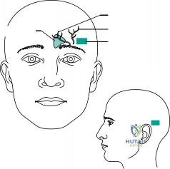
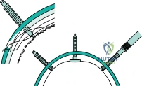
Describe the "safe zone" for halo pin placement to avoid major complications. What are the potential complications of halo immobilization in the elderly population?

Candidate: Anterior pins go in the forehead. I tighten them to 8 inch-pounds. In the elderly, it's dangerous because they might get pneumonia.
Vague anatomical descriptions ("forehead") are unacceptable. The candidate misses the supraorbital/supratrochlear neurovascular structures and fails to provide specific clinical risks for the elderly population, such as swallow dysfunction or mechanical falls.
Safe Zone: Anterior pins should be lateral to the middle third of the eyebrow (avoiding the supraorbital and supratrochlear nerves), below the equator of the skull, and above the orbital rim. Posterior pins are placed diagonally opposite, clearing the mastoid. Torque: 8 inch-pounds. Elderly Complications: Increased risk of respiratory failure, loss of balance/falls due to shifted center of gravity, pressure ulcers beneath the vest, and potential for pin loosening due to osteopenic bone.
