Failed Spine Surgery: Comprehensive Evaluation and Revision Strategies
Key Takeaway
Failed spine surgery, or Post-Laminectomy Syndrome, presents a complex clinical challenge characterized by persistent or recurrent pain following spinal intervention. Successful revision surgery depends entirely on identifying a distinct, surgically correctable anatomical lesion. This guide details the evidence-based evaluation of the failed spine, advanced diagnostic imaging, strict indications for reoperation, and meticulous surgical techniques required to navigate epidural fibrosis, correct pseudarthrosis, and manage adjacent segment pathology.
Comprehensive Introduction and Patho-Epidemiology
One of the most formidable and consistently humbling challenges in both orthopaedic spine surgery and neurosurgery is the management of failed spine surgery, clinically codified as Failed Back Surgery Syndrome (FBSS) or Post-Laminectomy Syndrome. Despite exponential advancements in minimally invasive techniques, high-resolution neuroimaging, and sophisticated spinal instrumentation, a substantial subset of patients continues to experience persistent, recurrent, or worsened pain following primary lumbar spine surgery. The nomenclature itself is somewhat of a misnomer, as "failure" may not necessarily imply a technical surgical error, but rather a failure to achieve the anticipated clinical resolution of the patient's complex pain syndrome. This condition represents a heterogeneous spectrum of pathophysiological states, ranging from discrete, correctable anatomical lesions to diffuse, intractable neuropathic pain syndromes completely devoid of a structural target.
The socioeconomic burden of FBSS is immense, precipitating prolonged functional disability, profound psychological distress, and exponentially escalating healthcare utilization costs. Epidemiological data suggest that the incidence of FBSS ranges from 10% to 40% following primary lumbar laminectomy or discectomy. The success rates for revision reoperation are historically variable and consistently inferior to primary interventions, reported in the literature to range widely from 31% to 80%. Crucially, the "law of diminishing returns" strictly applies to revision spine surgery: as the frequency of repeat back surgeries increases, the probability of a satisfactory clinical outcome decreases precipitously. A second operation may offer a 50% chance of success, a third 30%, and a fourth less than 15%.
The etiology of surgical failure is multifactorial and can be systematically categorized into preoperative, intraoperative, and postoperative domains. Preoperative failures are predominantly driven by improper patient selection, which landmark studies by Spengler et al. and Long et al. identified as the single major cause of primary surgical failure. Operating on patients with predominantly axial discogenic back pain without frank instability, or intervening in the presence of severe, unoptimized psychosocial comorbidities (e.g., major depressive disorder, active secondary gain, workers' compensation litigation), inevitably leads to suboptimal outcomes. Misdiagnosis, such as failing to recognize extra-spinal pain generators like sacroiliac joint dysfunction or hip osteoarthritis, further contributes to this preoperative failure cohort. Intraoperative errors include inadequate decompression (retained fragments, missed lateral recess stenosis), the catastrophic wrong-level surgery, or the creation of iatrogenic instability through excessive resection of the pars interarticularis or facet joints.
Patients undergoing revision spine surgery must be meticulously counseled to expect an improvement in the severity of their symptoms and an enhancement in functional capacity, rather than the complete and total relief of pain. Managing preoperative expectations is arguably as critical as the surgical execution itself. The decision to reoperate must be approached with extreme caution, guided by a rigorous diagnostic workup, a profound understanding of altered spinal biomechanics, and the absolute identification of a distinct, surgically correctable pathoanatomic lesion that correlates directly with the patient's clinical presentation.
Detailed Surgical Anatomy and Biomechanics
The anatomical landscape of the previously operated spine is fundamentally altered, presenting a hostile and disorienting environment for the revision surgeon. The pristine epidural fat that normally buffers the dura mater and neural elements is entirely replaced by dense, tenacious epidural fibrosis. This avascular scar tissue completely obliterates normal tissue planes, tethering the nerve roots to the anterior disc space, the posterior longitudinal ligament (PLL), and the residual bony elements. Consequently, the normal excursion of the nerve root during physiological movement is restricted, rendering it highly susceptible to tension-induced neuropraxia or frank axonotmesis during surgical mobilization. The loss of the midline ligamentous complex (supraspinous and interspinous ligaments) and the disruption of the paraspinal musculature further compromise the posterior tension band, altering the normal load-sharing biomechanics of the functional spinal unit.
Iatrogenic instability is a critical biomechanical consequence of overly aggressive primary decompression. The lumbar facet joints are highly specialized, diarthrodial joints that resist anterior shear forces and excessive axial rotation. Biomechanical studies have definitively demonstrated that bilateral resection of greater than 50% of the facet joints, or inadvertent violation of the pars interarticularis, critically destabilizes the segment. This leads to postoperative spondylolisthesis, dynamic foraminal narrowing, and accelerated degeneration of the intervertebral disc due to abnormal load transmission. The instantaneous axis of rotation (IAR) shifts aberrantly, increasing the mechanical stress on the annulus fibrosus and predisposing the patient to recurrent herniations or progressive disc space collapse.
Adjacent Segment Pathology (ASP) represents another profound biomechanical failure, typically observed following rigid spinal fusion. By eliminating motion at the fused segment, the kinematic requirements of the lumbar spine are disproportionately transferred to the adjacent cranial and caudal transitional zones. This hypermobility at the adjacent segments leads to increased intradiscal pressure, accelerated facet arthropathy, and hypertrophy of the ligamentum flavum. Over time, this compensatory mechanical stress culminates in adjacent segment stenosis or instability, often necessitating extension of the fusion construct. The biomechanical principle dictates that the longer the rigid lever arm (the fusion mass), the greater the stress concentrated at the adjacent mobile segments.
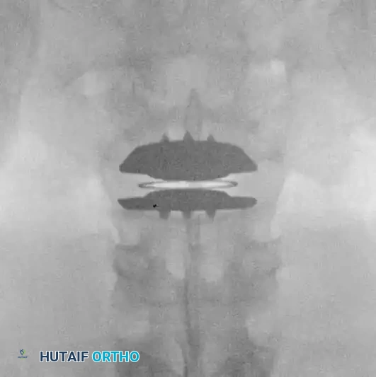
In cases where the primary cause of failure is internal disc derangement or adjacent segment disease without gross instability, motion-preserving technologies have been utilized as an alternative to rigid fusion. The goal is to restore disc height, decompress the neural elements indirectly via ligamentotaxis, and maintain segmental kinematics to protect adjacent levels. The Charité Total Disc Replacement (TDR) represents a historical milestone in this biomechanical approach. It consists of two metallic endplates and a mobile ultra-high-molecular-weight polyethylene (UHMWPE) sliding core. By allowing flexion, extension, lateral bending, and axial rotation, TDR theoretically reduces the abnormal stress transferred to adjacent segments.
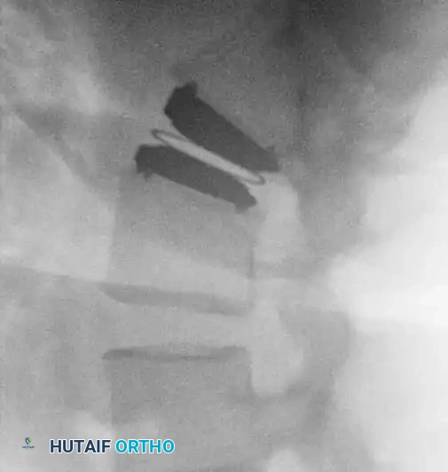
Fig. 39-47 A, Anteroposterior view of patient with internal disc derangement treated with Charité total disc replacement.
Fig. 39-47 B, Lateral view of patient with internal disc derangement treated with Charité total disc replacement.
Exhaustive Indications and Contraindications
The philosophical cornerstone of revision spine surgery is the absolute necessity of identifying a distinct, surgically correctable anatomical problem that corresponds flawlessly with the patient's radicular or mechanical symptoms. Operating on a patient with generalized, non-specific pain in the setting of an anatomically stable and adequately decompressed spine is a guaranteed pathway to further clinical deterioration. The recurrence or intensification of pain in the subacute or late period after primary surgery should initially be managed with aggressive conservative modalities. Only when targeted physical therapy, non-steroidal anti-inflammatory drugs (NSAIDs), membrane stabilizers (e.g., gabapentinoids), and fluoroscopically guided epidural steroid injections fail should surgical intervention be entertained.
Indications for revision surgery are strictly defined. A recurrent disc herniation, occurring in 5% to 15% of primary discectomies, is a prime indication, particularly if the patient experienced a distinct pain-free interval of greater than six months following the index procedure. Pseudarthrosis, defined as the failure of a fusion mass to consolidate solid trabecular bone across the intended arthrodesis site by one year postoperatively, requires revision if accompanied by hardware loosening, progressive deformity, or intractable mechanical back pain. Iatrogenic instability, wrong-level surgery, and symptomatic adjacent segment disease with objective neurological deficits or claudication also constitute definitive indications for re-exploration and stabilization.
Conversely, contraindications to revision surgery must be rigorously respected. Diffuse adhesive arachnoiditis, characterized by the clumping of nerve roots within the thecal sac on MRI, is an absolute contraindication to further decompressive surgery; intervention will only exacerbate the inflammatory cascade. Patients presenting with pure axial back pain without demonstrable instability, those with active, uncontrolled psychiatric disorders, or individuals exhibiting profound Waddell's signs of non-organic pain behavior should be diverted to multidisciplinary pain management programs. The presence of active infection without a plan for definitive source control and hardware removal (if applicable) also precludes elective revision for pain.
| Category | Specific Condition | Clinical Rationale |
|---|---|---|
| Absolute Indications | Progressive Neurological Deficit | Impending permanent nerve damage (e.g., foot drop, cauda equina syndrome) requires emergent decompression. |
| Absolute Indications | Symptomatic Pseudarthrosis | Hardware failure/loosening with mechanical pain requires revision arthrodesis and stabilization. |
| Absolute Indications | Wrong-Level Surgery | Catastrophic index error requiring immediate decompression of the correct symptomatic level. |
| Relative Indications | Recurrent Disc Herniation | Highly successful if radicular pain > axial pain and patient had >6 months pain-free interval. |
| Relative Indications | Adjacent Segment Pathology | Requires extension of fusion if conservative measures fail and claudication/radiculopathy is severe. |
| Relative Contraindications | Active Workers' Compensation | High correlation with poor postoperative outcomes and secondary gain behaviors; requires careful selection. |
| Absolute Contraindications | Diffuse Adhesive Arachnoiditis | Surgical intervention worsens the inflammatory scarring of the cauda equina; strictly medical management. |
| Absolute Contraindications | Pure Axial Pain without Instability | Surgery for nonspecific back pain in a previously operated spine yields dismal results. |
Pre-Operative Planning, Templating, and Patient Positioning
Preoperative planning for the failed spine is an exhaustive process that relies heavily on advanced, multi-modal diagnostic imaging. Magnetic Resonance Imaging (MRI) with and without intravenous gadolinium contrast is the undisputed gold standard. The contrast is critical for differentiating between retained/recurrent disc material and epidural fibrosis; vascularized scar tissue will enhance with gadolinium, whereas an avascular recurrent disc fragment will typically remain dark centrally, occasionally demonstrating a thin peripheral rim of enhancement. High-resolution Computed Tomography (CT) with sagittal, coronal, and 3D reconstructions is mandatory for evaluating the bony architecture. CT is essential for assessing the integrity of a previous fusion mass, identifying occult pseudarthrosis, measuring pedicle morphology for revision trajectory planning, and localizing retained osteophytes or areas of severe lateral recess stenosis. Dynamic flexion-extension radiographs remain vital for assessing gross translational or angular instability.
When non-invasive imaging is equivocal or reveals multi-level degenerative changes, diagnostic injections are deployed to isolate the specific pain generator. Selective Nerve Root Blocks (SNRB) under fluoroscopic guidance can confirm if a specifically compressed root is the source of the patient's radicular pain. Medial branch blocks are utilized to diagnose facet-mediated mechanical pain, while provocative discography—though controversial—may help identify symptomatic internal disc derangement at adjacent segments. A positive response to a differential spinal block can help rule out somatic pain, highlighting sympathetic or psychogenic pain syndromes that contraindicate surgical intervention.
Surgical templating is a critical phase of preparation. The surgeon must anticipate the need to remove stripped, broken, or cold-welded hardware. Specialized extraction sets must be available in the operating room. Planning for revision pedicle screw fixation requires templating for larger diameter "rescue" screws to achieve adequate purchase in previously tapped pedicles. Alternatively, the surgeon may plan for a Cortical Bone Trajectory (CBT) or extension of the construct to the pelvis (iliac or S2-alar-iliac screws) to bypass compromised bone and achieve rigid distal fixation. The use of intraoperative navigation or robotic assistance is increasingly utilized to safely navigate the distorted bony anatomy.
Patient positioning is executed with meticulous care to optimize surgical exposure and minimize physiological stress. The patient is typically positioned prone on a radiolucent Jackson table or Wilson frame. It is absolutely critical that the abdomen hangs completely free; any compression of the inferior vena cava will divert venous return through the epidural Batson's plexus, resulting in massive epidural venous engorgement and uncontrollable intraoperative hemorrhage. All pressure points must be heavily padded to prevent peripheral neuropathies. Given the high risk of iatrogenic nerve injury during dissection of epidural fibrosis, continuous intraoperative neuromonitoring—including Somatosensory Evoked Potentials (SSEPs), Motor Evoked Potentials (MEPs), and spontaneous Electromyography (sEMG)—is highly recommended and considered standard of care in complex revision scenarios.
Step-by-Step Surgical Approach and Fixation Technique
The surgical approach to the previously operated spine is technically demanding, fraught with peril, and requires a methodical, unhurried technique. The primary incision generally incorporates the previous surgical scar, excising the old cicatrix to promote optimal wound healing. However, the incision must be extended proximally and distally to expose "virgin," unoperated anatomy. Dissection is carried down sharply through the subcutaneous tissues. The fascial incision should ideally be made slightly off-midline or cleanly through the scarred midline, taking extreme care not to plunge, as the dura may be entirely exposed and tethered directly beneath the fascia due to a previous wide laminectomy.
Subperiosteal dissection must strictly adhere to the "Outside-In" principle. Muscle stripping must commence on normal, unoperated bone—typically the spinous process or intact lamina of the level immediately cranial or caudal to the previous surgical defect. Using Cobb elevators and electrocautery, the surgeon works laterally to identify the facet joints and the transverse processes. Only after the normal anatomical boundaries are established does the dissection proceed medially toward the scarred defect. The surgeon must absolutely avoid dissecting directly into the center of the epidural scar, as this is the most common mechanism for catastrophic dural tears or nerve root avulsions.
Navigating the epidural fibrosis requires identifying constant, reliable bony landmarks. The medial border of the pedicle and the pars interarticularis serve as the "lighthouses" of the lumbar spine. Once the normal dura is identified proximally and distally, micro-curettes, Penfield dissectors, and Kerrison rongeurs are used to carefully detach the scar tissue from the bony margins. The cardinal rule of revision decompression is: Do not pull on the scar. Epidural fibrosis is often densely adherent to the fragile dura and underlying nerve roots. Sharp dissection with a #15 blade or micro-scissors is vastly safer than blunt avulsion. If a recurrent disc herniation is present, it is often located ventral to the scarred nerve root. To mobilize the root safely, a wider bony decompression—often requiring a complete facetectomy—must be performed to allow medial retraction of the neural elements without inducing tension neuropraxia.
If the revision is indicated for pseudarthrosis, iatrogenic instability, or requires extensive facet resection for adequate decompression, a robust stabilization and fusion procedure is mandatory. Previous hardware is removed, and the pedicle tracts are meticulously evaluated. If the tracts are widened, larger diameter, longer "rescue" screws are inserted. To maximize fusion rates in the biologically compromised revision bed, an interbody fusion (PLIF or TLIF) is highly advantageous. This provides anterior column support, restores neuroforaminal height, and places the bone graft under compression, adhering to Wolff's Law. Autologous iliac crest bone graft (ICBG) remains the gold standard osteogenic material. However, given the hostile local environment, this is frequently supplemented with local bone and potent osteoinductive biologics, such as Recombinant Human Bone Morphogenetic Protein-2 (rhBMP-2), to ensure solid arthrodesis.
Complications, Incidence Rates, and Salvage Management
Revision spine surgery inherently carries a significantly higher complication profile compared to primary index procedures. The surgeon must be acutely aware of these risks and possess the technical armamentarium to manage them decisively. The incidence of complications scales linearly with the complexity of the revision, the number of previous operations, and the extent of the patient's medical comorbidities. Preoperative counseling must explicitly detail these elevated risks, ensuring true informed consent is obtained. The hostile biological environment, characterized by diminished local vascularity, dense scar tissue, and altered biomechanical loading, creates a "perfect storm" for both intraoperative misadventures and postoperative failures.
Incidental durotomy (dural tear) is the most frequent intraoperative complication, with incidence rates in revision surgery reported between 10% and 20%, compared to 2-4% in primary cases. The dura is often paper-thin and intimately fused with the overlying ligamentum flavum or epidural scar. If a durotomy occurs, it must be recognized immediately and repaired primarily using 4-0 or 5-0 non-absorbable sutures (e.g., Prolene, Nurolon, or Gore-Tex) in a running, locked, or interrupted fashion. The repair is typically augmented with a synthetic dural sealant (hydrogel) or an autologous fascial/muscle patch. Postoperatively, the patient may be managed with flat bed rest for 24-48 hours to reduce hydrostatic CSF pressure on the repair. In refractory cases presenting with a persistent CSF fistula or pseudomeningocele, the placement of a subarachnoid lumbar drain or a return to the operating room for definitive over-sewing is required.
Nerve root injury, resulting in transient neuropraxia or permanent motor/sensory deficits, is a devastating complication. The tethered nature of the nerve root makes it highly susceptible to stretch injury during retraction. Continuous intraoperative sEMG monitoring is critical for providing real-time feedback; sustained neurotonic discharges indicate excessive root irritation. If monitoring alerts occur, retraction must be immediately released, and further bony decompression (e.g., complete unroofing of the foramen) must be performed to allow tension-free mobilization. Postoperative management of a new neurological deficit requires emergent MRI to rule out an expanding epidural hematoma, followed by a high-dose corticosteroid protocol if mechanical compression is excluded.
Surgical Site Infection (SSI) and deep wound infections are markedly elevated in revision cases, approaching 5-8%. The prolonged operative time, extensive soft tissue dissection, and the presence of avascular scar tissue severely compromise local immune responses and antibiotic penetrance. Strict adherence to sterile technique, copious intraoperative irrigation (often with dilute betadine or antibiotic solutions), and the application of intrawound vancomycin powder are critical prophylactic measures. If a deep SSI develops, it requires aggressive management: emergent surgical irrigation and debridement (I&D), retention of the spinal hardware (unless grossly loose), potential placement of a negative pressure wound therapy (NPWT) device, and 6 to 12 weeks of culture-directed intravenous antibiotics managed in conjunction with an infectious disease specialist.
| Complication | Estimated Incidence (Revision) | Pathophysiology / Risk Factors | Salvage / Management Strategy |
|---|---|---|---|
| Incidental Durotomy | 10% - 20% | Thinning of dura, intimate adherence of epidural fibrosis, loss of anatomical planes. | Primary watertight suture repair, dural sealant, 24-48h flat bed rest, subarachnoid drain for refractory leaks. |
| Nerve Root Injury | 2% - 5% | Excessive medial retraction of a scarred, tethered root; thermal injury from electrocautery. | Prevent with wide bony decompression. Manage with immediate release of retraction, high-dose steroids, emergent MRI if post-op deficit. |
| Deep Surgical Site Infection | 5% - 8% | Prolonged operative time, avascular scar tissue, extensive dead space, compromised host immunity. | Emergent I&D, copious pulsatile lavage, retain stable hardware, NPWT, long-term IV culture-directed antibiotics. |
| Hardware Failure / Pullout | 5% - 10% | Poor bone stock (osteoporosis), failure to achieve anterior column support, pseudarthrosis. | Revision instrumentation with larger diameter screws, cortical bone trajectory, extension to pelvis, interbody structural support. |
| Epidural Hematoma | 1% - 3% | Venous oozing from Batson's plexus, inadequate hemostasis, coagulopathy. | Prevent with meticulous hemostasis and closed suction drains. Manage with emergent surgical evacuation if causing neurological deficit. |
Phased Post-Operative Rehabilitation Protocols
The postoperative management of a revision spine patient is as critical to the final clinical outcome as the surgical execution itself. It necessitates a highly structured, multidisciplinary approach involving the orthopaedic surgeon, physiatrists, physical therapists, and pain management specialists. The protocol must be phased, respecting the biological timeline of tissue healing and bone graft incorporation, while simultaneously combatting the deleterious effects of prolonged immobilization.
The immediate postoperative phase (0 to 2 weeks) focuses on medical stabilization, complication prevention, and early mobilization. Neurological checks are performed rigorously every 2 to 4 hours to detect early signs of epidural hematoma or evolving neuropraxia. A closed suction drain is typically left in the subfascial space for 24 to 48 hours, or until output is less than 30 cc per shift, to prevent hematoma accumulation. Deep vein thrombosis (DVT) prophylaxis is initiated with mechanical sequential compression devices immediately, and chemical prophylaxis (e.g., low molecular weight heparin) is carefully considered on postoperative day 1 or 2, balancing the risk of VTE against the risk of epidural hemorrhage. Patients are mobilized out of bed on postoperative day 1 with physical therapy. Depending on the complexity of the reconstruction and the patient's bone mineral density, a custom-molded rigid Thoracolumbosacral Orthosis (TLSO) may be prescribed to restrict gross motion and protect the instrumentation.
The early rehabilitation phase (2 to 6 weeks) transitions the patient to the outpatient setting. The primary focus is on wound healing, pain control, and progressive ambulation. Patients are instructed to adhere strictly to "BLT" restrictions—no Bending, Lifting (greater than 10 pounds), or Twisting. Formal physical therapy during this phase is gentle, emphasizing isometric core activation (e.g., transverse abdominis bracing), neural mobilization techniques (nerve gliding) to prevent recurrent tethering of the decompressed roots, and a progressive walking program to build cardiovascular endurance. Aggressive stretching or dynamic strengthening is strictly contraindicated during this vulnerable period of early osteogenesis.
The intermediate phase (6 to 12 weeks) marks the beginning of active functional restoration. Radiographic assessment (AP, lateral, and dynamic views) is performed at the 6-week mark to evaluate hardware integrity and early signs of fusion mass consolidation. If clinical and radiographic progress is satisfactory, the patient is gradually weaned from the TLSO brace. Physical therapy progresses to dynamic lumbar stabilization exercises, closed kinetic chain lower extremity strengthening, and aquatic therapy, which provides a buoyant environment to reduce axial loading while allowing active movement. The focus shifts toward correcting maladaptive movement patterns and restoring pelvic tilt and spinopelvic harmony.
The late rehabilitation phase (3 to 6+ months) is tailored to the patient's specific functional goals and occupational demands. Work hardening programs are initiated for patients returning to manual labor. Advanced core stabilization, proprioceptive training, and functional lifting mechanics are emphasized. It is during this phase that psychological support becomes paramount; patients with FBSS often harbor deep-seated fear-avoidance behaviors (kinesiophobia). Cognitive-behavioral therapy (CBT) integrated with physical rehabilitation yields the highest success rates in overcoming chronic pain behaviors and maximizing the patient's ultimate quality of life.
Summary of Landmark Literature and Clinical Guidelines
The contemporary treatment of failed spine surgery is heavily informed by decades of rigorous clinical research and landmark epidemiological studies. Understanding this literature is essential for the academic orthopaedic surgeon to practice evidence-based medicine and avoid repeating the historical errors of the past. The foundational work by Waddell et al. remains highly relevant today. Waddell established specific clinical criteria that portend a favorable outcome in revision discectomy: the patient must have experienced a complete, pain-free interval of at least six months following the index surgery; their primary complaint must be radicular leg pain rather than axial back pain; and a definitive, recurrent disc herniation must be unequivocally identified on advanced imaging. When these criteria are met, the success rate of revision discectomy approaches that of primary surgery.
Conversely, the literature is replete with warnings regarding poor prognostic indicators. Spengler, Long, and Finnegan independently demonstrated that improper patient selection is the genesis of the vast majority of failed back syndromes. Intervening surgically on patients with profound psychosocial distress, active litigation, or poorly localized axial pain consistently results in clinical failure, regardless of the technical perfection of the operation. Furthermore, Lehmann and LaRocca identified that extensive epidural scarring, a history of postoperative infection, and the need for repair of a pseudarthrosis significantly diminish the probability of a completely pain-free outcome.
Modern clinical guidelines, heavily influenced by data extrapolated from the Spine Patient Outcomes Research Trial (SPORT) and subsequent registry data, emphasize a highly conservative initial approach to the recurrently symptomatic postoperative spine. The consensus dictates that, in the absence of progressive neurological deficits or profound structural instability, a minimum of 6 to 12 weeks of exhaustive, multidisciplinary non-operative management is mandatory before revision surgery is contemplated. The literature strongly supports the use of targeted epidural steroid injections not only as a therapeutic modality but as a critical diagnostic tool to confirm the exact neural pain generator prior to committing to the knife.
In conclusion, the management of Failed Back Surgery Syndrome demands a rigorous, intellectually honest, and evidence-based approach. The fundamental tenet of revision surgery remains immutable: a distinct, surgically correctable anatomical lesion must be identified that correlates directly with the patient's clinical symptoms. Through meticulous preoperative evaluation, masterful surgical technique, and a profound respect for the altered biomechanics of the previously operated spine, the orthopaedic surgeon can navigate this hostile environment to restore stability and decompress neural elements. However, the ultimate clinical pearl is recognizing that conservative management, psychological optimization, and the wisdom to know when not to operate remain the surgeon's most powerful tools against the complexities of failed spine surgery.
