Orthopedic Pathology Review | Dr Hutaif Basic Science R -...
02 إبريل 2026
126 min read
76 Views
Key Takeaway
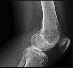
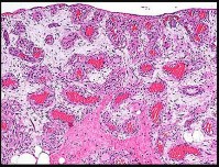
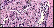
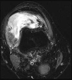
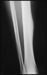
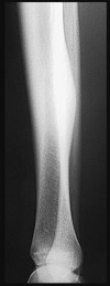
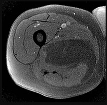
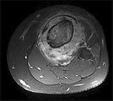
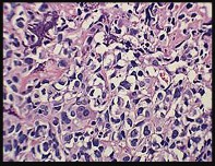
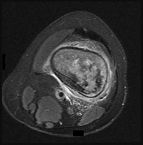
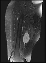
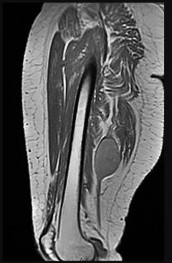
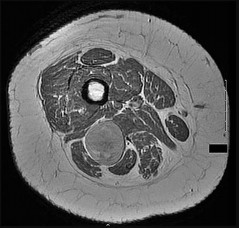
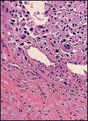
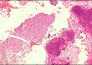
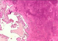
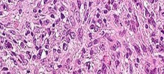
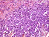
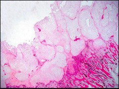
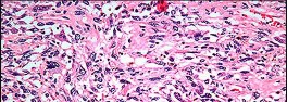
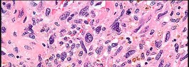
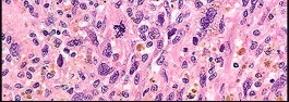
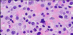
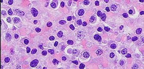
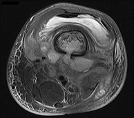
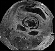
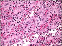
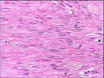
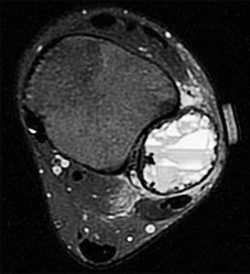
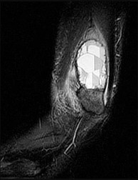
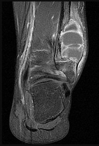
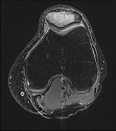
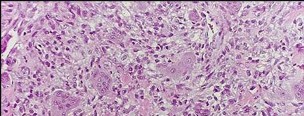
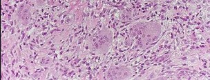
Discover the latest medical recommendations for ORTHOPEDIC MCQS ONLINE PATHOLOGY 017. Dedifferentiated liposarcoma is diagnosed via imaging and biopsy, often involving figures like those encountered in a pubmed question of figures. High-grade sarcomas require wide surgical resection. Radiation therapy reduces local recurrence, while chemotherapy remains investigational. This management contrasts with marginal resection for benign conditions like atypical lipomatous tumors, highlighting distinct treatment pathways based on pathology.
Score:
0
%
ORTHOPEDIC MCQS ONLINE PATHOLOGY 017
of 100
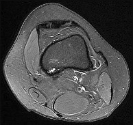
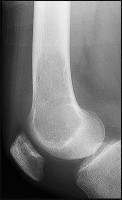
What is the diagnosis?
What is the diagnosis?
of 100
The role of surgery in this condition is best described as
The role of surgery in this condition is best described as
of 100
The role of radiation treatment for this lesion is
The role of radiation treatment for this lesion is
of 100
Chemotherapy for this condition is
Chemotherapy for this condition is
of 100
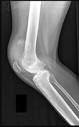
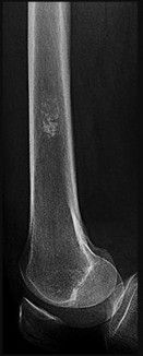
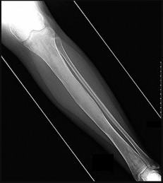
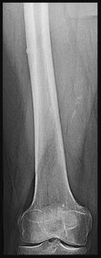
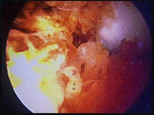
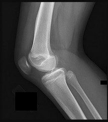
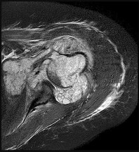
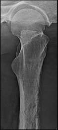
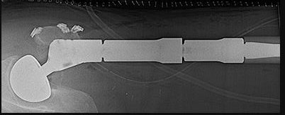
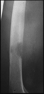
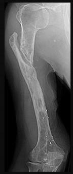
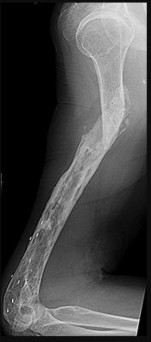
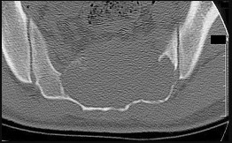
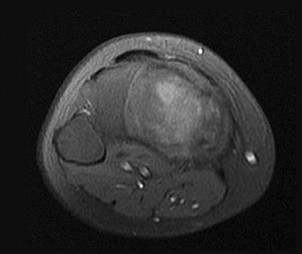
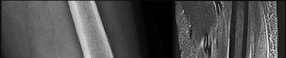
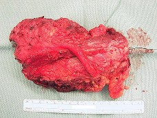
Figures 5a and 5b are the radiographs of a 74-year-old man with poorly differentiated squamous cell carcinoma of the lung. He has had an uneventful recovery after undergoing a wedge resection of his left upper lobe 6 months ago. He is experiencing left lateral knee pain, and a whole-body positron emission tomography/CT scan shows no avid area other than the lateral left distal femur. This patient has needed to use a wheelchair for 3 weeks because of his pain. You discuss these treatment options: aggressive curettage, local adjuvant treatment, cementation, and prophylactic fixation vs distal femoral resection and megaprosthesis total knee arthroplasty reconstruction. You should tell him that
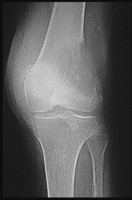
Figures 5a and 5b are the radiographs of a 74-year-old man with poorly differentiated squamous cell carcinoma of the lung. He has had an uneventful recovery after undergoing a wedge resection of his left upper lobe 6 months ago. He is experiencing left lateral knee pain, and a whole-body positron emission tomography/CT scan shows no avid area other than the lateral left distal femur. This patient has needed to use a wheelchair for 3 weeks because of his pain. You discuss these treatment options: aggressive curettage, local adjuvant treatment, cementation, and prophylactic fixation vs distal femoral resection and megaprosthesis total knee arthroplasty reconstruction. You should tell him that
of 100
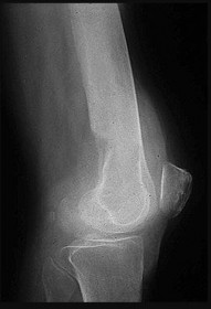
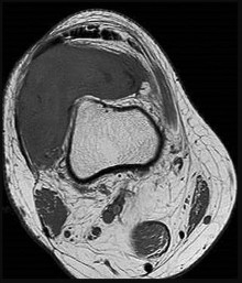
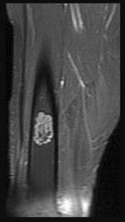
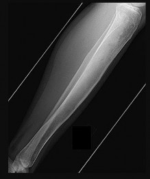
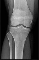
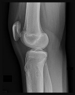
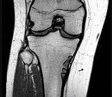
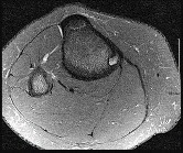
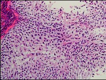
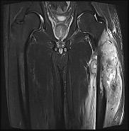
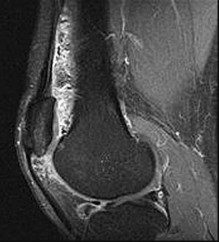
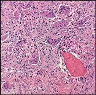
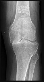
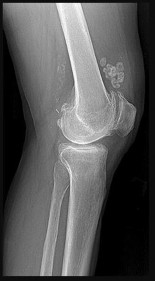
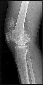
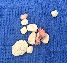
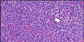
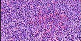
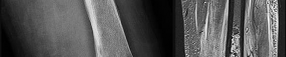
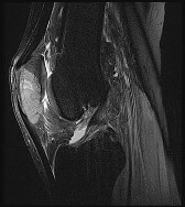
Figures 6a through 6d are the radiographs and T1-weighted sagittal and fat-saturated axial MR images of an otherwise healthy 56-year-old man who has anterior knee pain and intermittent swelling after sustaining a noncontact twisting injury. Low-power and high-power hematoxylin and eosin stained histologic specimens are shown in Figures 6e and 6f. Based on the history, radiographs, CT scan, MR imaging, and histologic findings, what is the most likely diagnosis?
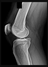
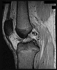
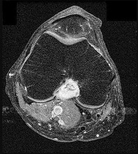
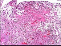
Figures 6a through 6d are the radiographs and T1-weighted sagittal and fat-saturated axial MR images of an otherwise healthy 56-year-old man who has anterior knee pain and intermittent swelling after sustaining a noncontact twisting injury. Low-power and high-power hematoxylin and eosin stained histologic specimens are shown in Figures 6e and 6f. Based on the history, radiographs, CT scan, MR imaging, and histologic findings, what is the most likely diagnosis?
of 100
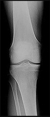
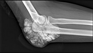
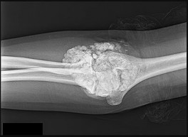
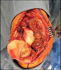
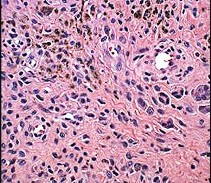
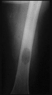
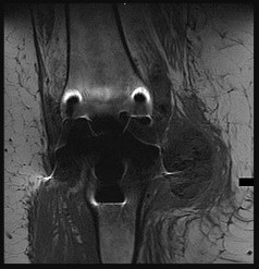
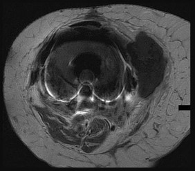
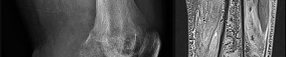
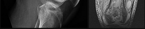
Figures 7a through 7d are the radiograph, MR images, and biopsy specimen of a 35-year-old man who has a painful, slowly enlarging knee mass. Which chromosomal translocation is characteristic of this pathology?
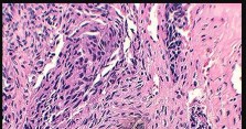
Figures 7a through 7d are the radiograph, MR images, and biopsy specimen of a 35-year-old man who has a painful, slowly enlarging knee mass. Which chromosomal translocation is characteristic of this pathology?
of 100
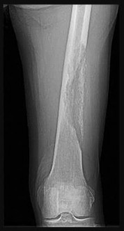
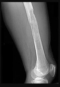
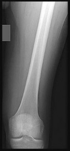
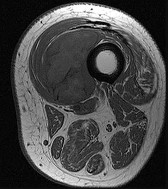
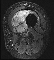
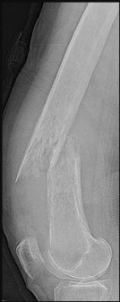
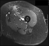
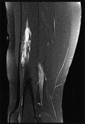
A 45-year-old woman has a painless thigh mass that is larger than 5 cm. What is the best next step?
A 45-year-old woman has a painless thigh mass that is larger than 5 cm. What is the best next step?
of 100
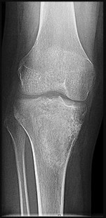
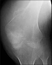
What is the most likely diagnosis?
What is the most likely diagnosis?
of 100
The most appropriate treatment of this lesion involves
The most appropriate treatment of this lesion involves
of 100
If this lesion occurred in the spine, which features would most likely be present?
If this lesion occurred in the spine, which features would most likely be present?
of 100
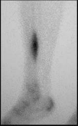
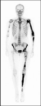
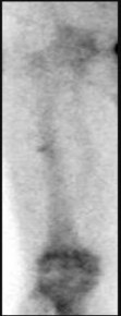
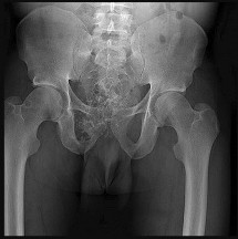
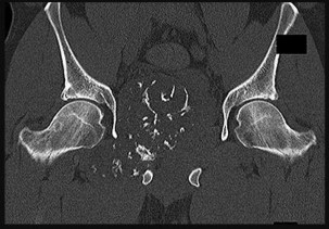
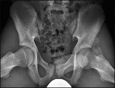
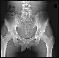
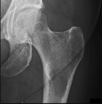
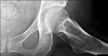
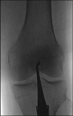
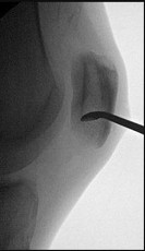
Figures 12a and 12b are a recent radiograph and a whole-body bone scan of an 81-year-old man who has hip pain and difficulty walking. His medical history is significant for obesity, hypertension, chronic kidney disease, and coronary artery disease. An examination demonstrates
moderate tenderness with passive range of motion of the left hip and an inability to actively flex the left hip against gravity. What is the best next step?
of 100
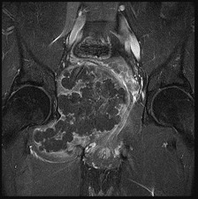
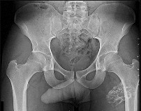
Figures 13a and 13b are the radiographs of a 57-year-old man who is seen in the emergency department. He has been experiencing left thigh pain for 2 month. Four years ago he underwent laparoscopic nephrectomy and states that he has been disease free since the resection (although he has not seen a doctor in 2 years). The pathogenesis of osteolysis in renal cell carcinoma metastatic to bone includes secretion of parathyroid hormone-related peptide (PTHrP), transforming growth factor-B (TGF-B), and vascular endothelial growth factor (VEGF), which directly cause overexpression receptor activation of nuclear factor kB ligand (RANKL) on which cells?
Figures 13a and 13b are the radiographs of a 57-year-old man who is seen in the emergency department. He has been experiencing left thigh pain for 2 month. Four years ago he underwent laparoscopic nephrectomy and states that he has been disease free since the resection (although he has not seen a doctor in 2 years). The pathogenesis of osteolysis in renal cell carcinoma metastatic to bone includes secretion of parathyroid hormone-related peptide (PTHrP), transforming growth factor-B (TGF-B), and vascular endothelial growth factor (VEGF), which directly cause overexpression receptor activation of nuclear factor kB ligand (RANKL) on which cells?
of 100
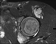
Figures 14a through 14c are the MR images of a 72-year-old man who has had a slow-growing asymptomatic mass in his thigh for more than 5 years. Cytogenetic testing on the mass reveals a ring chromosome and MDM2 expression with no 12;16 translocation. What is the most likely diagnosis?
Figures 14a through 14c are the MR images of a 72-year-old man who has had a slow-growing asymptomatic mass in his thigh for more than 5 years. Cytogenetic testing on the mass reveals a ring chromosome and MDM2 expression with no 12;16 translocation. What is the most likely diagnosis?
of 100
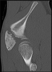
Figures 15a through 15c are the radiograph and MR images of a 16-year-old girl who experienced posterior knee pain after a dance recital 3 weeks ago; the pain resolved 1 week ago with ibuprofen use. What is the most appropriate treatment for this patient?
Figures 15a through 15c are the radiograph and MR images of a 16-year-old girl who experienced posterior knee pain after a dance recital 3 weeks ago; the pain resolved 1 week ago with ibuprofen use. What is the most appropriate treatment for this patient?
of 100
Figures 16a through 16c are the radiograph, MR image, and biopsy specimen of a 12-year-old boy who injured his leg during a soccer game. Assuming other staging study findings are negative, what is the Musculoskeletal Tumor Society (MSTS) stage of this lesion?
Figures 16a through 16c are the radiograph, MR image, and biopsy specimen of a 12-year-old boy who injured his leg during a soccer game. Assuming other staging study findings are negative, what is the Musculoskeletal Tumor Society (MSTS) stage of this lesion?
of 100
A 57-year-old man has a bone lesion that was identified on radiograph and MR imaging (Figures 17a and 17b) that were taken to evaluate anterior knee pain. An examination reveals a positive patellar apprehension test finding. The patient brings his imaging findings to his appointment, and you learn that an image-guided core needle biopsy was performed based upon the radiologist’s interpretation of the imaging. The core needle biopsy pathology interpretation text reads, “a low-
grade cartilage consistent with either enchondroma or low-grade chondrosarcoma. Clinical and imaging correlation is recommended.” What is the best next step?
of 100
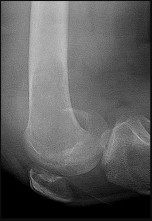
What is the most likely diagnosis?
What is the most likely diagnosis?
of 100
Despite adequate medical management, the patient continues to experience leg pain that interferes with even the lowest demands of daily living. You recommend prophylactic intramedullary nailing of the tibia with interlocking screws. Prior to the surgery, you should recommend
Despite adequate medical management, the patient continues to experience leg pain that interferes with even the lowest demands of daily living. You recommend prophylactic intramedullary nailing of the tibia with interlocking screws. Prior to the surgery, you should recommend
of 100
The most common extraskeletal manifestation of this disease is
The most common extraskeletal manifestation of this disease is
of 100
The underlying cause of the neoplasm is
The underlying cause of the neoplasm is
of 100
Figures 22a and 22b are the anteroposterior knee radiograph and an axial T2-weighted MR image of an 11-year-old boy who experienced knee pain following soccer practice. What is the best approach for biopsy?
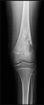
Figures 22a and 22b are the anteroposterior knee radiograph and an axial T2-weighted MR image of an 11-year-old boy who experienced knee pain following soccer practice. What is the best approach for biopsy?
of 100
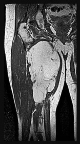
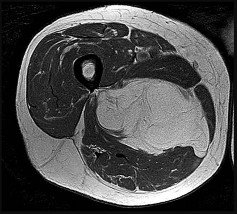
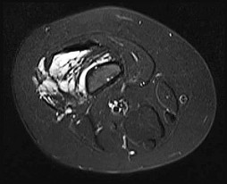
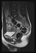
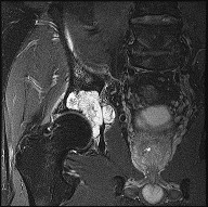
Figures 23a through 23c are the MR images of a 55-year-old woman who has experienced more than 10 years of right lower extremity pain that is radiating along the sciatic nerve distribution even though she has had multiple spine decompression procedures. She cannot sit comfortably in a chair and feels a fullness in the posterior aspect of her thigh. What is the most likely diagnosis?
Figures 23a through 23c are the MR images of a 55-year-old woman who has experienced more than 10 years of right lower extremity pain that is radiating along the sciatic nerve distribution even though she has had multiple spine decompression procedures. She cannot sit comfortably in a chair and feels a fullness in the posterior aspect of her thigh. What is the most likely diagnosis?
of 100
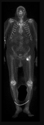
Figures 24a and 24b are the right femur radiograph and bone scan of a 71-year-old man with longstanding metastatic prostate cancer who has experienced increasing right thigh pain for 2 months. The pain is worse with activity and is alleviated with rest. He experienced similar pain in his left thigh 18 months ago and subsequently sustained a left subtrochanteric femur fracture after a low-energy twisting injury. He was successfully treated with an intramedullary nail. He had been receiving zoledronic acid for 4 years prior to the fracture. This patient’s history includes heavy steroid use. His current symptoms are most likely the result of
Figures 24a and 24b are the right femur radiograph and bone scan of a 71-year-old man with longstanding metastatic prostate cancer who has experienced increasing right thigh pain for 2 months. The pain is worse with activity and is alleviated with rest. He experienced similar pain in his left thigh 18 months ago and subsequently sustained a left subtrochanteric femur fracture after a low-energy twisting injury. He was successfully treated with an intramedullary nail. He had been receiving zoledronic acid for 4 years prior to the fracture. This patient’s history includes heavy steroid use. His current symptoms are most likely the result of
of 100
Denosumab, the human monoclonal antibody that specifically binds and inactivates receptor activator of nuclear factor-kB ligand (RANKL), is commonly used in the setting of metastatic disease. Its cell surface receptor is expressed by
Denosumab, the human monoclonal antibody that specifically binds and inactivates receptor activator of nuclear factor-kB ligand (RANKL), is commonly used in the setting of metastatic disease. Its cell surface receptor is expressed by
of 100
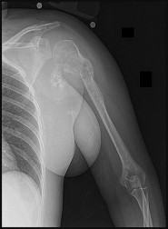
Figure 26 is the posteroranterior chest radiograph of a 76-year-old man with an atraumatic gradually enlarging mass overlying his left clavicle that has been present for 6 months. There are no changes in overlying skin. His only noteworthy medical history involves facial squamous cell carcinomas that have been successfully removed surgically.
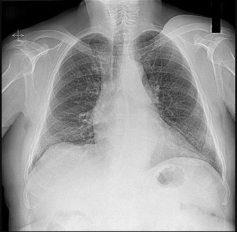
Figure 26 is the posteroranterior chest radiograph of a 76-year-old man with an atraumatic gradually enlarging mass overlying his left clavicle that has been present for 6 months. There are no changes in overlying skin. His only noteworthy medical history involves facial squamous cell carcinomas that have been successfully removed surgically.
of 100
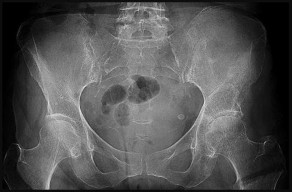
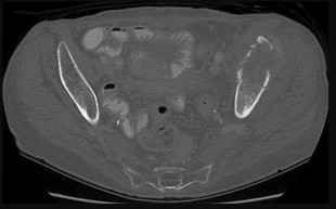
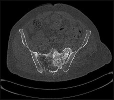
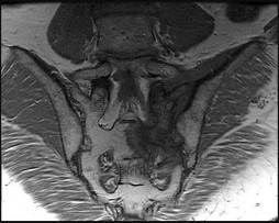
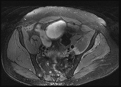
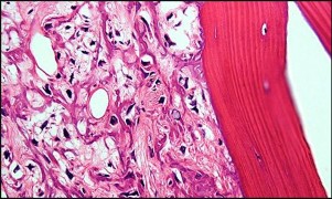
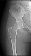
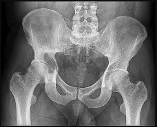
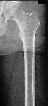
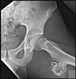
A 63-year-old man with right hip pain was followed 8 years ago for an incidental intraosseous lesion in the right periacetabular and ischial region that was isointense with fat on all images. He was discharged from follow-up after 3 years when no change was documented. He began experiencing pain in his hip, and a bone scan showed grade 3 uptake. New MR imaging was obtained, and an axial image at the level of the hip is shown in Figure 27. A PET/CT scan shows dramatic activity in the lesion without any other area of activity.
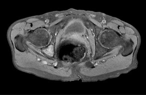
A 63-year-old man with right hip pain was followed 8 years ago for an incidental intraosseous lesion in the right periacetabular and ischial region that was isointense with fat on all images. He was discharged from follow-up after 3 years when no change was documented. He began experiencing pain in his hip, and a bone scan showed grade 3 uptake. New MR imaging was obtained, and an axial image at the level of the hip is shown in Figure 27. A PET/CT scan shows dramatic activity in the lesion without any other area of activity.
of 100
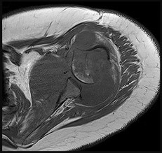
Figure 28 is the MR image of a 65-year-old man with an American Joint Committee on Cancer III anterior arm pleomorphic intermediate- to high-grade sarcoma. The patient is now considering treatment options. He underwent a wide excision at an outside hospital 2 years previously. The treating surgeon recommended an amputation, and the patient is now seeking a second opinion. Imaging studies reveal no other sites of disease.
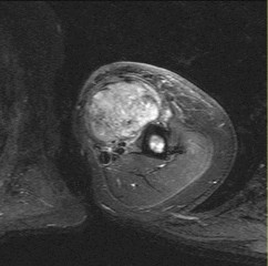
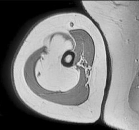
Figure 28 is the MR image of a 65-year-old man with an American Joint Committee on Cancer III anterior arm pleomorphic intermediate- to high-grade sarcoma. The patient is now considering treatment options. He underwent a wide excision at an outside hospital 2 years previously. The treating surgeon recommended an amputation, and the patient is now seeking a second opinion. Imaging studies reveal no other sites of disease.
of 100
Figure 30 is the MR image of a 29-year-old man with a large and enlarging thigh mass. Needle biopsy findings are inconclusive.
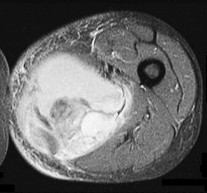
Figure 30 is the MR image of a 29-year-old man with a large and enlarging thigh mass. Needle biopsy findings are inconclusive.
of 100
Figure 31 is the sagittal MR image of a 30-year-old man with a clear-cell sarcoma of the foot. There is no evidence of disease elsewhere after standard staging of a soft-tissue sarcoma.
Figure 31 is the sagittal MR image of a 30-year-old man with a clear-cell sarcoma of the foot. There is no evidence of disease elsewhere after standard staging of a soft-tissue sarcoma.
of 100
Figures 32a through 32d are the radiographs and MR images of a 13-year-old girl with new posterolateral knee pain following a fall. What is the best next step?
Figures 32a through 32d are the radiographs and MR images of a 13-year-old girl with new posterolateral knee pain following a fall. What is the best next step?
of 100
Figures 33a through 33d are the radiograph, MR images, and biopsy specimen of a 66-year-old woman with a several-month history of an enlarging thigh mass after “pulling a muscle” while playing softball. Several weeks ago her physician aspirated the cyst, but the result is no change. Treatment of this lesion should include
Figures 33a through 33d are the radiograph, MR images, and biopsy specimen of a 66-year-old woman with a several-month history of an enlarging thigh mass after “pulling a muscle” while playing softball. Several weeks ago her physician aspirated the cyst, but the result is no change. Treatment of this lesion should include
of 100
A 79-year-old woman is seen for follow-up of a right arm posterior compartment high-grade soft-tissue sarcoma after undergoing wide resection of the tumor with preservation of the radial nerve
and minimal stripping of the posterior humeral periosteum. She then received 70 Gy of postsurgical radiation. Local recurrence occurred 4 years later, and she was treated with re-resection and adjuvant doxorubicin and ifosfamide chemotherapy. At that time, she learned she had osteoporosis and was treated with alendronate. She experienced an atraumatic fracture 2 years later without evidence of local recurrence. Which treatment poses highest risk for fracture in this scenario?
of 100
A neoplasm that involves rearrangements of 1p13 involving the colony-stimulating factor 1 (CSF1) gene which, when expressed, causes proliferation of neoplastic cells and the recruitment of monocyte-macrophage non-neoplastic cells is
A neoplasm that involves rearrangements of 1p13 involving the colony-stimulating factor 1 (CSF1) gene which, when expressed, causes proliferation of neoplastic cells and the recruitment of monocyte-macrophage non-neoplastic cells is
of 100
This tumor has been recently treated in phase 1 trials with molecularly targeted therapies including a conformation-specific inhibitor of CSF1 receptor (CSF1R), resulting in at least a 50% reduction of tumor volume in some patients. This type of inhibitor is further defined as
This tumor has been recently treated in phase 1 trials with molecularly targeted therapies including a conformation-specific inhibitor of CSF1 receptor (CSF1R), resulting in at least a 50% reduction of tumor volume in some patients. This type of inhibitor is further defined as
of 100
What is the neoplastic cell of origin for this tumor?
What is the neoplastic cell of origin for this tumor?
of 100
Figures 38a and 38b are the radiographs of a 12-year-old girl with a slowly enlarging mass on her posterior elbow. She has an unremarkable medical history. The mass is nontender, soft, and mobile with respect to the underlying bone. Her elbow motion is supple and unrestricted. What is the most likely cause of this condition?
Figures 38a and 38b are the radiographs of a 12-year-old girl with a slowly enlarging mass on her posterior elbow. She has an unremarkable medical history. The mass is nontender, soft, and mobile with respect to the underlying bone. Her elbow motion is supple and unrestricted. What is the most likely cause of this condition?
of 100
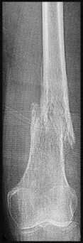
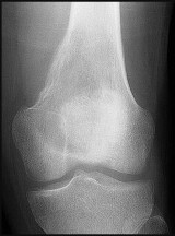
Figures 39a through 39c are the radiographs and MR image of a 14-year-old boy who has intermittent knee swelling and pain exacerbated by activity. What is the most likely diagnosis?
Figures 39a through 39c are the radiographs and MR image of a 14-year-old boy who has intermittent knee swelling and pain exacerbated by activity. What is the most likely diagnosis?
of 100
A 21-year-old man learned he had Ewing sarcoma of the right distal femur at age 13. He underwent treatment with neoadjuvant chemotherapy, surgical resection with distal femoral replacement, and adjuvant chemotherapy. He completed treatment at age 14, and subsequent surveillance imaging has shown no evidence of recurrence. He has noted increasing fatigue during the last 3 months, however, and bleeding when he brushes his teeth. An examination is unremarkable with the exception of bilateral pretibial bruising for which the patient does not recall trauma. Which tests will aid in diagnosis?
A 21-year-old man learned he had Ewing sarcoma of the right distal femur at age 13. He underwent treatment with neoadjuvant chemotherapy, surgical resection with distal femoral replacement, and adjuvant chemotherapy. He completed treatment at age 14, and subsequent surveillance imaging has shown no evidence of recurrence. He has noted increasing fatigue during the last 3 months, however, and bleeding when he brushes his teeth. An examination is unremarkable with the exception of bilateral pretibial bruising for which the patient does not recall trauma. Which tests will aid in diagnosis?
of 100
Figures 41a through 41d are the radiograph, MR images, and biopsy specimen of a 35-year-old woman with pain and progressive paresthesias in her left arm. Staging shows no other lesions. Appropriate local control for this condition requires
Figures 41a through 41d are the radiograph, MR images, and biopsy specimen of a 35-year-old woman with pain and progressive paresthesias in her left arm. Staging shows no other lesions. Appropriate local control for this condition requires
of 100
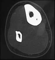
Figures 42a through 42d are the radiograph, CT scans, and biopsy specimen of a 38-year-old man who arrived at the emergency department with urinary retention. He has no other symptoms. What is the most appropriate treatment for this lesion?
Figures 42a through 42d are the radiograph, CT scans, and biopsy specimen of a 38-year-old man who arrived at the emergency department with urinary retention. He has no other symptoms. What is the most appropriate treatment for this lesion?
of 100
Which treatment regimen for Ewing sarcoma most effectively controls disease?
Which treatment regimen for Ewing sarcoma most effectively controls disease?
of 100
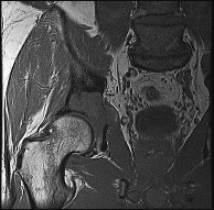
Figures 44a through 44c are the clinical photograph and radiographs of a 59-year-old man who has a 4-year history of metastatic renal cell carcinoma to the brain, lungs, and bones. He has been referred for a painful left proximal femur metastasis. Axial MR images are shown in Figures 44d and 44e. Presurgical embolization, en bloc resection, and proximal femur replacement are performed. The resection specimen and a postsurgical radiograph are shown in Figures 44f and 44g. Compared with intramedullary nail fixation, this treatment strategy
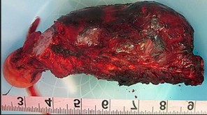
Figures 44a through 44c are the clinical photograph and radiographs of a 59-year-old man who has a 4-year history of metastatic renal cell carcinoma to the brain, lungs, and bones. He has been referred for a painful left proximal femur metastasis. Axial MR images are shown in Figures 44d and 44e. Presurgical embolization, en bloc resection, and proximal femur replacement are performed. The resection specimen and a postsurgical radiograph are shown in Figures 44f and 44g. Compared with intramedullary nail fixation, this treatment strategy
of 100
Figures 45a and 45b are the radiographs of a previously asymptomatic 10-year-old girl who fell off of her bike. Treatment of this lesion should consist of
Figures 45a and 45b are the radiographs of a previously asymptomatic 10-year-old girl who fell off of her bike. Treatment of this lesion should consist of
of 100
Figure 46a is the lateral radiograph of a 54-year-old man who has had a painless soft-tissue mass on his right foot that has been growing slowly for about 1 year. MR sequences are shown in Figures 46b through 46e. A biopsy is performed, and a low-power hematoxylin and eosin photomicrograph is seen in Figure 46f. The most appropriate treatment for this lesion is
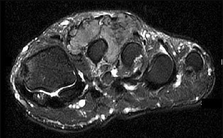
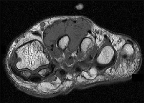
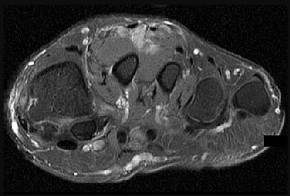
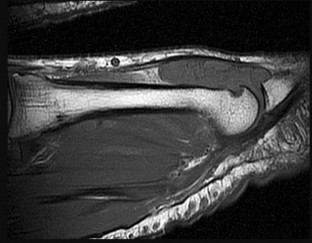
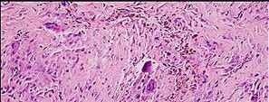
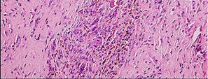
Figure 46a is the lateral radiograph of a 54-year-old man who has had a painless soft-tissue mass on his right foot that has been growing slowly for about 1 year. MR sequences are shown in Figures 46b through 46e. A biopsy is performed, and a low-power hematoxylin and eosin photomicrograph is seen in Figure 46f. The most appropriate treatment for this lesion is
of 100
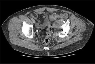
Figures 47a and 47b are the radiograph and axial CT section of a 73-year-old woman with metastatic lung cancer who has a painful left periacetabular lesion. She is a high-risk surgical candidate because of a prior pneumonectomy and progressive metastatic disease of her remaining lung. Palliative radiation is recommended. Two regimens are being considered: a single fraction of 8 Gy or 15 fractions of a 30-Gy cumulative dose over 3 weeks. Compared to the multifraction regimen, the 8-Gy single fraction is associated with
Figures 47a and 47b are the radiograph and axial CT section of a 73-year-old woman with metastatic lung cancer who has a painful left periacetabular lesion. She is a high-risk surgical candidate because of a prior pneumonectomy and progressive metastatic disease of her remaining lung. Palliative radiation is recommended. Two regimens are being considered: a single fraction of 8 Gy or 15 fractions of a 30-Gy cumulative dose over 3 weeks. Compared to the multifraction regimen, the 8-Gy single fraction is associated with
of 100
Figures 48a through 48e are the MR image, radiograph, CT scan, and biopsy specimens of a 14-year-old boy with right shoulder pain without antecedent trauma. What is the most likely diagnosis?
Figures 48a through 48e are the MR image, radiograph, CT scan, and biopsy specimens of a 14-year-old boy with right shoulder pain without antecedent trauma. What is the most likely diagnosis?
of 100
What is the likelihood of this patient’s children having a similar condition?
What is the likelihood of this patient’s children having a similar condition?
of 100
Germline alterations associated with this condition
Germline alterations associated with this condition
of 100
Based on imaging alone, what does this bone lesion most closely resemble?
Based on imaging alone, what does this bone lesion most closely resemble?
of 100
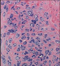
Figures 52a through 52c show the biopsy of this lesion. Based on the clinical history, radiograph, and biopsy, which diagnosis is most likely?
Figures 52a through 52c show the biopsy of this lesion. Based on the clinical history, radiograph, and biopsy, which diagnosis is most likely?
of 100
Staging for patients with this diagnosis necessitates which study or studies?
Staging for patients with this diagnosis necessitates which study or studies?
of 100
Which local treatment option is most appropriate?
Which local treatment option is most appropriate?
of 100
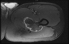
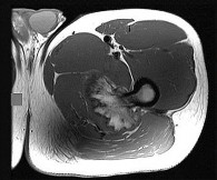
Figures 55a through 55d are the MR images, intraoperative photograph, and biopsy specimen of a 33-year-old man with progressive knee pain and recurrent effusions. What is the most likely diagnosis?
Figures 55a through 55d are the MR images, intraoperative photograph, and biopsy specimen of a 33-year-old man with progressive knee pain and recurrent effusions. What is the most likely diagnosis?
of 100
A disadvantage associated with presurgical (vs postsurgical) radiation therapy for soft-tissue sarcoma is a
A disadvantage associated with presurgical (vs postsurgical) radiation therapy for soft-tissue sarcoma is a
of 100
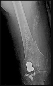
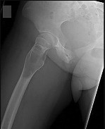
Figures 57a and 57b are the close-up femur radiographs of a 73-year-old man with nonsmall-cell carcinoma of the lung with visceral metastases. He has localized thigh pain at rest and with any
range of motion of the ipsilateral hip or knee. A bone scan shows multiple areas of increased uptake. Other femur images in 2 views show no other lesions. The medical oncologist predicts a survival of 3 months. What is the best next step?
of 100
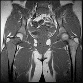
Figures 58a through 58c are the radiograph, MR image, and sagittal CT scan of a 13-year-old boy with a 5-month history of a left hip injury. He has no current pain, but substantial, progressive limitations in hip flexion are present. What is the most likely diagnosis?
Figures 58a through 58c are the radiograph, MR image, and sagittal CT scan of a 13-year-old boy with a 5-month history of a left hip injury. He has no current pain, but substantial, progressive limitations in hip flexion are present. What is the most likely diagnosis?
of 100
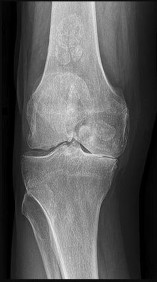
What is the diagnosis?
What is the diagnosis?
of 100
The best treatment is
The best treatment is
of 100
The prognosis for this condition is
The prognosis for this condition is
of 100
Figures 62a and 62b are the radiographs of a 69-year-old woman who is seen for follow-up of a right arm posterior compartment high-grade soft-tissue sarcoma. She previously underwent wide resection of the tumor with preservation of the radial. She experienced an atraumatic fracture 2 years after completion of all treatment. There is no evidence of recurrence on MR imaging. The patient desires treatment because of pain and instability at the fracture site. What is the most reliable treatment option?
Figures 62a and 62b are the radiographs of a 69-year-old woman who is seen for follow-up of a right arm posterior compartment high-grade soft-tissue sarcoma. She previously underwent wide resection of the tumor with preservation of the radial. She experienced an atraumatic fracture 2 years after completion of all treatment. There is no evidence of recurrence on MR imaging. The patient desires treatment because of pain and instability at the fracture site. What is the most reliable treatment option?
of 100
Figures 63a through 63d are the radiograph, CT scan, MR image, and biopsy specimen of a 20-year-old rower who has a several-month history of low-back pain. He has lost 10 pounds, but has no other constitutional symptoms. There is no bowel or bladder incontinence, and he does not have neurologic symptoms. Which medication can be used to treat this condition?
Figures 63a through 63d are the radiograph, CT scan, MR image, and biopsy specimen of a 20-year-old rower who has a several-month history of low-back pain. He has lost 10 pounds, but has no other constitutional symptoms. There is no bowel or bladder incontinence, and he does not have neurologic symptoms. Which medication can be used to treat this condition?
of 100
Which blastic metastases to bone are most common?
Which blastic metastases to bone are most common?
of 100
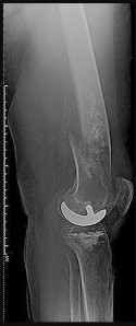
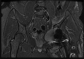
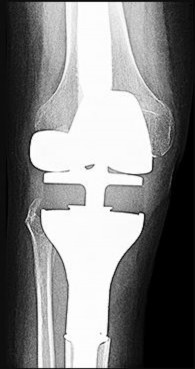
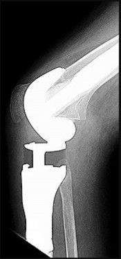
Figures 65a and 65b are the axial and coronal MR images of a 72-year-old woman who underwent a right knee arthroplasty 14 years ago. She has been referred by her primary care provider for evaluation and management of a right medial knee mass. What is the next best step?
Figures 65a and 65b are the axial and coronal MR images of a 72-year-old woman who underwent a right knee arthroplasty 14 years ago. She has been referred by her primary care provider for evaluation and management of a right medial knee mass. What is the next best step?
of 100
Figures 66a and 66b are the clinical photographs of an 86-year-old man with a high-grade undifferentiated pleomorphic sarcoma of the right thigh. He is pictured during preparations for wide surgical excision. What is a favorable prognostic factor?
Figures 66a and 66b are the clinical photographs of an 86-year-old man with a high-grade undifferentiated pleomorphic sarcoma of the right thigh. He is pictured during preparations for wide surgical excision. What is a favorable prognostic factor?
of 100
Figures 67a and 67b are the fused positron emission tomography/CT scans of a calf lesion at groin level in a 36-year-old woman with a soft-tissue mass. Her biopsy specimen is shown in Figure 67c. Staging studies reveal no other lesions. Using the American Joint Committee on Cancer (AJCC) staging system, the stage is most likely
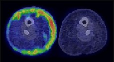
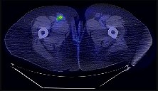
Figures 67a and 67b are the fused positron emission tomography/CT scans of a calf lesion at groin level in a 36-year-old woman with a soft-tissue mass. Her biopsy specimen is shown in Figure 67c. Staging studies reveal no other lesions. Using the American Joint Committee on Cancer (AJCC) staging system, the stage is most likely
of 100
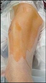
Figure 68a is the clinical photograph of a 59-year-old woman who has had a long-standing fungating ulcer on her left lower leg. She states that the ulcer began as a small reddened area and gradually enlarged during the last 4 years. Anteroposterior (AP) and lateral radiographs of her left leg are shown in Figures 68b and 68c. A whole-body bone scan is shown in Figure 68d. An axial T1-weighted MR image is shown in Figure 68e. A CT scan of the pelvis at the level of the groin is shown in Figure 68f. A histologic specimen is shown in Figure 68g. Based on the clinical, radiographic, and histologic information, the diagnosis is
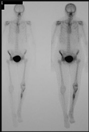
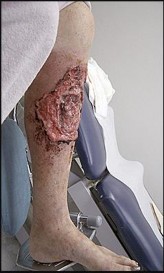

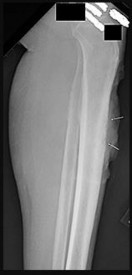
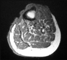
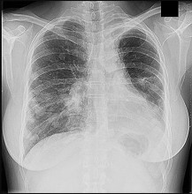
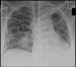
Figure 68a is the clinical photograph of a 59-year-old woman who has had a long-standing fungating ulcer on her left lower leg. She states that the ulcer began as a small reddened area and gradually enlarged during the last 4 years. Anteroposterior (AP) and lateral radiographs of her left leg are shown in Figures 68b and 68c. A whole-body bone scan is shown in Figure 68d. An axial T1-weighted MR image is shown in Figure 68e. A CT scan of the pelvis at the level of the groin is shown in Figure 68f. A histologic specimen is shown in Figure 68g. Based on the clinical, radiographic, and histologic information, the diagnosis is
of 100
Which treatment will most likely improve this patient’s condition?
Which treatment will most likely improve this patient’s condition?
of 100
Which prophylactic measures may decrease risk for this postsurgical complication?
Which prophylactic measures may decrease risk for this postsurgical complication?
of 100
Figures 71a through 71d are the radiographs, MR images, and biopsy specimen of a 15-year-old boy with a several-month history of right hip pain with no history of injury. This condition is associated with increased activity of which gene product?
Figures 71a through 71d are the radiographs, MR images, and biopsy specimen of a 15-year-old boy with a several-month history of right hip pain with no history of injury. This condition is associated with increased activity of which gene product?
of 100
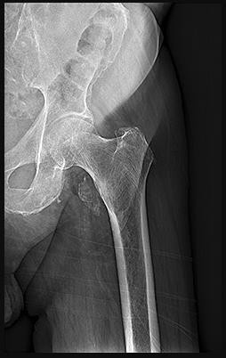
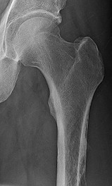
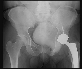
Figures 72a through 72d are the radiograph, MR images, and biopsy specimen of a 42-year-old man with an insidious onset of left hip pain. Further imaging reveals no other lesions. What is the most appropriate initial treatment?
Figures 72a through 72d are the radiograph, MR images, and biopsy specimen of a 42-year-old man with an insidious onset of left hip pain. Further imaging reveals no other lesions. What is the most appropriate initial treatment?
of 100
Figures 73a through 73c are the radiographs of a 68-year-old woman with a pathologic left femur fracture. A clinical examination demonstrates a large soft-tissue mass at the fracture site. CT scans of the chest, abdomen, and pelvis reveal numerous enlarged lymph nodes. Frozen section analysis at open biopsy reveals relapsed lymphoma. What is the most appropriate treatment?
Figures 73a through 73c are the radiographs of a 68-year-old woman with a pathologic left femur fracture. A clinical examination demonstrates a large soft-tissue mass at the fracture site. CT scans of the chest, abdomen, and pelvis reveal numerous enlarged lymph nodes. Frozen section analysis at open biopsy reveals relapsed lymphoma. What is the most appropriate treatment?
of 100
Survival estimation in guiding surgical decision-making in metastatic spine disease includes scores that include
Survival estimation in guiding surgical decision-making in metastatic spine disease includes scores that include
of 100
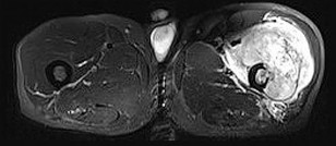
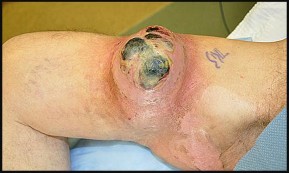
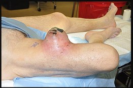
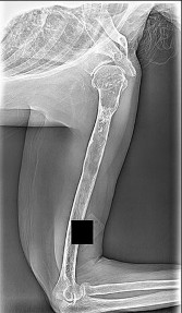
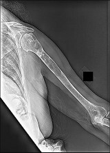
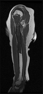
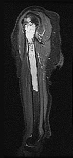
A 52-year-old woman with a medical history that includes type 1 diabetes mellitus and rheumatoid arthritis has a painless right thigh mass that increased in size during the preceding year. Ultrasound was “consistent with lipoma,” and the patient underwent uneventful resection. Final pathology revealed high-grade undifferentiated sarcoma. Figures 75a and 75b are the clinical photograph and postresection MR image. The treatment rendered prior to referral to a sarcoma center most likely will result in increased
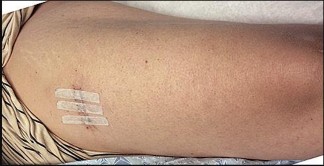
A 52-year-old woman with a medical history that includes type 1 diabetes mellitus and rheumatoid arthritis has a painless right thigh mass that increased in size during the preceding year. Ultrasound was “consistent with lipoma,” and the patient underwent uneventful resection. Final pathology revealed high-grade undifferentiated sarcoma. Figures 75a and 75b are the clinical photograph and postresection MR image. The treatment rendered prior to referral to a sarcoma center most likely will result in increased
of 100
A 16-year-old boy with a high-grade conventional osteosarcoma of the right proximal tibia has completed neoadjuvant chemotherapy. A restaging radiograph and MR image are shown in Figures 76a and 76b. Wide resection with limb salvage is planned. Which muscle will provide the primary protective margin for the tibial nerve and popliteal vessels?
A 16-year-old boy with a high-grade conventional osteosarcoma of the right proximal tibia has completed neoadjuvant chemotherapy. A restaging radiograph and MR image are shown in Figures 76a and 76b. Wide resection with limb salvage is planned. Which muscle will provide the primary protective margin for the tibial nerve and popliteal vessels?
of 100
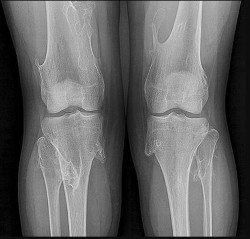
Figures 77a and 77b are the recent knee radiographs of a 53-year-old man whose history includes tobacco use and secondary polycythemia. He is now experiencing bilateral knee pain, knee swelling, and increasing discomfort with ambulation. All efforts at nonsurgical treatment have failed. What is the most reasonable next treatment option?
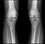
Figures 77a and 77b are the recent knee radiographs of a 53-year-old man whose history includes tobacco use and secondary polycythemia. He is now experiencing bilateral knee pain, knee swelling, and increasing discomfort with ambulation. All efforts at nonsurgical treatment have failed. What is the most reasonable next treatment option?
of 100
This patient should be told that
This patient should be told that
of 100
Definitive treatment for this tumor likely will necessitate
Definitive treatment for this tumor likely will necessitate
of 100
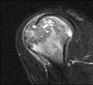
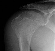
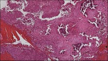
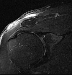
Figure 80a is the radiograph of a 5-year-old girl who has experienced 8 weeks of shoulder pain that is mostly relieved with nonsteroidal anti-inflammatory drugs. Her MR images are shown in Figures 80b through 80f. Histology is shown in Figures 80g through 80i. The most likely diagnosis is
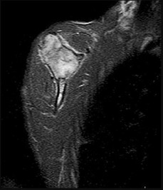
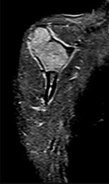
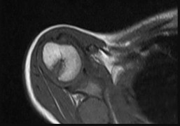
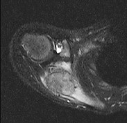
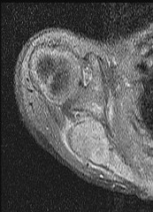
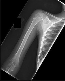
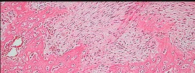
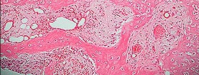
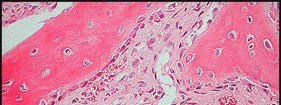
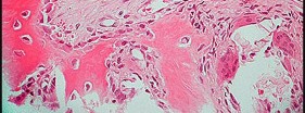
Figure 80a is the radiograph of a 5-year-old girl who has experienced 8 weeks of shoulder pain that is mostly relieved with nonsteroidal anti-inflammatory drugs. Her MR images are shown in Figures 80b through 80f. Histology is shown in Figures 80g through 80i. The most likely diagnosis is
of 100
A 32-year-old man underwent distal femur resection and endoprosthetic replacement at age 15 for high-grade conventional osteosarcoma. He was treated with neoadjuvant and adjuvant cisplatin, doxorubicin, and methotrexate. There has been no evidence of recurrent osteosarcoma, and he has been otherwise active and well. He is scheduled to undergo exchange of the polyethylene liner and bushings in his prosthesis because of wear that has caused recurrent effusions and a sensation of instability. Which study is most important to assess his perioperative medical risk?
A 32-year-old man underwent distal femur resection and endoprosthetic replacement at age 15 for high-grade conventional osteosarcoma. He was treated with neoadjuvant and adjuvant cisplatin, doxorubicin, and methotrexate. There has been no evidence of recurrent osteosarcoma, and he has been otherwise active and well. He is scheduled to undergo exchange of the polyethylene liner and bushings in his prosthesis because of wear that has caused recurrent effusions and a sensation of instability. Which study is most important to assess his perioperative medical risk?
of 100
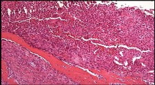
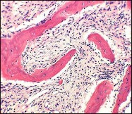
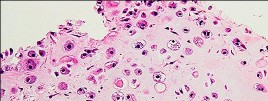
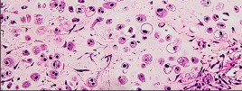
Figures 82a and 82b are the histopathology of an otherwise healthy 50-year-old man who had a growing mass marginally excised from his shoulder with local anesthetic and no presurgical imaging. The mass includes the deep fascia and muscle and is larger than 5 cm. Postsurgical MR imaging was obtained, and T2 and T1+C images are shown in Figures 82c and 82d. What is the best local treatment option?

Figures 82a and 82b are the histopathology of an otherwise healthy 50-year-old man who had a growing mass marginally excised from his shoulder with local anesthetic and no presurgical imaging. The mass includes the deep fascia and muscle and is larger than 5 cm. Postsurgical MR imaging was obtained, and T2 and T1+C images are shown in Figures 82c and 82d. What is the best local treatment option?
of 100
The optimal initial treatment of this pathologic fracture is
The optimal initial treatment of this pathologic fracture is
of 100
A series of axial T1-weighted MR images is shown in Figure 84. The postfracture MRI reveals tissue contamination by fracture hematoma. Based upon the MRI findings, which aspect of limb-sparing resection and reconstruction will be most challenging?
A series of axial T1-weighted MR images is shown in Figure 84. The postfracture MRI reveals tissue contamination by fracture hematoma. Based upon the MRI findings, which aspect of limb-sparing resection and reconstruction will be most challenging?
of 100
The patient ultimately undergoes rotationplasty. What is the most likely etiology of early limb loss after rotationplasty for sarcoma?
The patient ultimately undergoes rotationplasty. What is the most likely etiology of early limb loss after rotationplasty for sarcoma?
of 100
When compared to patients who undergo endoprosthetic reconstruction, patients who undergo rotationplasty are more likely to
When compared to patients who undergo endoprosthetic reconstruction, patients who undergo rotationplasty are more likely to
of 100
Figures 87a through 87d are radiographs of both knees of a 75-year-old man who is experiencing bilateral knee pain and stiffness. The gross appearance of the lesions in the suprapatellar pouch is seen in Figure 87e. The histologic finding that would best determine this process is osteoarthritic in nature and not neoplastic is the absence of
Figures 87a through 87d are radiographs of both knees of a 75-year-old man who is experiencing bilateral knee pain and stiffness. The gross appearance of the lesions in the suprapatellar pouch is seen in Figure 87e. The histologic finding that would best determine this process is osteoarthritic in nature and not neoplastic is the absence of
of 100
Figures 88a through 88d are the radiographs and biopsy specimens of a 65-year-old woman with a history of breast cancer who has been experiencing 6 weeks of increasing left hip pain. She denies any injury. What is the most likely diagnosis?
Figures 88a through 88d are the radiographs and biopsy specimens of a 65-year-old woman with a history of breast cancer who has been experiencing 6 weeks of increasing left hip pain. She denies any injury. What is the most likely diagnosis?
of 100
Figures 89a through 89g are the radiographs, MR images, and biopsy specimen of a 32-year-old man who has chronic left thigh and knee pain and recent knee swelling with no specific trauma or injury. The pain is exacerbated with activity but is also present at rest and at night. What is the most appropriate treatment?
Figures 89a through 89g are the radiographs, MR images, and biopsy specimen of a 32-year-old man who has chronic left thigh and knee pain and recent knee swelling with no specific trauma or injury. The pain is exacerbated with activity but is also present at rest and at night. What is the most appropriate treatment?
of 100
What is the most likely diagnosis?
What is the most likely diagnosis?
of 100
Immunohistochemical analysis of this lesion will be characterized by expression of
Immunohistochemical analysis of this lesion will be characterized by expression of
of 100
Figures 92a and 92b are the reconstruction radiographs of a 16-year-old boy with a high-grade conventional osteosarcoma of his right proximal tibia. He has completed neoadjuvant
chemotherapy. Wide resection, endoprosthetic proximal tibia replacement, a medial gastrocnemius flap, and a split-thickness skin graft were performed. The preferred postsurgical knee rehabilitation regimen is
of 100
Figures 93a through 93f are radiographs, selected MR imaging sequences, and biopsy specimens of the left humerus of a 76-year-old woman who has experienced long-term left arm pain. She has received previous treatment for osteoarthritis of her left shoulder with nonsteroidal anti-inflammatory drugs and an intra-articular corticosteroid injection for her rotator cuff arthropathy. Recent staging studies show no evidence of metastatic disease. What is the most appropriate next treatment?
Figures 93a through 93f are radiographs, selected MR imaging sequences, and biopsy specimens of the left humerus of a 76-year-old woman who has experienced long-term left arm pain. She has received previous treatment for osteoarthritis of her left shoulder with nonsteroidal anti-inflammatory drugs and an intra-articular corticosteroid injection for her rotator cuff arthropathy. Recent staging studies show no evidence of metastatic disease. What is the most appropriate next treatment?
of 100
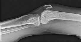
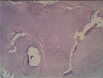
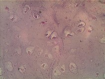
Figures 94a through 94f are the radiographs and MR images of a 16-year-old boy who experiences left ankle pain with activity. What is the most likely diagnosis?
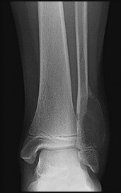
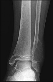

Figures 94a through 94f are the radiographs and MR images of a 16-year-old boy who experiences left ankle pain with activity. What is the most likely diagnosis?
of 100
A patient undergoes excision of a presumed lipoma of the superficial thigh. Final pathology reveals synovial sarcoma without reference to the margins. What is the recommendation for definitive treatment?
A patient undergoes excision of a presumed lipoma of the superficial thigh. Final pathology reveals synovial sarcoma without reference to the margins. What is the recommendation for definitive treatment?
of 100
Figures 96a and 96b are the MRI sections of the symptomatic left knee of a 28-year-old man with left anterior knee pain 18 months after undergoing an allogenic bone marrow transplant for acute myelogenous leukemia. His intraoperative fluoroscopy images are shown in Figures 96c and 96d. What is most critical when obtaining a diagnosis for this patient?
Figures 96a and 96b are the MRI sections of the symptomatic left knee of a 28-year-old man with left anterior knee pain 18 months after undergoing an allogenic bone marrow transplant for acute myelogenous leukemia. His intraoperative fluoroscopy images are shown in Figures 96c and 96d. What is most critical when obtaining a diagnosis for this patient?
of 100
Figures 97a through 97d are the radiographs and MR images of a 21-year-old man with symptoms of a left medial thigh mass. Upon examination, you palpate a firm, fixed, deep, nontender mass of the medial proximal left thigh. No other masses are found during the examination. The patient fears metastatic disease. What is the risk for malignant transformation throughout this patient’s lifetime?
Figures 97a through 97d are the radiographs and MR images of a 21-year-old man with symptoms of a left medial thigh mass. Upon examination, you palpate a firm, fixed, deep, nontender mass of the medial proximal left thigh. No other masses are found during the examination. The patient fears metastatic disease. What is the risk for malignant transformation throughout this patient’s lifetime?
of 100
Figures 98a and 98b are the radiograph and biopsy specimen of a 20-year-old man who is being evaluated for the first time for foot pain. Treatment should include
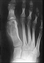
Figures 98a and 98b are the radiograph and biopsy specimen of a 20-year-old man who is being evaluated for the first time for foot pain. Treatment should include
of 100
What are the most common primary sites for carcinomas metastatic to bone?
What are the most common primary sites for carcinomas metastatic to bone?
of 100
Figures 100a through 100c are the select T1 and T2 axial and T2 sagittal MR images of a 26-year-old-woman who has had intermittent left thigh pain for 6 months that is exacerbated by activity. Left femur radiographs are unremarkable. Initial treatment should include
Figures 100a through 100c are the select T1 and T2 axial and T2 sagittal MR images of a 26-year-old-woman who has had intermittent left thigh pain for 6 months that is exacerbated by activity. Left femur radiographs are unremarkable. Initial treatment should include
You Might Also Like


Previous ChapterOrthopedic Basic Review | Dr Hutaif Basic Science Revie -...
Next Chapter Orthopedic Pathology Review | Dr Hutaif Basic Science R -...

Medically Verified Content by
Prof. Dr. Mohammed Hutaif
Consultant Orthopedic & Spine Surgeon