Cerebral Palsy of the Hand: Comprehensive Evaluation and Surgical Management
Key Takeaway
Surgical management of the cerebral palsy hand requires meticulous patient selection, as fewer than 4% of patients are operative candidates. Success depends on differentiating static contractures from dynamic spasticity, confirming adequate proximal motor control, and assessing hand sensibility. Operative interventions aim to balance muscle forces, correct pronation, wrist flexion, and thumb-in-palm deformities, ultimately improving grasp, release, and overall upper extremity function.
Introduction to Cerebral Palsy of the Upper Extremity
Cerebral palsy (CP) represents a heterogeneous group of nonprogressive, nonhereditary encephalopathies occurring in the prenatal or perinatal period. It is characterized by altered motor, sensory, and frequently intellectual function. With an approximate annual incidence of 7 per 1,000 live births in the United States, CP presents a complex reconstructive challenge for the orthopedic surgeon.
The neurologic manifestations of CP are broadly classified into two primary categories:
* Pyramidal (Spastic): Includes spastic hemiplegia, diplegia, paraplegia, and quadriplegia. These patients exhibit velocity-dependent increased muscle tone and are the primary candidates for surgical intervention.
* Extrapyramidal (Non-spastic): Includes athetoid, choreoathetoid, and ataxic patterns. These patients exhibit involuntary movements and fluctuating tone.
* Mixed: A combination of spasticity and athetosis.
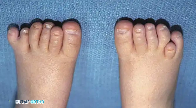
Hand function is impaired to varying degrees in virtually all types of CP, with the possible exception of pure spastic paraplegia. The classic upper extremity posture in a spastic patient consists of shoulder adduction and internal rotation, elbow flexion, forearm pronation, wrist and finger flexion, thumb-in-palm deformity, and swan-neck deformities of the digits.
Historically, surgical interventions for the cerebral palsied hand yielded unpredictable and often disappointing results, primarily due to inappropriate patient selection. However, the foundational and extensive works of Green, Goldner, Swanson, Zancolli, and Hoffer have established rigorous, evidence-based principles for the evaluation and management of these complex deformities.
Comprehensive Patient Evaluation
The cornerstone of managing the cerebral palsied hand is exhaustive, repeated patient evaluation. It is estimated that fewer than 4% of patients with hands disabled by cerebral palsy will benefit from surgical reconstruction. Consequently, the surgeon’s primary role is often to identify the vast majority of patients for whom surgery should be discouraged, while meticulously optimizing the small cohort who stand to gain meaningful functional improvement.
History and Developmental Milestones
Evaluation must occur over a considerable length of time. A detailed history should capture birth or perinatal medical complications, the achievement of developmental milestones, and the degree to which the child spontaneously incorporates the affected hand into daily activities.
Clinical Pearl: The early development of handedness (preference for one hand before the age of 3 years) is highly abnormal. It frequently represents a degree of weakness, spasticity, or incoordination in the contralateral (less preferred) extremity and is a classic early indicator of spastic hemiplegia.
If the child completely ignores the affected hand during bimanual play, it is highly doubtful that surgical intervention will restore or improve function. Surgery cannot create central motor planning or cortical integration where none exists.
Neurologic and Motor Assessment
The specific cerebral lesion must be identified and characterized. Children with extrapyramidal (athetoid or ataxic) patterns are generally not surgical candidates due to the unpredictable nature of their involuntary movements, which doom tendon transfers and releases to failure.
The persistence of infantile postural reflexes (e.g., asymmetric tonic neck reflex, Moro reflex) must be documented, as their obligate presence severely limits voluntary motor control.
Deformities must be strictly classified into two categories:
1. Dynamic Deformities: Spastic, velocity-dependent posturing that is slowly correctable with gentle, sustained passive stretching. Most children exhibit dynamic deformities early in life.
2. Static Contractures: Fixed myostatic contractures or joint capsular contractures that do not correct with compensatory positioning of adjacent joints. Untreated dynamic spasticity inevitably progresses to static contracture over time.
Muscle examination must quantify the degree of spasticity (using the Modified Ashworth Scale), voluntary strength, and coordination of each major muscle group. Special attention must be given to the child’s ability to perform pinch, grasp, and release.
Surgical Warning (The Proximal Control Test): The patient must possess sufficient proximal control of the upper extremity to voluntarily place the hand on top of their head, and subsequently on the opposite knee, within 5 to 10 seconds. If a child lacks this degree of proximal shoulder and elbow control, they will not utilize the hand enough in space to justify distal reconstructive surgery.
Sensibility Evaluation
The sensibility pattern of the hand is arguably the most critical prognostic factor for postoperative functional use. While most patients possess intact epicritic sensation (the ability to discern pinprick, heat, and cold), approximately 50% suffer from profound impairments in cortical sensory modalities, including diminished two-point discrimination, stereognosis, and proprioception.
Because sensibility dictates whether a hand will be integrated into bimanual tasks or ignored, its status must be evaluated meticulously.
* Observation: An ignored hand, in the presence of adequate motor coordination, strongly suggests an absence of functional sensibility.
* Basic Testing (Ages 4+): Ask a blindfolded child to differentiate between a sphere and a cube, or to identify whether the examiner has placed their palm facing upward or downward (proprioception).
* Advanced Testing: Test recognition of blunt versus sharp points, identification of familiar objects (coins, keys), and temperature differentiation.
Advanced Diagnostic Modalities
Dynamic Electromyography (EMG)
As described by Hoffer, Perry, and Melkonian, dynamic EMG is invaluable for determining the firing phase of spastic muscles during attempted grasp and release. Tendon transfers in CP do not undergo cortical "re-education" as they do in peripheral nerve palsies. Therefore, a donor muscle must be selected that is already firing in phase with the desired augmented function (e.g., using a muscle that fires during finger extension to augment wrist extension).
Diagnostic Neuromuscular Blocks
Neuromuscular blocking agents are highly effective in differentiating dynamic spasticity from static contracture, and for unmasking the strength of antagonist muscles.
* Agents: 1% Lidocaine or 0.25% Bupivacaine (temporary), or 45% Ethanol / Phenol (prolonged effect).
* Application: Blocking a spastic flexor carpi ulnaris (FCU) eliminates its overbearing flexor effect. If the patient can subsequently extend the wrist and fingers, it confirms that the extensor musculature is functional and that a flexor lengthening or transfer will likely yield a successful outcome.
Operative Management: Goals and Principles
Surgical Goals
Surgical intervention in the cerebral palsied hand is broadly divided into two distinct goals:
1. Functional Improvement: Aimed at patients with good voluntary control, intact sensibility, and specific dynamic imbalances. The goal is to improve grasp, release, and pinch kinematics.
2. Hygiene and Cosmesis (Salvage): Aimed at severely involved, non-functional hands with severe static contractures. The goal is to release severe flexion deformities to prevent palmar maceration, facilitate dressing, and improve the aesthetic appearance of the limb.
Core Surgical Principles
- Release before Augmentation: Spastic antagonists must be lengthened or released before weak agonists are augmented via tendon transfer.
- Avoid Over-lengthening: Fractional lengthening at the musculotendinous junction is preferred over Z-lengthening of the tendon to prevent devastating over-lengthening and loss of grip strength.
- Simultaneous Correction: Address all levels of deformity (forearm, wrist, fingers, thumb) in a single surgical setting when possible, as uncorrected proximal deformities will compromise distal reconstructions.
Surgical Management of Specific Deformities
Pronation Contracture of the Forearm
Pronation deformity is driven primarily by spasticity of the pronator teres (PT) and, to a lesser extent, the pronator quadratus. It severely limits the patient's ability to supinate the hand to accept objects.
Operative Technique: Pronator Teres Rerouting
1. Incision: A longitudinal incision is made over the middle third of the volar-radial forearm.
2. Exposure: The superficial radial nerve and radial artery are identified and protected. The insertion of the PT on the lateral radius is exposed.
3. Detachment: The PT tendon is detached from the radius along with a strip of periosteum to maximize length.
4. Rerouting: The tendon is passed through the interosseous membrane (or routed subcutaneously around the radius) from volar to dorsal.
5. Reattachment: The tendon is reattached to its original insertion site while the forearm is held in maximum supination. This converts the PT from a pronator into a weak supinator, or at least neutralizes its pronating force while maintaining its stabilizing effect on the radius.
Flexion Deformities of the Wrist and Fingers
Wrist flexion deformities are typically caused by spasticity of the FCU, flexor carpi radialis (FCR), and fractional tightness of the flexor digitorum superficialis (FDS) and profundus (FDP).
Operative Technique: Fractional Lengthening of Finger Flexors
* Indication: Dynamic finger flexion spasticity with preserved voluntary extension when the wrist is flexed.
* Procedure: Through a volar forearm incision, the musculotendinous junctions of the FDS and FDP are identified. Transverse incisions are made through the aponeurotic fascia of the muscle bellies, leaving the underlying muscle fibers intact. The wrist and fingers are passively extended, allowing the aponeurosis to slide and lengthen by 1 to 2 cm. This preserves the continuity of the muscle while reducing resting tension.
Operative Technique: FCU to ECRB Transfer (Green Transfer)
* Indication: Severe dynamic wrist flexion with weak active wrist extension.
* Procedure:
1. The FCU is harvested through a volar-ulnar incision, detached from the pisiform.
2. The muscle belly is mobilized proximally to the mid-forearm, taking care to protect the ulnar nerve and artery.
3. A second incision is made dorsally over the extensor carpi radialis brevis (ECRB) insertion.
4. The FCU tendon is routed subcutaneously around the ulnar border of the forearm to the dorsal wrist.
5. It is woven into the ECRB tendon using a Pulvertaft weave under appropriate tension (wrist in 30 degrees of extension).
Pitfall: Never transfer the FCU if it is the sole active wrist flexor, as this will result in an irreversible hyperextension deformity of the wrist. Ensure the FCR is functional before sacrificing the FCU.
Thumb-in-Palm Deformity
The thumb-in-palm deformity is one of the most disabling conditions in the CP hand, as it completely blocks the entry of objects into the palm and prevents pinch. It is caused by spasticity of the adductor pollicis, first dorsal interosseous, flexor pollicis longus (FPL), and thenar intrinsics, combined with weakness of the abductor pollicis longus (APL) and extensor pollicis longus (EPL).
Operative Management:
Treatment requires a multi-level approach tailored to the specific spastic components:
1. Intrinsic Release: Through a palmar incision parallel to the thenar crease, the adductor pollicis transverse and oblique heads are released from the third metacarpal. The first dorsal interosseous may be stripped from the first metacarpal.
2. Extrinsic Lengthening: If the FPL is spastic (interphalangeal joint acutely flexed), it is fractionally lengthened at the distal forearm.
3. Extensor Augmentation: To maintain the thumb out of the palm, the EPL can be rerouted from Lister's tubercle to the radial aspect of the wrist, increasing its abduction moment. Alternatively, a tendon transfer (e.g., Brachioradialis to APL) may be performed to augment thumb abduction.
4. Joint Stabilization: If the metacarpophalangeal (MCP) joint is highly unstable and hyperextends during pinch, an MCP joint arthrodesis is highly effective in providing a stable post for pinch.
Swan-Neck Deformity
Swan-neck deformity in CP is characterized by proximal interphalangeal (PIP) joint hyperextension and distal interphalangeal (DIP) joint flexion. It is driven by spastic intrinsic muscles pulling excessively on the central slip, combined with volar plate laxity at the PIP joint.
Operative Management:
* Mild/Flexible Deformity: Intrinsic release (sliding the interosseous muscles distally) or lateral band rerouting.
* Moderate Deformity with Volar Plate Laxity: Flexor digitorum superficialis (FDS) tenodesis. One slip of the FDS is divided proximally, left attached to the middle phalanx, and sutured to the A2 pulley or proximal phalanx with the PIP joint held in 20 degrees of flexion. This creates a volar tether that prevents PIP hyperextension.
* Severe/Fixed Deformity: PIP joint arthrodesis in a functional position of flexion.
Postoperative Protocol and Rehabilitation
The success of surgical intervention in the cerebral palsied hand relies heavily on rigorous postoperative rehabilitation.
* Immobilization: Following tendon transfers and lengthenings, the extremity is typically immobilized in a long-arm cast for 4 weeks. The wrist and digits are positioned to protect the transfers (e.g., wrist in extension, thumb abducted).
* Orthoses: Upon cast removal, custom thermoplastic splints are fabricated. A resting hand splint is used at night to maintain muscle length and prevent recurrence of contractures. Dynamic or functional daytime splints may be utilized to assist with grasp and release.
* Therapy: Intensive occupational therapy is initiated to train the newly transferred muscles and integrate the hand into bimanual activities. Cortical adaptation takes months, and therapy should be maintained for at least 6 to 12 months postoperatively.
In conclusion, the operative management of the cerebral palsied hand is a highly specialized endeavor. By adhering to strict patient selection criteria, utilizing advanced diagnostic modalities like dynamic EMG, and applying sound biomechanical principles to balance spastic and weak forces, the orthopedic surgeon can achieve profound improvements in the functional capacity and quality of life for these complex patients.
You Might Also Like