Principles of Hand Fracture Fixation: A Comprehensive Surgical Guide
Key Takeaway
Successful management of hand fractures requires meticulous surgical technique and a profound understanding of small bone biomechanics. While conservative management suffices for many injuries, operative intervention is mandated for displaced intra-articular fractures, multiple unstable fractures, and irreducible dislocations. This guide details the critical principles of Kirschner wire placement, minifragment screw fixation, and soft-tissue management to optimize functional outcomes and minimize complications.
PRINCIPLES OF HAND FRACTURE MANAGEMENT
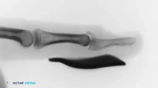
The fundamental objective in the management of hand and small tubular bone fractures is the restoration of anatomy to achieve early, pain-free mobilization. The hand is an intricate biomechanical marvel, relying on the seamless gliding of tendons over osseous structures. Consequently, the physician’s judgment is paramount when selecting between conservative management, percutaneous techniques, and open reduction with internal fixation (ORIF).
The soft-tissue envelope of the hand is unforgiving. Unlike long bone fractures where bulky implants can be easily buried beneath thick muscle bellies, the phalanges and metacarpals possess minimal soft-tissue coverage. Ill-advised plating of small tubular bones—particularly the phalanges—frequently precipitates catastrophic complications, including skin sloughs, extensor tendon adherence, tendon ruptures, and severe joint contractures. Similarly, while external fixators are valuable in specific trauma settings, they can impinge on delicate tendons or ligaments, severely interfering with postoperative function. Even with minimally invasive percutaneous or closed pinning, the surgeon must remain vigilant; neurovascular bundles can easily become wound around a rotating pin, or tendons and ligaments may be inadvertently pinned directly to the bone, tethering their excursion.
Surgical Warning: The decision to operate on a hand fracture must always balance the mechanical need for skeletal stability against the biological cost of soft-tissue dissection. Stiffness is the most common and debilitating complication of hand fractures, often exacerbated by overzealous surgical intervention.
INDICATIONS FOR OPERATIVE INTERVENTION
While the vast majority of hand fractures can be managed non-operatively with external splinting, buddy taping, and early mobilization, specific fracture patterns demand surgical stabilization. Operative intervention is strictly indicated in the following clinical scenarios:
- Displaced Intra-articular Fractures: Exact anatomical reduction is mandatory to restore joint kinematics and prevent post-traumatic osteoarthritis. Any articular step-off greater than 1 mm or involvement of more than 25% of the articular surface generally warrants fixation.
- Major Ligamentous or Tendinous Avulsions: Fractures representing the avulsion of critical stabilizing structures (e.g., bony mallet finger, collateral ligament avulsions) require fixation to restore joint stability and tendon function.
- Irreducible Fractures with Soft-Tissue Interposition: When a fracture is so severely displaced that the interposition of tendons, volar plate, or intrinsic muscles prevents realignment by closed manipulation, open reduction is required.
- Multiple Unstable Fractures: In the polytraumatized hand, multiple fractures often result in the collapse of the hand's architectural arches. If the hand cannot be maintained in the intrinsic-plus "position of function" without internal fixation, operative stabilization is mandated.
- Open Fractures: Internal fixation provides the rigid skeletal stability necessary to facilitate aggressive postoperative wound care, soft-tissue reconstruction, and early mobilization without the loss of fracture reduction.
Clinical Pearl: Severely comminuted closed fractures of the phalanges and metacarpals should rarely be opened. The periosteal stripping required to expose and internally fix multiple small fragments devitalizes the bone, virtually guaranteeing nonunion and profound stiffness. In these cases, limited percutaneous pinning or external fixation is the treatment of choice.
BIOMECHANICS AND IMPLANT SELECTION
Rarely is more fixation required than that afforded by external splinting, Kirschner wires (K-wires), and minifragment screws. The overarching goal is to achieve close approximation and apposition of the fractured bone ends to promote primary or secondary bone healing, depending on the construct's rigidity.
Kirschner Wire Biomechanics
Kirschner wires remain the workhorse of hand fracture fixation. However, their application requires a nuanced understanding of small bone biomechanics.
- Single Oblique vs. Crossed Wires: One obliquely placed K-wire is frequently preferable to two crossed wires. A single oblique wire allows for dynamic impaction at the fracture site during axial loading, promoting secondary bone healing. Conversely, two crossed wires, while rotationally stable, can act as a rigid strut that holds the fragments apart, leading to distraction and subsequent delayed union or nonunion.
- Rotational Control: If a single oblique wire provides insufficient rotational stability, a second wire is necessary. Two parallel, longitudinal K-wires can effectively control rotation while avoiding the distraction phenomenon associated with crossed wires.
- Wire Tip Morphology: A trocar-pointed wire is vastly superior to a diamond or diagonally cut wire. The trocar tip provides greater initial holding power, cuts through cortical bone more efficiently (generating less thermal necrosis), and allows for significantly easier initial bone engagement when placed at an acute angle.
Minifragment Screws and Wiring Techniques
- Interfragmentary Screw Fixation: Unstable, long oblique, or spiral fractures of the metacarpals and phalanges are often best treated with interfragmentary lag screw fixation alone. This technique provides absolute stability and dynamic compression across the fracture plane, allowing for immediate postoperative mobilization.
- Tension Band and 90/90 Wiring: Certain transverse or short oblique fracture patterns, particularly at the metaphyseal-diaphyseal junction, benefit from tension band wiring or 90/90 intraosseous wiring. These techniques convert tensile distraction forces on the convex side of the bone into compressive forces at the fracture site.
SURGICAL EQUIPMENT AND INSTRUMENTATION
Except for dedicated minifragment plate and screw sets or specialized external fixators, remarkably little equipment is required for the operative management of hand fractures. The minimalist approach is often the most effective.
Reduction Instruments
The same standard instruments utilized for soft-tissue handling can be ingeniously repurposed to manipulate small bones.
* A straight Kocher clamp or a standard towel clip is usually sufficient to provisionally reduce and hold a metacarpal shaft fracture.
* A fine hemostat is excellent for manipulating smaller phalangeal fragments prior to definitive fixation.
Drilling and Wire Insertion
- Double-Ended K-wires: Kirschner wires should be sharpened on both ends. This allows the surgeon to drill the wire antegrade through the fracture and out the skin, and then drill it retrograde back across the fracture site if necessary.
- Power Equipment: A small, hand-held power driver or a lightweight battery-driven drill is essential. Cumbersome air supply lines or heavy orthopedic drills compromise the delicate tactile feedback required for accurate drilling and placement of K-wires in small bones.
- Insertion Technique: A slow insertion speed is highly recommended to prevent thermal necrosis of the bone, which can lead to pin loosening and infection.
Pitfall: The Kirschner wire should project no more than 5 cm from the drill chuck. Excessive length allows the wire to bend, wobble, and walk along the cortex during insertion. If a longer trajectory is required, insert the first 5 cm, halt, advance the drill chuck closer to the skin, and proceed in increments.
STEP-BY-STEP SURGICAL TECHNIQUES
Closed Reduction and Percutaneous Pinning (CRPP)
- Positioning and Assessment: The patient is positioned supine with the arm on a radiolucent hand table. Fluoroscopy is positioned parallel to the table.
- Reduction: Closed reduction is performed using longitudinal traction and manipulation.
- Rotational Alignment: Crucial Step: When possible, the fractured finger must be flexed fully at the metacarpophalangeal (MCP) and interphalangeal (IP) joints. This utilizes the tenodesis effect to assess rotational alignment. All fingers should point toward the scaphoid tubercle in flexion; any overlapping or "scissoring" indicates a rotational malreduction that must be corrected prior to pinning.
- Pin Placement: Using a trocar-tipped K-wire on a slow-speed drill, the wire is introduced percutaneously. Care is taken to avoid the extensor mechanism dorsally and the neurovascular bundles laterally.
- Pin Management: After insertion and fluoroscopic confirmation, the wires may be cut off flat and the ends left protruding through the skin for easy removal, or they may be cut short and worked well beneath the skin to prevent pin-tract infections. End-cutting wire cutters are mandatory for this step to ensure a clean, flat cut without sharp burrs.
Interfragmentary Lag Screw Fixation
- Approach: A dorsal or mid-axial approach is utilized depending on the fracture geometry. The periosteum is elevated minimally—only enough to visualize the fracture lines.
- Reduction: The fracture is anatomically reduced and held with a reduction forceps or a provisional 0.035-inch K-wire.
- Glide Hole: A glide hole (equal to the outer thread diameter of the screw) is drilled in the near cortex.
- Thread Hole: A drill sleeve is inserted into the glide hole, and the thread hole (equal to the core diameter of the screw) is drilled through the far cortex.
- Countersinking: The near cortex is countersunk to prevent the screw head from creating a stress riser or irritating the overlying extensor tendon.
- Measurement and Insertion: The tract is measured, and the appropriate length screw is inserted, achieving interfragmentary compression.
MANAGEMENT OF DISLOCATIONS AND LIGAMENTOUS INJURIES
While fractures often dominate the radiographic picture, pure dislocations and fracture-dislocations of the hand represent severe soft-tissue injuries that can permanently compromise function if mismanaged.
The majority of simple dislocations can be managed by prompt closed manipulation, splinting in a stable position for a brief period, and early functional mobilization. Many simple interphalangeal dislocations are self-reduced by the patient at the scene of the injury. Functional motion through “buddy taping” to an adjacent, uninjured finger generally provides an excellent clinical result by utilizing the uninjured digit as a dynamic splint.
However, the surgeon must meticulously examine every dislocation for associated ligamentous instability or tendon avulsion. Operative intervention is most often required for the following complex conditions:
1. Unstable Carpometacarpal (CMC) Joint Dislocations
Dislocations of the thumb CMC joint (Bennett or Rolando fracture-dislocations) or the finger CMC joints (often involving the mobile 4th and 5th rays) are notoriously unstable due to the deforming pull of the extrinsic tendons (e.g., Abductor Pollicis Longus, Extensor Carpi Ulnaris). Closed reduction followed by percutaneous pinning (CRPP) to the adjacent stable metacarpals or the carpus is usually required to maintain anatomical alignment.
2. Thumb Metacarpophalangeal (MCP) Joint Injuries
Complete rupture of the ulnar collateral ligament (UCL) of the thumb MCP joint (Skier's or Gamekeeper's thumb) frequently requires surgery. If the torn ligament displaces superficial to the adductor aponeurosis (a Stener lesion), healing is anatomically impossible without surgical exploration, reduction, and repair using suture anchors or transosseous sutures.
3. Irreducible Dislocations (Trapped Tendons/Soft Tissue)
Complex dislocations of the MCP joints (most commonly the index finger) often present with a puckered skin dimple on the volar aspect. This indicates that the metacarpal head has buttonholed through the volar plate, and the lumbrical or flexor tendons are tightly wrapped around the metacarpal neck. Manipulative reduction is impossible and contraindicated, as it will only tighten the noose. Open reduction via a volar or dorsal approach is mandatory to extricate the trapped structures.
4. Chronic Undiagnosed Dislocations
Dislocations that present weeks or months post-injury are accompanied by severe soft-tissue contracture and fibrosis. Closed reduction is impossible. Open reduction, extensive capsulotomy, and temporary transarticular K-wire fixation are required to restore joint congruency.
5. “Buttonhole” (Boutonniere) Dislocations
Central slip ruptures or avulsions at the proximal interphalangeal (PIP) joint lead to volar subluxation of the lateral bands, resulting in PIP flexion and distal interphalangeal (DIP) hyperextension. While acute closed injuries can be splinted, open injuries, large avulsion fractures, or chronic deformities require surgical reconstruction of the extensor mechanism.
POSTOPERATIVE PROTOCOLS AND REHABILITATION
The success of hand fracture fixation is ultimately determined by the postoperative rehabilitation protocol. The goal of internal fixation is to provide sufficient stability to allow for early, protected range of motion.
- Pin Care and Removal: Protruding Kirschner wires require meticulous daily pin-site care with chlorhexidine or saline to prevent superficial infections. K-wires can usually be removed in the outpatient clinic under local anesthesia at 3 to 6 weeks, depending on radiographic evidence of clinical union. A pointed extractor with grooved, corrugated, and parallel jaws is ideal for gripping pins. For smaller, buried, or difficult-to-grip K-wires, a diamond or carbide-tipped needle holder provides superior traction.
- Mobilization: If rigid internal fixation (e.g., lag screws) is achieved, active range of motion under the guidance of a certified hand therapist should commence within 24 to 48 hours. If K-wires cross a joint, that joint must remain immobilized until pin removal, but all adjacent unpinned joints must be mobilized immediately to prevent cascading stiffness throughout the hand.
- Edema Control: Aggressive edema control using compressive dressings, elevation, and active finger motion is critical. Persistent edema leads to protein-rich exudate organization, resulting in irreversible fibrotic contractures of the delicate intrinsic musculature and gliding planes of the hand.
You Might Also Like