Pediatric Scoliosis: Advanced Surgical Management, Anatomy & Indications

Key Takeaway
Surgical intervention for pediatric scoliosis is indicated for progressive curves unresponsive to conservative management. Key thresholds include AIS >45-50° in immature patients, EOS >40-50° despite bracing, or neuromuscular scoliosis >40-50° with functional impairment. The decision balances risks against the natural history and impact on quality of life.
A 14-year-old female presents with a progressive thoracic curvature. Physical examination reveals a right-sided rib prominence. You are presented with her standing PA spine radiograph.
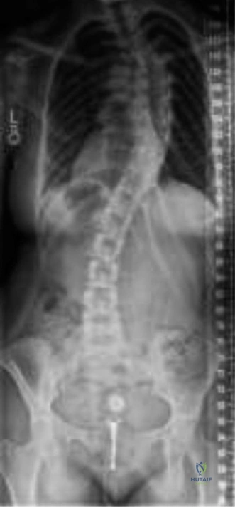
How do you classify this curve according to the Lenke system, and what does this classification tell you about the surgical plan?
Candidate: I would classify this using the Lenke system. I need to determine the curve type (1-6) based on flexibility films, the lumbar modifier (A, B, or C) based on the center sacral vertical line (CSVL) relation to the lumbar apical vertebrae, and the thoracic sagittal modifier (hypo, normal, or hyperkyphotic). The classification helps identify which curves are structural and dictate the levels for fusion.
Failing to mention the "flexibility films" (supine side-bending) as the primary determinant for distinguishing structural from non-structural curves. Candidates often forget to mention the sagittal thoracic modifier, which is essential for determining the appropriate rod contouring strategy.
A structured response: 1) State the curve type (1-6) determined by structural criteria on side-bending films (Cobb >25°). 2) Assign the lumbar modifier (A, B, C) based on the distance of the apical lumbar pedicle to the CSVL. 3) Define the sagittal thoracic modifier (T5-T12). The classification allows for "selective fusion," sparing motion segments by only fusing the structural curves, while providing a framework for 3D correction.
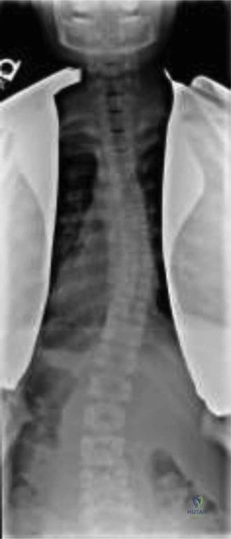
During the planning of the posterior approach, you are evaluating the pedicle anatomy at the apex of the thoracic deformity. You observe the following radiographic anatomy.
What are the specific risks during screw insertion at this level, and how do you mitigate them?
Candidate: The apex of the deformity, particularly the concave side, presents with thin, sclerotic, and dysplastic pedicles. The primary risk is medial or lateral pedicle wall breach, which could injure the spinal cord or pleura/aorta, respectively. I use the freehand technique with tactile "gear-shifting" to feel the walls, confirm with a ball-tipped sounder, and verify position.
Ignoring the "medial angulation" of the thoracic pedicles. Candidates often treat all pedicles as if they have the same anatomy, failing to mention the difference in trajectory between upper and mid-thoracic levels or the specific risk to the aorta on the left side.
Candidates must describe the "five-point check" (medial, lateral, superior, inferior walls and floor). Emphasize the anatomical danger zones: the aorta located anterior to the left mid-thoracic vertebrae and the concave apex pedicle dysplasia. Mentioning intraoperative navigation or fluoroscopic verification as a backup to freehand techniques demonstrates high-level surgical awareness.
You are performing the deformity correction. You have applied the rod to the concave side. Look at this image regarding the derotation maneuver.
Explain the biomechanics of the "Rod Derotation" maneuver and the risks associated with it.
Candidate: Rod derotation involves engaging the pre-contoured rod into the concave screws and rotating it. This converts the coronal deformity into a sagittal shape—effectively fixing the kyphosis. The risk is spinal cord injury due to traction or stretch as the spine is straightened, which is why neuromonitoring is essential.
Failing to discuss the "Direct Vertebral Rotation" (DVR) as a separate, more controlled technique. Relying solely on rod rotation can lead to "flatback" syndrome if the sagittal contouring is not meticulous.
The perfect answer highlights the 3D nature of the correction: coronal (Cobb angle), axial (rib rotation), and sagittal (kyphosis/lordosis). Mention the role of IONM (SSEP/MEP) as the "safety net." Explain that rod derotation is a powerful force that must be balanced by DVR to achieve true 3D correction while respecting the spinal cord's vascularity (Artery of Adamkiewicz).
Intraoperatively, after the derotation maneuver, the neuromonitoring team reports a 60% drop in MEP amplitude. What is your immediate, systematic action plan?
Candidate: I would immediately alert the team. I would check the MAP and increase it to >85mmHg, check for patient blood loss/hypovolemia, and reverse any recent correction maneuvers. If it persists, I would consider a wake-up test.
Panicking or jumping to remove all hardware immediately. The candidate must present a logical, tiered approach that rules out "systemic" causes (anesthesia, blood pressure) before assuming a mechanical cause.
The "Rule of 4": 1) **Systemic:** Check MAP, hemoglobin/hematocrit, oxygenation, and temperature. 2) **Technical:** Verify monitoring leads/probes. 3) **Surgical:** Reverse the most recent maneuver (e.g., untighten rod, back off distraction). 4) **Neurological:** If no improvement, perform Stagnara wake-up test. Emphasize that in AIS, this is a "do-not-ignore" event requiring complete, calm transparency with the theater team.
