Septic Arthritis: Pathophysiology, Diagnosis, and Emergency Management
Key Takeaway
Septic arthritis is an acute, critical joint infection, an orthopedic emergency that rapidly destroys cartilage. Often caused by Staphylococcus aureus, it presents with severe pain, swelling, and warmth. Diagnosis relies on urgent arthrocentesis for joint fluid analysis to identify the causative pathogen.
Introduction & Epidemiology
Septic arthritis (SA) represents a true orthopedic emergency characterized by an acute infection within the synovial space, leading to rapid and irreversible destruction of articular cartilage if not promptly diagnosed and aggressively treated. The devastating potential for long-term joint morbidity, including progressive osteoarthritis, ankylosis, and limb-threatening sepsis, underscores the imperative for immediate intervention.
Pathophysiology
Infection typically gains access to the joint space via three primary mechanisms:
*
Hematogenous spread:
The most common route, particularly in children and immunocompromised adults. Bacteria disseminate from a distant primary site (e.g., skin infection, UTI, pneumonia, endocarditis) through the bloodstream and colonize the highly vascularized synovial membrane.
*
Direct inoculation:
Occurs secondary to penetrating trauma (e.g., open fractures, animal bites), iatrogenic complications (e.g., intra-articular injections, arthroscopy, reconstructive surgery), or spread from adjacent soft tissue infections.
*
Contiguous spread:
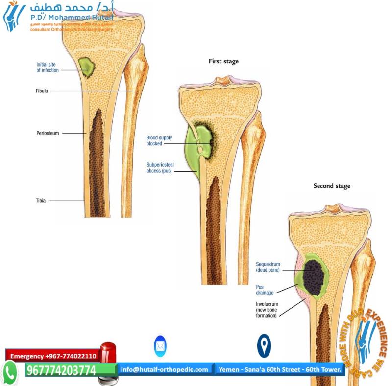
Less common, but can occur from adjacent osteomyelitis, particularly relevant in children where the metaphysis lies within the joint capsule (e.g., proximal femur, proximal humerus, radial neck, distal fibula). In infants, the transphyseal vessels allow infection to cross the physis directly into the joint.
Once established, bacteria proliferate within the synovial fluid, triggering an intense inflammatory response. Polymorphonuclear leukocytes release proteolytic enzymes (e.g., collagenase, elastase) and cytokines (e.g., IL-1, TNF-alpha) that directly degrade the avascular articular cartilage. Elevated intra-articular pressure from the effusion can further compromise cartilage nutrition and contribute to avascular necrosis (AVN), particularly in enclosed joints like the hip in children.
Etiology & Microbiology
The causative organisms vary with age, risk factors, and geographic location:
*
Staphylococcus aureus
:
The predominant pathogen across all age groups, accounting for 50-70% of cases in native joints. Methicillin-resistant
S. aureus
(MRSA) incidence is increasing and demands specific antibiotic considerations.
*
Streptococci (Groups A, B, C, G,
S. pneumoniae
):
* Account for 10-20% of infections, often seen in patients with comorbidities or skin infections.
*
Neisseria gonorrhoeae
:
Common in sexually active young adults (under 40 years old), often presenting as migratory polyarthralgia or tenosynovitis before evolving into oligo- or monoarticular arthritis. Generally less destructive to cartilage than staphylococcal infections.
*
Gram-negative bacilli (e.g.,
E. coli
,
Pseudomonas aeruginosa
,
Klebsiella spp.
):
More prevalent in intravenous drug users, immunocompromised individuals, elderly patients, or those with underlying genitourinary/gastrointestinal pathology.
*
Coagulase-negative Staphylococci:
Common in prosthetic joint infections but can occasionally cause native joint SA, especially after instrumentation.
*
Propionibacterium acnes
(now
Cutibacterium acnes*):
Rarely causes acute septic arthritis in native joints but has been implicated in chronic, low-grade infections and is a significant pathogen in shoulder prosthetic joint infections. Its role as an antigenic trigger in SAPHO syndrome is distinct from acute SA.
*
Fungi (e.g.,
Candida spp.
):
Rare, usually seen in immunocompromised hosts, those on prolonged broad-spectrum antibiotics, or with indwelling catheters.
*
Mycobacteria (e.g.,
M. tuberculosis
):
Presents as a chronic monoarthritis, often mimicking inflammatory or degenerative conditions.
Risk Factors
Recognized risk factors include:
* Pre-existing joint disease (rheumatoid arthritis, osteoarthritis, gout).
* Immunosuppression (diabetes mellitus, renal failure, liver disease, HIV, malignancy, systemic corticosteroid use, TNF-alpha inhibitors, other biologics).
* Advanced age.
* Intravenous drug use.
* Prosthetic joints (distinct entity but shared risk factors).
* Recent joint surgery, arthroscopy, or intra-articular injections.
* Skin infections or open wounds near the joint.
* Trauma.
Clinical Presentation & Diagnosis
A high index of suspicion is paramount.
*
History:
Acute onset of severe, progressive joint pain, swelling, warmth, and erythema in a monoarticular distribution (knee > hip > shoulder > ankle > elbow > wrist). Polyarticular involvement is less common (10-20%), often associated with gonococcal arthritis or underlying inflammatory conditions. Patients typically report significant loss of joint function and pain with any motion or loading of the joint. Systemic symptoms like fever, chills, and malaise are common but not universally present, particularly in immunocompromised individuals.
*
Physical Examination:
Affected joint is swollen, warm, erythematous, exquisitely tender to palpation, and held in a position of maximum comfort (e.g., slight flexion for the knee) to minimize capsular tension. Both active and passive range of motion are severely restricted and painful.
*
Laboratory Investigations:
*
Complete Blood Count (CBC):
Leukocytosis with a left shift is common but not always present.
*
Erythrocyte Sedimentation Rate (ESR) & C-reactive protein (CRP):
Usually elevated (>90% sensitivity for both), but non-specific. Baseline values and trend are more informative.
*
Procalcitonin:
Can be elevated in bacterial infections, potentially more specific than ESR/CRP, but its routine use in SA is still debated.
*
Blood Cultures:
Positive in approximately 30-50% of cases, crucial for identifying the causative organism, especially if joint fluid cultures are negative. Draw two sets from different sites prior to antibiotic administration.
*
Arthrocentesis (Diagnostic Gold Standard):
Urgent aspiration of synovial fluid is mandatory for any suspected septic joint.
*
Gross Appearance:
Turbid, purulent fluid.
*
Cell Count with Differential:
White blood cell (WBC) count typically >50,000 cells/mm³ (often >100,000 cells/mm³), with >90% polymorphonuclear leukocytes (PMNs). Lower counts do not rule out infection, especially early or in immunocompromised patients.
*
Gram Stain:
Positive in 30-70% of cases, providing crucial empiric antibiotic guidance. A negative Gram stain does not exclude infection.
*
Cultures:
Aerobic and anaerobic cultures are essential. Crystal analysis (polarized light microscopy) to rule out gout/pseudogout.
*
Other:
Glucose (low, <50% of serum glucose), protein (high), lactate (high).
*
Imaging:
*
Plain Radiographs:
Typically normal in early stages. Later findings include soft tissue swelling, joint effusion, joint space narrowing, periarticular osteopenia, and eventual erosions or subchondral lucencies indicating cartilage and bone destruction.
*
Ultrasound:
Highly sensitive for detecting joint effusions, particularly in difficult-to-examine joints like the hip or shoulder, and can guide arthrocentesis.
*
Magnetic Resonance Imaging (MRI):
Highly sensitive for identifying effusions, synovitis, capsular distention, cartilage erosions, and concomitant osteomyelitis or periarticular abscesses. Also useful for assessing surrounding soft tissue involvement.
*
Bone Scan:
Increased uptake, but non-specific and lacks diagnostic specificity for infection vs. inflammation.
Differential Diagnosis of Acute Monoarthritis
Promptly distinguishing septic arthritis from other causes of acute monoarthritis is critical:
*
Gout/Pseudogout:
Crystal-induced arthropathies. Joint fluid aspiration with crystal analysis is diagnostic. History of recurrent attacks, specific joint involvement (e.g., MTP joint for gout), and dietary triggers may be suggestive. However, septic arthritis can coexist with crystal arthropathy.
*
Inflammatory Arthritis Flare:
Rheumatoid arthritis, psoriatic arthritis, ankylosing spondylitis. History of chronic disease, but an acute flare can mimic infection.
*
Reactive Arthritis:
Follows an infection elsewhere (e.g., genitourinary or gastrointestinal), typically sterile joint fluid.
*
Lyme Arthritis:
Usually large joint, monoarticular, with migratory arthralgias, history of tick exposure.
*
Transient Synovitis of the Hip:
Common in children, typically viral, self-limiting. Less severe pain, child can usually bear weight.
*
Traumatic Hemarthrosis:
History of significant trauma, blood on aspiration.
*
Osteomyelitis (adjacent):
Can mimic SA, but infection is primarily bone-confined. Imaging (MRI) helps differentiate and identify contiguous spread.
*
SAPHO Syndrome (Synovitis, Acne, Pustulosis, Hyperostosis, Osteitis):
An inflammatory disorder that can cause bone pain and joint involvement, sometimes mistaken for chronic osteomyelitis or septic arthritis.
* Also known as acquired hyperostosis syndrome.
* Typically affects young to middle-aged adults with bone pain and characteristic skin involvement (palmoplantar pustulosis, severe acne, hidradenitis suppurativa).
* Suspicion exists that
Propionibacterium acnes
(now
Cutibacterium acnes
) may serve as an antigenic trigger, inducing a sterile inflammatory response. Cultures are typically sterile or occasionally yield
P. acnes
.
* Characterized by humoral induction of sclerosis and erosions.
* Sternoclavicular region is most commonly involved, followed by the axial skeleton (vertebrae, sacroiliac joints, especially unilateral sacroiliitis).
* Laboratory findings: ESR, CRP moderately elevated, but typically lower than in acute septic arthritis.
* Imaging: Bone scan ("bull's head sign" at the sternoclavicular region, increased sacroiliac joint uptake) is considered the gold standard for identifying affected areas. MRI can show erosion of vertebral body corners and synovitis.
* Pathology: Sterile neutrophilic pseudoabscesses within bone, characteristic of an inflammatory, not infective, process.
* Treatment: NSAIDs, rheumatology consultation, methotrexate, sulfasalazine, and biologics (e.g., anti-TNF agents) are the mainstays, not surgical drainage and prolonged antibiotics as in septic arthritis.
It is critical to reiterate that the "treatment: symptomatic; resolves spontaneously; NSAIDs help" mentioned in the seed content applies to conditions like transient synovitis or possibly a mild SAPHO flare, and categorically does NOT apply to septic arthritis , which demands aggressive surgical and antimicrobial management.
Surgical Anatomy & Biomechanics
Understanding the intricate anatomy and biomechanics of various joints is paramount for effective surgical management of septic arthritis. The susceptibility of articular cartilage to rapid destruction mandates immediate and thorough intervention.
Articular Cartilage
- Composition: Articular cartilage is a specialized connective tissue composed primarily of chondrocytes, extracellular matrix (collagen type II, proteoglycans), and water. It is avascular, aneural, and alymphatic.
- Nutrition: Relies entirely on the synovial fluid for nutrient supply and waste removal.
- Vulnerability: The avascular nature makes it highly susceptible to enzymatic degradation. Bacterial toxins and host inflammatory mediators (proteases, cytokines from PMNs) directly attack chondrocytes and the cartilage matrix. Prolonged exposure to purulent synovial fluid, even for a few hours, can cause irreparable damage. Increased intra-articular pressure also compromises nutrient flow, contributing to chondrocyte necrosis.
Synovial Membrane and Joint Capsule
- Synovium: The highly vascularized synovial membrane lines the non-articular surfaces of the joint capsule. Its rich blood supply makes it the primary site of hematogenous bacterial seeding.
- Capsule: The joint capsule encloses the joint space. While it forms a barrier preventing infection spread, it also sequesters the infection, creating a closed space under pressure.
Specific Joint Considerations
- Knee: The largest synovial joint, most commonly affected by septic arthritis. Its superficial location and large synovial volume allow for relatively easy aspiration and arthroscopic access, but the extensive cartilage surface area is vulnerable.
-
Hip:
The second most common site in adults, but the most common in infants and young children.
- Children: The hip joint capsule in infants extends to include the metaphysis of the proximal femur. This anatomical peculiarity makes it highly susceptible to septic arthritis as a direct extension of metaphyseal osteomyelitis. Furthermore, the tenuous blood supply to the femoral head, particularly the retinacular vessels, is easily compromised by elevated intra-articular pressure, leading to a high risk of avascular necrosis (AVN) and subsequent femoral head collapse or coxa vara. Urgent decompression is critical.
- Adults: Deep-seated, making aspiration challenging without imaging guidance. Open or arthroscopic drainage is often required due to its depth and the difficulty of complete evacuation via needle.
- Shoulder: Also a deep joint with a complex capsule. Often requires arthroscopic or open drainage.
- Ankle/Elbow/Wrist: Less common than knee/hip/shoulder, but equally susceptible to cartilage destruction. Arthroscopy is often feasible.
- Sternoclavicular (SC) / Acromioclavicular (AC) Joints: Less common but can occur, especially in IV drug users or with local trauma/infection. Anatomical proximity to vital structures (subclavian vessels, pleura) demands careful surgical planning. These joints can also be involved in sterile inflammatory conditions like SAPHO syndrome.
Intra-articular Physis
As noted in the seed content, the "extension of metaphyseal osteomyelitis at intraarticular physis" is a critical consideration in children. This occurs in specific locations:
*
Proximal Femur:
The metaphysis of the femoral neck is intra-articular in infants and young children, making hip septic arthritis a common sequela of femoral neck osteomyelitis.
*
Proximal Humerus:
Similar anatomy, the metaphyseal bone is partially intra-articular.
*
Radial Neck:
The metaphysis of the radial head is largely intra-articular.
*
Distal Fibula:
The distal fibular metaphysis is within the ankle joint capsule.
Understanding these anatomical relationships underscores the importance of urgent surgical drainage and thorough debridement to prevent severe joint destruction and long-term functional impairment.
Indications & Contraindications
The decision-making process for managing septic arthritis centers on prompt and effective source control and antimicrobial therapy. Surgical intervention is the mainstay for most cases.
Operative Indications
- Confirmed or Highly Suspected Septic Arthritis: Especially in large, weight-bearing joints (hip, knee) or joints with significant functional importance (shoulder, elbow).
- Failure of Serial Aspiration/Non-Operative Management: If initial aspirations and empiric intravenous (IV) antibiotics do not lead to rapid clinical improvement (e.g., persistent fever, increasing pain, signs of ongoing inflammation, lack of fluid sterility on repeat aspiration after 24-48 hours).
- Inadequate Drainage by Aspiration: If the joint fluid is thick, loculated, or cannot be fully evacuated via needle aspiration. This is common in the hip and shoulder due to their deep anatomy or in more advanced infections with significant fibrin and debris.
- Concomitant Osteomyelitis: If imaging (especially MRI) reveals adjacent bone infection, surgical debridement of both the joint and the involved bone is necessary.
- Presence of Foreign Material: Intra-articular foreign bodies (e.g., retained hardware, traumatic debris) require surgical removal.
- Delayed Presentation/Advanced Disease: Cases presenting late with extensive synovitis, pannus formation, cartilage erosion, or loculated pus.
- Specific Organisms: Certain highly virulent organisms (e.g., S. aureus , Gram-negative rods) often necessitate immediate surgical drainage to minimize cartilage damage.
- Pediatric Hip Septic Arthritis: Considered an absolute surgical emergency due to the high risk of AVN of the femoral head.
Non-Operative Indications (Adjunctive to IV Antibiotics)
It is crucial to emphasize that "non-operative" in this context refers to aspiration-based management
instead of arthrotomy/arthroscopy
for definitive source control, but
not
the absence of treatment. All patients with septic arthritis require intravenous antibiotics.
*
Early, Uncomplicated Gonococcal Arthritis:
Often responds well to IV antibiotics alone. Serial aspirations may be sufficient if effusion persists or to confirm sterility. Surgical drainage is typically reserved for cases that fail to respond or have persistent large effusions.
*
Small, Easily Accessible Joints:
For small joints (e.g., digits) where repeated aspirations can reliably achieve complete drainage and clinical response is rapid.
*
Very Early Stage Disease:
Rare, but if the diagnosis is made within hours of symptom onset, and the fluid is not overtly purulent, a trial of serial aspirations (e.g., every 12-24 hours) may be considered
only if
the patient shows rapid clinical improvement and fluid analysis rapidly normalizes. This is a high-risk strategy and surgical drainage remains the safer default for large joints.
*
Severe Medical Comorbidities:
In patients with overwhelming comorbidities precluding anesthesia, serial aspirations may be the only option, but this is a relative contraindication to surgery rather than a preferred treatment for septic arthritis.
Contraindications to Operative Management
There are no absolute contraindications to operative drainage if septic arthritis is diagnosed and requires surgical intervention. However, relative contraindications may necessitate delaying surgery for patient stabilization:
*
Patient Instability/Severe Sepsis:
Patients in septic shock or with uncontrolled medical comorbidities should be resuscitated and stabilized in an intensive care setting before surgery, if possible. Delaying source control, however, worsens outcomes. The decision for immediate versus delayed surgery must weigh the risks of anesthesia against the urgency of joint decompression.
*
Coagulopathy:
Should be corrected prior to surgery to minimize bleeding complications.
Table: Operative vs. Non-Operative Indications for Septic Arthritis
| Feature | Operative Management (I&D via Arthrotomy/Arthroscopy) | Non-Operative Management (Serial Aspiration + IV ABX) |
|---|---|---|
| Joint Involvement | Large joints (hip, shoulder), joints difficult to drain (e.g., deep-seated) | Small joints (e.g., wrist, ankle, elbow), readily aspirable large joints (e.g., knee) |
| Joint Fluid Characteristics | Frank pus, thick fluid, loculated fluid, inability to fully aspirate | Serous/less purulent fluid, successful complete aspiration |
| Causative Organism | S. aureus , Gram-negative rods, fungal, polymicrobial, highly virulent organisms | N. gonorrhoeae (often responds to ABX alone), low virulence organisms (rare) |
| Response to Initial Rx | Persistent infection, worsening symptoms, fever, increasing pain, non-sterile fluid | Rapid clinical improvement, resolution of systemic symptoms, sterile fluid on repeat aspiration |
| Concomitant Pathology | Adjacent osteomyelitis, foreign body, extensive cartilage damage, chronic synovitis | None |
| Patient Factors | Immunocompromised, diabetic, pediatric hip septic arthritis | Healthy, young, early presentation (rare for large joints) |
| Severity | Moderate to severe, delayed presentation | Mild, very early presentation |
Pre-Operative Planning & Patient Positioning
Thorough pre-operative planning is essential to ensure a safe and effective surgical intervention for septic arthritis.
Assessment & Resuscitation
- Rapid Medical Assessment: Evaluate for signs of systemic sepsis (fever, tachycardia, hypotension, altered mental status). Initiate sepsis protocol if indicated (fluid resuscitation, vasopressors as needed).
-
Laboratory Work-up:
- Complete blood count (CBC) with differential.
- ESR, CRP.
- Renal and liver function tests.
- Coagulation profile (PT/INR, PTT).
- Type and Screen/Crossmatch if significant blood loss is anticipated (rare in routine SA drainage).
- Blood cultures (already drawn, but ensure results are pending or available).
- Imaging Review: Review plain radiographs, ultrasound, and MRI (if obtained) to identify potential concomitant osteomyelitis, abscesses, or the extent of joint destruction.
Antibiotics
-
Empiric IV Antibiotics:
Initiate broad-spectrum intravenous antibiotics immediately after joint fluid and blood cultures are drawn. This is critical and should not await surgical drainage.
-
Common Empiric Regimens:
- Adults: Vancomycin (for MRSA coverage) + a third-generation cephalosporin (e.g., ceftriaxone or cefotaxime) or an antipseudomonal beta-lactam (e.g., piperacillin-tazobactam) if Gram-negative or Pseudomonas is suspected (e.g., IV drug users, immunocompromised).
- Children: Vancomycin + cefotaxime/ceftriaxone.
- Gonococcal arthritis: Ceftriaxone.
-
Common Empiric Regimens:
- Antibiotic Adjustment: Once Gram stain results are available, adjust antibiotics accordingly. Definitive narrowing of the antibiotic spectrum occurs when culture sensitivities are known.
Informed Consent
Thorough discussion with the patient (or legal guardian) regarding:
* Diagnosis and its urgency.
* Proposed surgical procedure (e.g., arthroscopic vs. open I&D).
* Risks: Persistent infection, joint stiffness, progressive cartilage damage, need for repeat surgery, avascular necrosis (especially hip), DVT/PE, anesthesia risks, wound infection.
* Expected post-operative course and rehabilitation.
Anesthesia
- General Anesthesia: Typically preferred for optimal muscle relaxation and patient comfort during surgical drainage and extensive lavage.
- Regional Anesthesia: Can be used as an adjunct for post-operative pain control, but typically not as the sole anesthetic for definitive surgical management.
Patient Positioning & Surgical Preparation
Meticulous attention to positioning and sterile preparation is essential.
*
Knee:
*
Position:
Supine. The knee is often flexed to 90 degrees over a bolster or a leg holder. A footrest may be used.
*
Tourniquet:
Applied to the proximal thigh, inflated after exsanguination for a bloodless field, which is critical for arthroscopy.
*
Hip:
*
Position:
Supine for anterior approaches (e.g., Smith-Petersen, modified Watson-Jones). Lateral decubitus for lateral or posterior approaches. A fracture table or traction is often not required for drainage but may be used for distraction to aid arthroscopy in some cases.
*
Fluoroscopy:
Often useful to guide arthrocentesis or confirm drain placement if not performing an open arthrotomy.
*
Shoulder:
*
Position:
Beach chair position or lateral decubitus.
*
Arm Preparation:
Free-draped to allow full range of motion during arthroscopy.
*
Other Joints (Ankle, Elbow, Wrist):
Positioned to allow maximal access and visualization of the joint.
*
Skin Preparation:
Wide sterile prep and drape using an antiseptic solution (e.g., chlorhexidine-alcohol or povidone-iodine). Include a large area to allow for potential extension of the incision or placement of additional portals/drains.
Detailed Surgical Approach / Technique
The primary goals of surgical intervention for septic arthritis are thorough removal of pus, debridement of infected and necrotic tissue (including fibrin and synovium), copious irrigation/lavage of the joint, and obtaining tissue for culture and histopathology. The choice between open arthrotomy and arthroscopic debridement depends on the joint, surgeon's expertise, and the extent of the infection.
General Principles of Surgical Drainage & Debridement
- Incision/Portal Placement: Adequate access for thorough visualization and debridement.
- Initial Aspiration/Culture: Before extensive irrigation, obtain additional fluid for Gram stain and culture directly from the joint through the surgical opening.
- Synovial Biopsy: Obtain samples of inflamed synovium for routine cultures (aerobic, anaerobic, fungal, mycobacterial) and histopathology. This is especially important for chronic infections or atypical presentations.
- Debridement: Remove all fibrin, purulent material, necrotic tissue, and any synovial pannus. A power shaver is useful during arthroscopy; curettes and rongeurs are used in open procedures. Ensure no loculations of pus remain.
- Copious Irrigation/Lavage: The cornerstone of mechanical debridement. Use large volumes (typically 6-12 liters or more) of sterile isotonic saline. Pulsatile lavage may enhance debris removal.
- Inspection: Thoroughly inspect all joint compartments, articular cartilage, and surrounding structures for additional infection, cartilage damage, or signs of osteomyelitis.
-
Drainage:
- Drains: Placement of an intra-articular suction drain (e.g., Hemovac, Jackson-Pratt) is common practice for 24-48 hours to remove residual exudate and prevent hematoma formation, which can act as a nidus for recurrent infection.
- Closure: Capsule may be loosely closed or left open depending on joint distension and surgeon preference. Subcutaneous and skin layers are closed in a standard fashion. In severely contaminated or necrotic wounds, delayed primary closure may be considered.
Specific Joint Approaches
1. Knee (Most Common Joint for Septic Arthritis)
- Preferred Method: Arthroscopic irrigation and debridement.
- Patient Positioning: Supine, knee flexed to 90 degrees over a bolster. Tourniquet.
-
Technique:
- Standard arthroscopic portals: Anterolateral, anteromedial. A suprapatellar portal is often used for inflow.
- Initial diagnostic arthroscopy: Systematically inspect all compartments (suprapatellar pouch, medial/lateral gutters, medial/lateral compartments, intercondylar notch).
- Synovectomy: Use a full-radius or aggressive shaver to debride all purulent, necrotic, and hypertrophied synovium and fibrin. Address any loculations.
- Lavage: Copious irrigation with 6-12 liters of saline using high-flow pumps.
- Drainage: A suction drain may be placed, often through a separate small stab incision or existing portal, and brought out inferomedially.
- Open Arthrotomy (Alternative/Failure Salvage): If arthroscopy is not feasible (e.g., severe contracture, massive swelling, limited equipment) or fails to adequately clear the infection. A medial or lateral parapatellar approach provides excellent visualization.
2. Hip (Surgical Emergency, Especially in Children)
- Preferred Method: Open arthrotomy is often favored due to the deep nature of the joint, difficulty with complete visualization and debridement arthroscopically in acute purulent conditions, and critical need for complete decompression. Arthroscopy is an option for certain cases and surgeons.
- Patient Positioning: Supine for anterior approach, lateral decubitus for lateral/posterior approaches.
-
Anterior Approach (Smith-Petersen or Modified Watson-Jones):
- Internervous Plane: Between the sartorius (femoral nerve) and tensor fascia lata (superior gluteal nerve).
- Incision: Longitudinal incision over the anterior aspect of the hip.
- Dissection: Superficial dissection to the fascia lata. Develop the interval between the sartorius (retracted medially) and tensor fascia lata (retracted laterally). Identify and ligate ascending branches of the lateral femoral circumflex artery and vein.
- Deep Dissection: Identify the rectus femoris. The direct head (originating from AIIS) can be retracted medially or released.
- Capsulotomy: A T-shaped or longitudinal capsulotomy is performed.
- Debridement & Lavage: Evacuate pus, obtain cultures/biopsy, thoroughly debride the synovium, and perform copious lavage. Inspect the femoral head and acetabulum.
- Closure: Capsule may be partially closed or left open. Drain placement is critical.
- Lateral/Posterior Approaches: May be used if extensive involvement is suspected posteriorly or if prior attempts via anterior approach failed.
- Arthroscopy (Advanced Cases): Requires specialized equipment and expertise. May be performed using standard anterior, anterolateral, or posterolateral portals with traction.
3. Shoulder
- Preferred Method: Arthroscopic irrigation and debridement.
- Patient Positioning: Beach chair position or lateral decubitus.
-
Technique:
- Standard arthroscopic portals: Posterior (viewing), anterior (working), lateral (working). An inflow cannula via an anterior portal is often used.
- Debridement: Use a shaver to remove purulent material and inflamed synovium from the entire joint, including the subacromial space if bursitis is present.
- Lavage: Copious saline irrigation.
- Drainage: Suction drain, typically through a separate lateral stab incision.
- Open Arthrotomy (Alternative/Failure Salvage): Deltopectoral approach is commonly used. Requires division or retraction of the pectoralis major and deltoid, identification of the cephalic vein, and then incision through the subscapularis and capsule.
4. Ankle, Elbow, Wrist
- Preferred Method: Arthroscopic irrigation and debridement for most cases, given the joint size and accessibility.
- Open Arthrotomy: Reserved for severe cases, inability to achieve adequate arthroscopic debridement, or specific anatomic considerations.
Post-Operative Antibiotics
- Continuation of IV Antibiotics: Continue culture-directed IV antibiotics. The duration is organism-specific and typically ranges from 2 to 6 weeks.
- Transition to Oral: Transition to oral antibiotics is considered when the patient is afebrile, clinically improving, and inflammatory markers (ESR/CRP) are trending down. The total duration of antibiotics (IV + oral) is typically 4-6 weeks for most cases, longer for more virulent organisms or immunocompromised hosts. Close collaboration with an Infectious Disease specialist is essential.
Complications & Management
Septic arthritis is associated with significant morbidity despite prompt treatment. Understanding potential complications and their management is crucial for optimizing patient outcomes.
Common Complications
-
Persistent Infection (5-20%):
- Description: Failure to eradicate the infection, often due to inadequate debridement, loculated pus, resistant organisms, or underlying host factors.
- Clinical Presentation: Persistent fever, elevated inflammatory markers (ESR/CRP), continued joint pain/effusion.
-
Management:
- Repeat I&D: Often required. May necessitate a more aggressive open approach if initial arthroscopy was performed.
- Antibiotic Adjustment: Re-evaluate and adjust antibiotic regimen based on new cultures and sensitivities, often requiring consultation with Infectious Disease.
- Imaging: MRI to rule out loculated abscesses or concomitant osteomyelitis.
-
Cartilage Destruction / Post-Septic Osteoarthritis:
- Description: Irreversible damage to articular cartilage due to enzymatic degradation and mechanical wear, leading to progressive osteoarthritis. Inevitable if diagnosis and treatment are delayed.
- Clinical Presentation: Chronic joint pain, stiffness, crepitus, reduced range of motion, eventually leading to end-stage joint disease.
-
Management:
- Prevention: The best treatment is prompt and aggressive initial management.
- Symptomatic Management: NSAIDs, physical therapy, intra-articular injections (corticosteroids, hyaluronic acid, carefully considering infection risk).
- Joint Salvage Procedures: Osteotomies to realign the joint.
- End-Stage Treatment: Total joint arthroplasty (TJA) or arthrodesis. TJA in a previously infected joint carries a higher risk of recurrent infection and requires careful planning.
-
Joint Stiffness / Arthrofibrosis (30-50%):
- Description: Formation of intra-articular adhesions and capsular contracture, limiting joint range of motion.
- Clinical Presentation: Restricted active and passive ROM, pain with movement.
-
Management:
- Early Mobilization: Aggressive, early post-operative physical therapy is paramount.
- Manipulation Under Anesthesia (MUA): Can be performed for acute-onset stiffness if infection is deemed resolved.
- Arthroscopic Lysis of Adhesions / Open Capsular Release: For recalcitrant stiffness.
-
Avascular Necrosis (AVN) of the Femoral Head (Hip in Children):
- Description: Ischemic death of osteocytes in the femoral head due to compromised blood supply, often secondary to elevated intra-articular pressure and capsular distension in pediatric hip septic arthritis.
- Incidence: High, up to 15-50% in pediatric hip SA.
- Clinical Presentation: Persistent pain, limping, deformities (e.g., coxa plana, coxa magna), eventual collapse of the femoral head.
-
Management:
- Prevention: Urgent surgical decompression of the hip.
- Observation: Initial management, especially in early stages.
- Core Decompression: For early AVN.
- Containment Procedures: Osteotomies (e.g., varus derotation osteotomy) to protect the collapsing femoral head.
- Future Arthroplasty: For severe, end-stage AVN and osteoarthritis.
-
Sepsis / Systemic Complications (5-10% of SA patients):
- Description: Systemic inflammatory response syndrome (SIRS) progressing to severe sepsis, septic shock, multi-organ dysfunction syndrome (MODS), acute respiratory distress syndrome (ARDS), renal failure.
-
Management:
- ICU Care: Aggressive medical management in an intensive care unit.
- Hemodynamic Support: Fluid resuscitation, vasopressors.
- Source Control: Immediate and definitive surgical drainage is critical.
- Antibiotics: Broad-spectrum IV antibiotics.
-
Recurrence of Infection (2-5%):
- Description: Re-establishment of infection in the same joint after apparent resolution.
- Management: Re-evaluation with joint aspiration, imaging, and repeat surgical debridement and lavage, followed by prolonged culture-directed antibiotics.
-
Growth Plate Arrest / Deformity (Children):
- Description: Damage to the physis (growth plate) by infection can lead to premature physeal closure, limb length discrepancy, or angular deformities.
- Management: Long-term monitoring, corrective osteotomies, or epiphysiodesis for limb length discrepancies.
-
Wound Complications (5-10%):
- Description: Superficial or deep wound infection, dehiscence, hematoma formation.
- Management: Local wound care, debridement of necrotic tissue, appropriate antibiotics, delayed primary closure if necessary.
Table: Common Complications, Incidence, and Salvage Strategies
| Complication | Incidence (approximate) | Salvage Strategies |
|---|---|---|
| Persistent Infection | 5-20% | Repeat I&D, antibiotic adjustment, conversion to open surgery if arthroscopy fails, consider PJI protocols if prosthetic |
| Cartilage Destruction/OA | High with delayed treatment, esp. hip; ~30-50% long-term | Symptomatic treatment, joint salvage procedures (osteotomy), eventual arthroplasty/arthrodesis |
| Joint Stiffness/Arthrofibrosis | Common, up to 30-50% in various joints | Early ROM, PT, MUA, arthroscopic lysis of adhesions, open capsular release |
| Avascular Necrosis (AVN) | Pediatric Hip: 15-50% | Observation, core decompression, containment osteotomies, eventual arthroplasty |
| Sepsis/Systemic Complications | 5-10% (patients with SA who progress to severe sepsis/shock) | ICU care, hemodynamic support, aggressive source control, broad-spectrum ABX |
| Recurrence of Infection | 2-5% | Re-evaluation, repeat surgical intervention, prolonged/adjusted antibiotics |
| Growth Plate Arrest/Deformity | 5-10% (pediatric cases with physeal involvement) | Long-term monitoring, corrective osteotomies, epiphysiodesis |
| Wound Complications (infection/dehiscence) | 5-10% | Local wound care, debridement, antibiotics, delayed primary closure |
| Deep Vein Thrombosis (DVT) | Varies, ~1-5% without prophylaxis | Prophylactic anticoagulation, early mobilization, therapeutic anticoagulation for confirmed DVT |
Post-Operative Rehabilitation Protocols
Post-operative rehabilitation is a critical component of successful septic arthritis management, focusing on restoring joint function, preventing stiffness, and ensuring complete eradication of infection. The protocol must be individualized, considering the affected joint, patient's age, and overall medical status. Close collaboration between the surgeon, physical therapist, and infectious disease specialist is essential.
General Principles
- Pain Management: Adequate pain control is fundamental for successful participation in rehabilitation. Multimodal analgesia (opioids, NSAIDs if appropriate, acetaminophen, regional nerve blocks) should be employed.
- Infection Control: Continuation of appropriate antibiotic therapy as guided by Infectious Disease.
- Early Mobilization: Rapid, controlled initiation of range of motion (ROM) exercises is paramount to prevent arthrofibrosis and cartilage nutritional compromise, once immediate post-operative pain allows.
- Gradual Progression: Exercises should progress from passive to active-assisted to active ROM, followed by strengthening and functional activities.
- Weight Bearing Restrictions: Often necessary for lower extremity joints, especially if there's significant cartilage damage or risk of AVN.
Phase 1: Acute Post-Operative Phase (Days 0-14)
- Goals: Control pain and swelling, protect surgical repair, initiate gentle ROM, monitor for signs of persistent infection.
-
Immobilization:
- Brief periods of immobilization (e.g., in a brace or splint) may be used for comfort or to protect a very unstable joint, but prolonged immobilization is generally avoided.
- For the knee, a hinged knee brace locked in extension for transfers, but removed for exercises.
- For the hip, an abduction pillow/splint for comfort may be used.
-
Range of Motion:
- Continuous Passive Motion (CPM): Particularly beneficial for the knee, initiated as early as post-operative day 1, to prevent adhesions and promote synovial fluid circulation.
- Gentle Passive & Active-Assisted ROM: Under the guidance of a physical therapist. Emphasis on pain-free motion within protected arcs.
-
Joint-Specific ROM:
- Knee: Flexion/extension to tolerance.
- Hip: Flexion, abduction, rotation (carefully).
- Shoulder: Pendulum exercises, passive external rotation, flexion.
-
Weight Bearing (Lower Extremity Joints):
- Non-Weight Bearing (NWB) or Touch-Down Weight Bearing (TDWB): Typically prescribed for the affected lower extremity joint (hip, knee, ankle) to protect the healing joint surfaces and minimize stress on inflamed cartilage.
- Use of crutches or a walker for ambulation.
- Pain & Edema Management: Cryotherapy, elevation, gentle compression.
- Wound Care: Monitor incision sites for signs of infection (erythema, drainage, warmth).
- Antibiotics: Continue IV antibiotics as prescribed. Monitor inflammatory markers (ESR, CRP) for trending down.
Phase 2: Intermediate Recovery Phase (Weeks 2-6)
- Goals: Increase active ROM, begin strengthening, progress weight-bearing, normalize gait.
-
Range of Motion:
- Progress from active-assisted to full active ROM as tolerated.
- Gentle stretching exercises to improve flexibility and address any developing contractures.
-
Strengthening:
- Isometrics: Initiate pain-free isometric contractions around the joint.
- Progressive Isotonic Exercises: With light resistance, gradually increasing as tolerated. Focus on major muscle groups surrounding the joint (e.g., quadriceps, hamstrings, gluteals for lower limb; deltoid, rotator cuff for shoulder).
-
Weight Bearing:
- Gradual progression from TDWB to Partial Weight Bearing (PWB), then to Full Weight Bearing (FWB) as pain allows and imaging (if obtained) shows no adverse changes. This progression can be slow and may extend beyond 6 weeks, especially for pediatric hips or joints with significant cartilage damage.
- Gait training with appropriate assistive devices.
- Proprioception and Balance: Incorporate exercises (e.g., single-leg stance, wobble board) to improve joint stability and balance, particularly for lower extremity joints.
- Antibiotics: Transition from IV to oral antibiotics based on Infectious Disease recommendations and clinical response.
Phase 3: Advanced Recovery & Return to Activity (Weeks >6)
- Goals: Restore full functional strength, endurance, and agility. Prepare for return to activities of daily living (ADLs), work, and potentially sports.
-
Intensified Strengthening:
- Progressive resistance exercises, functional movements, plyometrics (if appropriate).
- Endurance training.
- Sport-Specific Training: For athletes, a structured return-to-sport program is initiated, focusing on agility, jumping, landing mechanics, and sport-specific drills.
-
Long-Term Monitoring:
- Regular follow-up appointments to monitor for late complications such as osteoarthritis, persistent pain, or recurrence of infection.
- Annual radiographs may be considered for joints with significant cartilage damage.
- Patients should be educated on the long-term potential for post-septic arthritis osteoarthritis.
Summary of Key Literature / Guidelines
The management of septic arthritis is guided by a robust body of literature and consensus guidelines from various orthopedic, infectious disease, and rheumatology societies. The overarching principles emphasize urgency and a multidisciplinary approach.
Consensus Guidelines
Key organizations providing guidelines include:
*
Infectious Diseases Society of America (IDSA):
Publishes comprehensive guidelines for the diagnosis and management of various infectious diseases, including bone and joint infections.
*
American Academy of Orthopaedic Surgeons (AAOS):
Provides clinical practice guidelines relevant to orthopedic surgical management.
*
National Institute for Health and Care Excellence (NICE) (UK):
Offers evidence-based recommendations for healthcare.
Key Principles of Management
The current understanding of septic arthritis management can be distilled into several core tenets:
-
Prompt Diagnosis:
Urgent joint aspiration is the gold standard for diagnosis. Delay significantly correlates with worse outcomes due to rapid cartilage destruction.
- Literature Support: Numerous studies confirm that delay in diagnosis beyond 24-48 hours dramatically increases the risk of permanent joint damage, emphasizing the "septic joint emergency" dictum.
-
Empiric Broad-Spectrum Intravenous Antibiotics:
Initiate immediately after joint fluid and blood cultures are obtained. This bridges the gap until definitive culture and sensitivity results are available.
- Rationale: Early antibiotic penetration into the synovial fluid helps control bacterial proliferation and mitigate cartilage degradation.
-
Urgent Surgical Debridement and Lavage:
For most cases of septic arthritis in native joints, particularly large, weight-bearing joints (hip, knee) and deep joints (shoulder, hip), surgical debridement and copious lavage is the definitive treatment.
- Evidence: Meta-analyses and systematic reviews consistently demonstrate superior outcomes (lower rates of persistent infection, less joint destruction) with surgical debridement compared to serial aspirations alone, especially for purulent effusions. Arthroscopic debridement has largely replaced open arthrotomy for many joints due to its less invasive nature, superior visualization, and similar efficacy, but open arthrotomy remains essential for hips, complex cases, or failed arthroscopy.
- Culture-Directed Antibiotic Therapy: Antibiotic regimen must be narrowed and adjusted based on Gram stain results and subsequent bacterial culture and sensitivity reports to ensure optimal efficacy and minimize resistance.
- Appropriate Duration of Antibiotics: The total duration of antibiotic therapy (initial IV phase followed by oral) is typically 2-6 weeks, individualized based on the pathogen, joint involved, patient's immune status, and clinical response. Longer courses (e.g., 6 weeks) are common for S. aureus or Gram-negative infections.
- Early Mobilization: Post-operative rehabilitation should prioritize early, controlled range of motion to prevent arthrofibrosis, promote cartilage healing, and improve long-term joint function.
Recent Advances and Debates
- Role of Arthroscopy vs. Open Arthrotomy: While arthroscopy is favored for knees and shoulders, the optimal approach for the hip remains debated. Many argue for open arthrotomy in the hip, especially in children, to ensure complete decompression and debridement, given the high risk of AVN.
- Predictors of Poor Outcome: Factors consistently associated with worse outcomes include delayed diagnosis, older age, Staphylococcus aureus infection, immunocompromised status, and involvement of the hip joint in children.
- Biomarkers: The utility of procalcitonin and other inflammatory biomarkers in differentiating septic arthritis from other inflammatory arthropathies and in guiding antibiotic duration is an ongoing area of research.
- Management of Prosthetic Joint Infections (PJI): While distinct from native joint septic arthritis, the principles of source control and prolonged antibiotics are shared. PJI management often involves complex algorithms of debridement, antibiotics, and implant retention (DAIR), one-stage, or two-stage revision arthroplasty, depending on acuity, organism, and host factors. These specialized protocols are beyond the scope of acute native joint septic arthritis but highlight the ongoing evolution of infection management in orthopedics.
In conclusion, septic arthritis remains a formidable challenge in orthopedic surgery. Adherence to established principles of urgent diagnosis, appropriate empiric and culture-directed antibiotic therapy, and definitive surgical source control, followed by aggressive rehabilitation, offers the best opportunity to preserve joint function and prevent devastating long-term sequelae.
You Might Also Like