Deep Posterior Thigh Liposarcoma: An Orthopedic Oncology Case Study & Diagnostic Approach
Key Takeaway
Diagnosing deep posterior thigh liposarcoma (ALT) involves patient history of an enlarging painless mass and clinical exam of a firm, deep tumor. MRI is critical, revealing a fatty mass with thick enhancing septations and non-lipomatous nodules, vital for distinguishing it from benign lipomas and guiding treatment.
A 62-year-old male presents with a large, slowly enlarging, painless mass in the posterior thigh. He reports a "rapid growth phase" over the last six months. On examination, it is a firm, deep, subfascial mass measuring approximately 10cm. What is your differential diagnosis and how would you approach the initial investigation?
Candidate: I would be concerned about a soft tissue sarcoma. My differential includes a liposarcoma—likely well-differentiated or dedifferentiated given the size and growth—but also undifferentiated pleomorphic sarcoma (UPS), synovial sarcoma, or potentially an intramuscular hemangioma. My initial investigation would start with plain radiographs to check for calcification or bone involvement, followed by an MRI of the thigh with gadolinium to characterize the mass. I would then plan an image-guided core biopsy once I have staging imaging.
Failing to emphasize the "Rule of 5cm"—any deep soft tissue mass >5cm in an adult is a sarcoma until proven otherwise. Candidates often jump to biopsy before adequate MRI, risking biopsy tract contamination in an unplanned anatomical plane. Failing to mention staging (e.g., CT Chest) is a major oversight for potential metastatic disease.
Start with the clinical urgency: "This is a soft tissue sarcoma until proven otherwise." Structure the workup: 1. Staging: Plain films (rule out bone/matrix), MRI (local anatomy, neurovascular involvement, fat content), and Chest CT (lung metastasis). 2. Biopsy: Emphasize longitudinal biopsy placement, core needle approach, and planning the biopsy to be resected in the definitive incision. 3. Multidisciplinary: Mentioning the Musculoskeletal Tumor Board and avoiding any surgical incision before the histopathology is finalized.
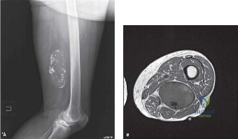
You obtain the following MRI of the thigh. Based on the imaging appearance, what is the most likely diagnosis, and what are the specific features influencing your decision?
Candidate: The MRI shows a large, heterogeneous mass with significant fatty content (high T1, suppression on STIR). However, the thick, irregular internal septations and nodular non-fatty components that enhance avidly are highly suggestive of a dedifferentiated liposarcoma (DDLPS).
Calling it a "lipoma." Even if it looks fatty, a deep mass >5cm with thick septations (>2mm) is a red flag for malignancy. Never label a deep lipomatous mass as benign without pathological confirmation.
Discuss the "biphasic" appearance. Identify that the lipomatous component indicates WDLPS, while the nodular, non-fatty, enhancing areas suggest dedifferentiation. Mention that you would target these specific nodular regions during the core biopsy to avoid a false-negative result, as they represent the highest-grade component of the tumor.
The biopsy confirms Grade 2 Dedifferentiated Liposarcoma. You are planning surgery. The mass is closely abutting the sciatic nerve. How do you approach the resection, and what is your strategy regarding the nerve?
Candidate: My goal is an R0 resection. If the nerve is not grossly invaded, I would perform an epimesial dissection (nerve-peeling). I would dissect the tumor capsule off the epineurium, effectively taking the outer layer but preserving the functional nerve fascicles. I would have already considered neoadjuvant radiotherapy to improve local control in this scenario.
Suggesting a "radical" resection that involves sacrificing the sciatic nerve without discussing the functional consequences (foot drop, sensory loss). Failing to mention that if the nerve *were* invaded, the decision to resect would involve a difficult discussion with the patient regarding limb function vs. oncological necessity.
Discuss the multidisciplinary decision-making process. Emphasize "Neoadjuvant RT" to create a distinct pseudocapsule, which makes the epimesial plane easier to define. Clearly differentiate between R0 (marginal vs. wide) and explain that nerve-sparing surgery, when combined with adjuvant/neoadjuvant therapy, provides acceptable oncological outcomes without the morbidity of nerve sacrifice.
