Patellofemoral Pain Syndrome (Runner's Knee): Epidemiology, Surgical Anatomy & Biomechanics
Key Takeaway
Patellofemoral Pain Syndrome (PFPS), or runner's knee, is a common knee pain condition influenced by multifactorial etiology, including biomechanical imbalances, overuse, and anatomical variations. Key structures like the patella, femoral trochlea, and MPFL, alongside dynamic quadriceps, govern its complex biomechanics and patellar tracking, crucial for effective diagnosis.
Introduction & Epidemiology
Patellofemoral pain syndrome (PFPS), colloquially known as "runner's knee," represents a ubiquitous and often debilitating orthopedic condition characterized by retropatellar or peripatellar pain exacerbated by activities that load the patellofemoral joint. While commonly associated with athletic populations, particularly runners, its incidence extends to individuals engaged in activities involving repetitive knee flexion and extension, such as stair climbing, cycling, and prolonged sitting.
Epidemiologically, PFPS is the most prevalent knee pathology in adolescents and young adults, accounting for an estimated 20-25% of all knee pain presentations in sports medicine clinics. Its incidence is higher in females, with ratios varying from 2:1 to 5:1 compared to males, particularly in specific athletic cohorts. This sex disparity is often attributed to differences in pelvic width, Q-angle, ligamentous laxity, and quadriceps-to-hamstring strength ratios. Peak incidence typically occurs between 10 and 35 years of age. While often self-limiting, a significant proportion of patients experience chronic or recurrent symptoms, with up to 70-90% reporting persistent pain at 5-year follow-up, underscoring the chronic nature and challenges in management. Understanding the multifactorial etiology—encompassing biomechanical imbalances, anatomical variations, overuse, and training errors—is paramount for appropriate diagnosis and tailored intervention.
Surgical Anatomy & Biomechanics
A thorough understanding of the intricate anatomy and complex biomechanics of the patellofemoral joint is fundamental to diagnosing and managing PFPS, particularly when considering surgical intervention for refractory cases or associated patellofemoral instability.
Surgical Anatomy
- Patella: The largest sesamoid bone, positioned within the quadriceps tendon. Its posterior surface is covered with thick articular cartilage and articulates with the femoral trochlea. Facet morphology (medial, lateral, odd facet) is critical for tracking. Variations in patellar height (patella alta, patella baja) significantly influence joint kinematics and contact forces. The patellar tendon connects the patella to the tibial tuberosity.
- Femoral Trochlea: The V-shaped groove on the distal femur that guides patellar tracking. Its morphology is highly variable; a shallow trochlea, flattening, or even convex shape (trochlear dysplasia) are significant risk factors for patellar instability. The lateral trochlear facet is typically longer and more prominent than the medial, contributing to lateral patellar stability at knee flexion >20-30 degrees. The trochlear sulcus angle and depth are key anatomical parameters.
-
Quadriceps Femoris Muscle:
Comprises four heads: rectus femoris, vastus lateralis (VL), vastus medialis (VM), and vastus intermedius.
- Vastus Medialis Obliquus (VMO): The most distal and oblique fibers of the VM, originating from the adductor magnus tendon and inserting onto the superomedial patella. Its primary role is to stabilize the patella medially and contribute to the terminal 10-15 degrees of knee extension. Atrophy or delayed activation of the VMO is frequently implicated in patellar maltracking.
- Vastus Lateralis: A powerful lateral stabilizer, if overactive or tight, can contribute to lateral patellar tilt and shift.
-
Patellar Retinacula and Ligaments:
- Medial Patellofemoral Ligament (MPFL): The primary static restraint to lateral patellar translation, contributing 50-60% of total resistance. It originates from the adductor tubercle/medial epicondyle and inserts onto the superomedial patella. Injury or laxity of the MPFL is the hallmark of recurrent lateral patellar instability.
- Lateral Patellar Retinaculum: A complex fibrous structure with transverse and oblique fibers, providing lateral stability. Tightness can contribute to lateral patellar compression and tilt.
- Medial Patellotibial and Medial Patellofemoral Ligaments: Secondary medial stabilizers.
- Lateral Patellotibial Ligament: A secondary lateral stabilizer.
- Tibial Tuberosity (TT): The insertion site of the patellar tendon. The relationship between the TT and the deepest point of the trochlear groove (TT-TG distance) is a critical measure for assessing patellar tracking. An elevated TT-TG (>15-20 mm) indicates a more lateralized TT, predisposing to lateral patellar subluxation/dislocation.
- Lower Extremity Alignment: Global alignment parameters, including Q-angle (quadriceps angle), femoral anteversion, tibial torsion, and foot pronation, can significantly influence patellofemoral mechanics. A dynamic Q-angle, assessed during functional activities, may be more relevant than a static measurement.
Biomechanics
The patellofemoral joint functions as a fulcrum, increasing the effective moment arm of the quadriceps, thus enhancing its mechanical efficiency. Normal patellar tracking involves a complex interaction of bony geometry, soft tissue restraints, and dynamic muscular forces throughout the range of motion.
-
Kinematics:
- Extension (0-20° flexion): Patella is largely superior to the trochlea, with minimal bony engagement. Soft tissues (MPFL, retinacula) and quadriceps balance are crucial.
- Early Flexion (20-30°): Patella begins to engage the trochlea, typically entering the groove slightly laterally, then translating medially as flexion increases. The MPFL is taut.
- Mid-Flexion (30-90°): Patella tracks centrally within the trochlear groove. Joint contact area increases progressively.
- Deep Flexion (>90°): Patella engages the intercondylar notch. The odd facet and lateral facets bear increasing load.
-
Forces:
- Patellofemoral Joint Reaction Forces (PFJRF): Increase exponentially with knee flexion angle and quadriceps contraction force. Activities like deep squatting or stair climbing impose forces many times body weight.
- Quadriceps Pull: The net vector of the quadriceps muscle pull tends to be slightly lateral, requiring counterbalancing by the VMO and medial retinacular structures.
- Q-angle: Represents the angle between the vector of the quadriceps femoris (from ASIS to patella mid-point) and the vector of the patellar tendon (from patella mid-point to tibial tuberosity). An increased Q-angle (>15-20 degrees) correlates with increased lateral patellar tracking forces.
- Maltracking and Pain: Aberrant patellar kinematics, such as lateral tilt, lateral shift, and excessive rotation, lead to altered stress distribution on the articular cartilage, potentially causing localized overload, cartilage degeneration, and nociceptive input from the subchondral bone and synovium. Factors contributing to maltracking include VMO weakness or delayed activation, vastus lateralis tightness/dominance, tight lateral retinaculum, femoral trochlear dysplasia, patella alta, and increased TT-TG distance. These factors disrupt the delicate balance of forces, leading to patellofemoral pain.
Indications & Contraindications
Management of PFPS primarily centers on non-operative strategies. Surgical intervention is reserved for a select subset of patients with specific, identifiable anatomical derangements or chronic, debilitating symptoms refractory to exhaustive conservative treatment, particularly those with recurrent patellofemoral instability.
Non-Operative Indications (Primary Management for PFPS)
The vast majority of patients presenting with PFPS are initially, and often definitively, managed non-operatively.
- Acute Presentation: Initial onset of symptoms without a history of recurrent instability or traumatic dislocation.
- Mild to Moderate Symptoms: Pain effectively managed with activity modification, analgesia, and physiotherapy.
- Absence of Gross Instability: No clinical evidence of recurrent patellar subluxation or dislocation.
-
Responsiveness to Conservative Measures:
Improvement in pain and function with structured physical therapy focusing on:
- Activity Modification: Limiting aggravating activities, pacing, gradual return to activity.
- Quadriceps Strengthening: Emphasizing VMO activation and eccentric loading.
- Hip Abductor and External Rotator Strengthening: Addressing proximal kinematic chain deficiencies.
- Core Stability Training: Enhancing lumbopelvic control.
- Flexibility Training: Addressing hamstring, gastrocnemius, iliotibial band (ITB), and quadriceps tightness.
- Foot Orthotics: For individuals with excessive foot pronation.
- Taping/Bracing: Patellar taping (McConnell taping) or knee braces to improve patellar tracking acutely.
- Pharmacotherapy: NSAIDs for pain and inflammation.
Operative Indications (for Refractory PFPS or Associated Pathology)
Surgical intervention for PFPS is indicated in a carefully selected patient population where a clear anatomical pathology contributes to the symptoms and has proven recalcitrant to prolonged, appropriately directed non-operative management. It is more commonly indicated for recurrent patellofemoral instability than isolated PFPS.
-
Recurrent Patellar Instability/Dislocation:
- Documented episodes of lateral patellar dislocation or symptomatic subluxation, especially with identifiable anatomical risk factors (e.g., trochlear dysplasia, elevated TT-TG distance, patella alta, MPFL insufficiency). This is the most common indication for patellofemoral surgery.
-
Severe Patellofemoral Maltracking:
- Persistent, debilitating pain and functional limitation despite extensive conservative treatment (>6-12 months), with objective evidence of severe patellar tilt, shift, or excessive lateral pressure, often associated with a tight lateral retinaculum or significant muscle imbalance refractory to rehabilitation. Lateral retinacular release as a standalone procedure for isolated pain remains highly controversial.
-
Significant Osteochondral Lesions:
- Symptomatic full-thickness cartilage defects on the patellar or trochlear articular surfaces, typically identified on MRI or during diagnostic arthroscopy, especially if causing mechanical symptoms (e.g., catching, locking).
-
Patellofemoral Arthroplasty/Replacement:
- End-stage, debilitating patellofemoral osteoarthritis refractory to all other interventions, typically in older patients. This is distinct from PFPS but represents a sequela in some chronic cases.
-
Specific Anatomical Conditions:
- Symptomatic plica syndrome (e.g., medial plica) documented to impinge, refractory to conservative measures.
- Symptomatic patellar tendinopathy (Jumper's Knee) or quadriceps tendinopathy refractory to conservative measures (though usually managed with rehabilitation).
- Rarely, saphenous nerve entrapment causing anterior knee pain.
Contraindications
- Active Infection: Absolute contraindication.
- Unrealistic Patient Expectations: Poor understanding of surgical outcomes, recovery period, or potential complications.
- Skeletally Immature Patients: Relative contraindication for procedures involving osteotomies across physes (e.g., tibial tubercle osteotomy) unless growth plates are fused or specific techniques are used to avoid growth disturbance.
- Absence of Clearly Identifiable and Surgically Correctable Pathology: Surgery without a specific anatomical target for addressing the symptoms is unlikely to be successful.
- Poor General Health/Coagulopathy: Conditions that increase surgical risk.
- Unwillingness to Participate in Post-Operative Rehabilitation: Critical for surgical success.
Table: Operative vs. Non-Operative Indications
| Feature / Indication | Non-Operative Management | Operative Management |
|---|---|---|
| Primary Symptom | Peripatellar/retropatellar pain, crepitus. | Recurrent patellar dislocation/subluxation, severe debilitating pain refractory to 6-12 months of conservative care, mechanical symptoms due to osteochondral lesions. |
| History | First presentation, gradual onset, absence of clear instability episodes. | Documented recurrent lateral patellar dislocations, chronic subluxation, significant traumatic patellar dislocation with risk factors, failed prolonged conservative treatment. |
| Physical Exam | Tenderness, crepitus, subtle maltracking, muscle imbalances (VMO weakness, hip abductor weakness, hamstring/ITB tightness). | Apprehension sign, J-sign, hyperlaxity, significant patellar maltracking, evidence of lateral patellar tilt/shift, focal osteochondral tenderness/defects. |
| Imaging Findings | Normal radiographs, mild cartilage signal changes on MRI, minimal to no significant anatomical risk factors. | Radiographs: Patella alta, increased sulcus angle. MRI: MPFL rupture/laxity, trochlear dysplasia (Dejour classification B-D), increased TT-TG distance (>15-20mm), patella alta, significant osteochondral lesions (full-thickness, large, symptomatic). CT: Precise TT-TG measurement, 3D trochlear morphology. |
| Surgical Procedures | N/A | Soft Tissue: MPFL reconstruction, lateral retinacular release (controversial as standalone), VMO advancement. Bony: Tibial Tubercle Osteotomy (medialization, anteromedialization, distalization), Trochleoplasty (deepening), Osteochondral Autograft/Allograft Transplantation (OATS) for lesions, Patellofemoral Arthroplasty (end-stage OA). |
Pre-Operative Planning & Patient Positioning
Meticulous pre-operative planning and appropriate patient positioning are critical for optimizing outcomes and minimizing complications in patellofemoral surgery.
Pre-Operative Planning
-
Comprehensive Patient Evaluation:
- Detailed History: Elucidate the duration, frequency, severity, and aggravating/alleviating factors of pain. Quantify episodes of instability (number, direction, reduction method). Document previous conservative treatments and their efficacy. Assess functional limitations and patient goals.
-
Physical Examination:
- Gait Analysis: Observe lower extremity kinematics.
- Alignment: Assess Q-angle (static and dynamic), femoral anteversion, tibial torsion, foot posture (pronation/supination).
- Palpation: Patellar facets, retinacula, quadriceps and patellar tendons, tibial tuberosity, pes anserine.
- Range of Motion (ROM): Knee flexion/extension, hip, ankle.
- Stability Testing: Patellar apprehension test, J-sign, medial/lateral patellar glide test, ligamentous stability of the knee.
- Muscle Strength and Flexibility: Quadriceps (especially VMO), hip abductors/external rotators, hamstrings, ITB, gastrocnemius.
- Neurovascular Status: Essential baseline.
-
Imaging Review:
- Standard Radiographs: AP, lateral (with specific attention to patellar height—Insall-Salvati, Caton-Deschamps indices), and Merchant (sunrise) or Laurin views (patellar tilt, subluxation, trochlear morphology).
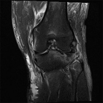
- Magnetic Resonance Imaging (MRI): Gold standard for assessing soft tissue pathology (MPFL integrity, retinacular tears), articular cartilage status (patellar and trochlear), bone edema, and trochlear morphology. Provides a good estimate of TT-TG distance.
- Computed Tomography (CT) Scan: The most accurate modality for measuring the TT-TG distance (axial cuts from tibial tuberosity to trochlear groove) and for detailed 3D assessment of trochlear dysplasia. Crucial for planning tibial tubercle osteotomies and trochleoplasties.
- Dynamic Imaging (Fluoroscopy/Dynamic MRI): Occasionally useful to visualize patellar tracking patterns during knee flexion/extension if static imaging is inconclusive.
-
Surgical Decision-Making:
- Integrate clinical findings with imaging results to identify specific anatomical abnormalities contributing to pain or instability.
- Formulate a personalized surgical plan, often involving a combination of procedures (e.g., MPFL reconstruction + tibial tubercle osteotomy for patella alta and increased TT-TG).
- Discuss the chosen procedure(s), expected outcomes, potential complications, and post-operative rehabilitation protocol thoroughly with the patient to ensure informed consent and realistic expectations.
-
Pre-operative Optimization:
- Smoking cessation.
- Management of comorbidities.
- Pre-operative physical therapy to optimize strength and ROM (if not already completed).
- Prophylactic antibiotics, DVT prophylaxis.
Patient Positioning
- Supine Position: The most common position for patellofemoral surgery.
- Tourniquet Placement: A pneumatic tourniquet is typically applied to the proximal thigh, providing a bloodless field, which is often crucial for delicate soft tissue and articular cartilage work. The tourniquet is inflated after limb elevation and exsanguination.
- Operating Table: A radiolucent table is preferred, especially if intra-operative fluoroscopy is anticipated (e.g., for MPFL femoral tunnel placement or tibial tubercle osteotomy fixation).
-
Leg Holder vs. Free Draping:
- Leg Holder: Provides stable positioning for osteotomies or longer procedures. Often used with a foot support.
- Free Draping: Allows for intra-operative assessment of patellar tracking throughout the range of motion during procedures like lateral retinacular release or MPFL reconstruction, which is critical for achieving appropriate tension. The limb can be manipulated by an assistant or with a specialized table-mounted limb positioner.
- Padded Bony Prominences: Ensure all pressure points (heel, calf, fibular head, sacrum, ulnar nerve) are adequately padded to prevent nerve injury or pressure sores.
- Sterile Prep and Drape: Standard sterile preparation of the knee and entire lower extremity, ensuring adequate exposure for incisions and potential graft harvest sites. Draping should allow full range of motion of the knee if free-draping is utilized.
- Equipment Placement: Ensure ready access to arthroscopy tower, instrument sets, fluoroscopy unit, and graft harvesting instruments (if autograft is planned).
Detailed Surgical Approach / Technique
Surgical intervention for patellofemoral pain syndrome and instability is highly individualized, addressing specific anatomical derangements. The following techniques represent common approaches.
1. Arthroscopic Procedures
Diagnostic arthroscopy is often performed initially to assess articular cartilage, meniscal integrity, and loose bodies, and to confirm patellar tracking.
-
Lateral Retinacular Release (LRR):
- Indications: Purely for painful patellar tilt/compression (lateral patellar hyperpressure syndrome) refractory to conservative care, without significant patellar instability. Controversial as a standalone procedure; high failure rate if not combined with other procedures addressing underlying malalignment.
-
Technique:
- Standard anteromedial and anterolateral portals.
- Identify the lateral patellar retinaculum, located just lateral to the patella.
- Carefully release the retinaculum, typically from the patellar border distally towards the patellar tendon and proximally up to the vastus lateralis border, using an arthroscopic shaver or electrocautery.
- Crucially, ensure the vastus lateralis fascia is not released, as this can lead to quadriceps weakness and iatrogenic medial instability. The release should be sufficient to reduce lateral patellar tilt and improve patellar centralization, which can be assessed dynamically under arthroscopic visualization.
- Avoid injury to the lateral geniculate artery and nerve branches.
-
Chondroplasty / Microfracture / Osteochondral Autograft/Allograft Transplantation (OATS):
- Indications: Symptomatic focal articular cartilage defects (chondromalacia) on the patellar or trochlear surfaces.
-
Technique:
- Chondroplasty: Debridement of unstable cartilage flaps to a stable rim using a shaver or radiofrequency ablator.
- Microfracture: For full-thickness defects down to subchondral bone, perforating the bone plate to allow marrow elements (stem cells) to form a fibrocartilaginous repair tissue. Requires strict non-weight-bearing post-operatively.
- OATS: For larger, symptomatic full-thickness defects. Cartilage-bone cylinders are harvested from a less weight-bearing area (e.g., lateral femoral condyle) and transplanted into the prepared defect. Allograft options are available for larger defects or when autograft sources are limited.
2. Medial Patellofemoral Ligament (MPFL) Reconstruction
- Indications: Recurrent lateral patellar dislocation with demonstrated MPFL insufficiency, often in conjunction with other predisposing factors (e.g., mild trochlear dysplasia, patella alta, borderline TT-TG).
- Graft Choice: Autografts (gracilis, semitendinosus) are most common. Allografts (tibialis anterior, semitendinosus) are alternatives.
-
Technique (Autograft):
- Graft Harvest: A small incision over the pes anserine for semitendinosus/gracilis harvest. The tendon is stripped, prepared, and folded to create a double-bundle graft.
-
Patellar Fixation:
- Transosseous tunnels: Two parallel tunnels drilled from the superomedial patella to exit anteromedially. The graft limbs are passed through.
- Suture anchors: One or two anchors placed in the superomedial patella.
- Cortical button fixation: Less common.
-
Femoral Fixation:
- Femoral tunnel placement: Critical for functional outcomes. The anatomical insertion is typically slightly anterior and proximal to the adductor tubercle, often referred to as Schöttle's point. Intra-operative fluoroscopy is essential to confirm placement (anterior to the posterior femoral cortex extension line, distal to the physis, proximal to the cartilage, and proximal to Blumensaat's line).
- A small incision is made over the medial femoral epicondyle. A guide pin is inserted, and a tunnel is drilled, typically 4.5-6 mm.
-
Graft Passage and Tensioning:
- The graft limbs are passed through the patellar tunnels/suture anchors and then through the femoral tunnel.
- The knee is typically taken to 30-45 degrees of flexion (where the MPFL is isometric), and the graft is tensioned to allow 1-2 quadrants of lateral patellar translation, preventing over-constraining the patella. The graft is secured with an interference screw or cortical button on the femoral side.
- Closure: Standard layered closure.
3. Tibial Tubercle Osteotomy (TTO)
- Indications: Excessive lateralization of the tibial tuberosity (TT-TG > 15-20 mm), patella alta (Insall-Salvati > 1.2-1.3, Caton-Deschamps > 1.3), or significant lateral patellar compression/tilt, often combined with MPFL reconstruction.
-
Types:
- Medialization (Elmslie-Trillat): For lateralization.
- Anteromedialization (Fulkerson): For lateralization and patellofemoral contact pressure reduction (anteriorization).
- Distalization (Maquet): For patella alta.
-
Technique (Fulkerson Anteromedialization Osteotomy):
- Incision: A longitudinal incision approximately 10-15 cm long, centered over the tibial tuberosity.
- Dissection: Subcutaneous dissection to expose the tibial tuberosity and patellar tendon insertion. Periosteum is incised and elevated.
-
Osteotomy:
- A sagittal saw is used to make a proximal cut from the anterolateral aspect of the tibial tuberosity, extending distally and slightly medially to the anterior tibial crest. The cut should be parallel to the patellar tendon, typically 1.5-2 cm medial to the lateral border of the tibia.
- A second cut is made obliquely from distal to proximal, connecting to the first cut, creating a wedge-shaped osteotomy. The distal portion of the osteotomy is left intact as a hinge or fully detached for greater mobility.
- A chisel or osteotome is used to mobilize the tibial tubercle fragment, including its periosteal attachment and the patellar tendon.
-
Repositioning:
- Medialization: The fragment is shifted medially to normalize the TT-TG distance, assessed visually or with intra-operative fluoroscopy/measurement.
- Anteriorization: The fragment is elevated anteriorly using a bone graft or by tilting the fragment to offload the patellofemoral joint.
- Distalization: If patella alta is present, the fragment can be repositioned distally.
- Fixation: The repositioned fragment is secured with two or three cancellous screws (e.g., 4.5 mm or 6.5 mm fully threaded cancellous screws) directed obliquely from the anterior fragment into the posterior tibial cortex. The fragment should be firmly secured to prevent non-union or displacement.
- Closure: Layered closure.
4. Trochleoplasty
- Indications: Severe trochlear dysplasia (Dejour Type B, C, D) with recurrent patellar instability, particularly when other factors like TT-TG and patellar height have been addressed or are normal. A technically demanding procedure.
-
Types:
- Deepening Trochleoplasty: Reshaping the bone underneath the trochlear cartilage to deepen the sulcus.
- Recession Trochleoplasty: Excise a wedge of bone to recess the trochlea.
-
Technique (Sulcus Deepening Trochleoplasty):
- Incision: Midline longitudinal incision.
- Exposure: The articular cartilage of the trochlea is carefully elevated as a full-thickness chondral flap or incised to create a hinge.
- Bone Reshaping: An osteotome or burr is used to remove subchondral bone from the femoral trochlea, deepening the sulcus and creating a more prominent lateral trochlear wall. The goal is to create a new, anatomically appropriate trochlear groove.
- Cartilage Re-lay: The elevated chondral flap is carefully repositioned into the newly deepened trochlear groove.
- Fixation: The cartilage flap is secured with bioabsorbable pins, sutures, or bone anchors.
- Closure: Standard layered closure.
Complications & Management
Patellofemoral surgery, while often effective, is associated with a spectrum of potential complications. Vigilant pre-operative assessment, meticulous surgical technique, and comprehensive post-operative care are crucial for mitigation.
Table: Common Complications, Incidence, and Salvage Strategies
| Complication | Incidence (Approximate) | Salvage Strategies |
|---|---|---|
| Persistent Pain / Failed Surgery | 10-30% | Thorough re-evaluation for missed pathology, inadequate correction, or new pathology. Aggressive non-operative management (PT, injections). Revision surgery (e.g., further osteotomy, MPFL revision, arthroplasty), chronic pain management referral. |
| Infection (Superficial/Deep) | <1-2% | Superficial: Oral antibiotics, local wound care. Deep: Urgent surgical irrigation & debridement (I&D), parenteral antibiotics (IV), hardware removal (if applicable and infection controlled), debridement, tissue reconstruction, two-stage revision. |
| Stiffness / Arthrofibrosis | 5-15% | Aggressive, supervised physical therapy focusing on ROM. Manipulation Under Anesthesia (MUA) if unresponsive. Arthroscopic lysis of adhesions (LOA) for refractory cases, especially in the suprapatellar pouch or lateral gutter. |
| Recurrent Instability/Dislocation | 5-15% | Occurs after stabilization procedures. Re-evaluate anatomical risk factors (e.g., insufficient TT-TG correction, persistent trochlear dysplasia, MPFL graft failure/laxity, inadequate MPFL femoral tunnel placement). Revision MPFL reconstruction, additional/revision osteotomy (TTO, trochleoplasty). |
| Neurovascular Injury | <1% | Nerve (saphenous, peroneal): Careful dissection, nerve blocks, neurolysis for entrapment, observation for neuropraxia. Surgical exploration and repair for transection. Vascular (geniculates): Ligation for minor bleeds. Surgical repair/reconstruction for major vessel injury (rare). |
| Hardware-Related Issues | 5-10% | Prominence/Irritation: Symptomatic hardware removal after bone healing (e.g., TTO screws, MPFL femoral screw). Breakage/Migration: Removal, possible re-fixation if stability compromised. |
| Fracture (Patella, Tibia) | <1-2% | Patellar (post-LRR): Avoid over-releasing the lateral retinaculum. Manage non-operatively with bracing or operatively with ORIF. Tibial (post-TTO): Inadequate fixation, early weight-bearing. Manage with cast/brace, delayed weight-bearing, or revision ORIF. |
| Patella Baja/Alta (Post-TTO) | <1% | Patella Baja (over-distalization): Increased patellofemoral contact pressure, pain. Salvage is challenging, may require revision TTO for proximalization. Patella Alta (under-distalization/iatrogenic): Continue instability. Revision TTO for distalization. |
| Overcorrection / Medialization (TTO) | Rare | Excessive medialization causing iatrogenic medial patellar instability or increased medial patellofemoral contact pressure/pain. Salvage involves revision TTO for lateralization, often with hardware removal. |
| Graft Failure/Laxity (MPFL recon) | 5-10% | Re-evaluation of femoral tunnel placement, graft tensioning, healing. Revision MPFL reconstruction, potentially with different graft choice or fixation method. May require augmentation with other bony procedures if primary instability factors were not adequately addressed. |
| Deep Vein Thrombosis (DVT) / PE | <1-2% | Prophylaxis (mechanical/chemical) as per institutional guidelines. Management with anticoagulation. Surgical thrombectomy for massive PE. |
| Compartment Syndrome | Rare | Urgent fasciotomy. |
Post-Operative Rehabilitation Protocols
Post-operative rehabilitation is as crucial as the surgical technique itself for achieving optimal outcomes in patellofemoral surgery. Protocols are typically structured in phases, progressively advancing activities while protecting the surgical repair. Individualization based on the specific procedure(s) performed, patient healing capacity, and pain tolerance is paramount.
General Principles
- Protection: Safeguard the surgical repair (e.g., graft, osteotomy fixation, cartilage repair).
- Gradual Progression: Slowly increase weight-bearing, range of motion, and strengthening.
- Pain and Swelling Management: Crucial for early mobility and compliance.
- Restoration of ROM: Achieve full, pain-free knee flexion and extension.
- Strength Training: Focus on quadriceps (especially VMO), hip abductors/external rotators, and core musculature.
- Neuromuscular Control: Proprioception, balance, and dynamic stability.
- Functional Progression: Return to activities of daily living (ADLs), work, and sport-specific movements.
Example Protocol (Combined MPFL Reconstruction and Tibial Tubercle Osteotomy)
Phase I: Protection & Early Motion (Weeks 0-6)
- Goals: Protect surgical site, control pain/swelling, achieve early controlled ROM, initiate muscle activation.
-
Weight-Bearing:
- Weeks 0-2: Touch-down weight-bearing (TDWB) or non-weight-bearing (NWB) with crutches, knee brace locked in full extension (especially for TTO or cartilage procedures).
- Weeks 2-6: Progress to partial weight-bearing (PWB) with brace unlocked for controlled ROM (e.g., 0-90°), as tolerated.
-
Range of Motion (ROM):
- Weeks 0-2: Passive ROM (CPM machine encouraged), focus on full extension. Flexion limited to 0-30° initially, gradually progressing to 0-60° by week 2 to protect MPFL graft.
- Weeks 2-6: Active-assisted ROM. Progress flexion to 90-100° by week 6. Maintain full extension.
-
Therapeutic Exercises:
- Quadriceps sets (isometric), gluteal sets, ankle pumps.
- Straight Leg Raises (SLR) in all planes, initially with brace locked, progressing to unlocked/off brace as quad control improves.
- Gentle hamstring curls (avoiding excessive pull on tibial tuberosity if TTO performed).
- Core stability exercises (e.g., abdominal bracing, pelvic tilts).
- Cryotherapy, elevation, compression for swelling management.
- Scar massage once incision is healed.
Phase II: Strength & Neuromuscular Control (Weeks 6-12)
- Goals: Restore full pain-free ROM, improve quadriceps and hip strength, enhance neuromuscular control, progress weight-bearing.
- Weight-Bearing: Gradually progress to full weight-bearing (FWB) and discontinue crutches by week 8-10, as tolerated. Wean off brace by week 10-12 if stability is good and ROM is sufficient.
- ROM: Achieve full, pain-free ROM (0-135° or equivalent).
-
Therapeutic Exercises:
- Closed Chain Strengthening: Mini-squats (0-60°), wall slides, leg press (light resistance), step-ups/downs (initially small steps).
- Open Chain Strengthening: Hamstring curls, hip abduction/adduction/extension (resistance bands). Caution with open-chain quadriceps extension (knee extension machines) as they can place high stress on the patellofemoral joint and TTO site. If used, limit range (e.g., 90-45°).
- Proprioception/Balance: Single-leg stance, balance board/foam pad.
- Core Strengthening: Continue progression.
- Low-impact cardio (stationary bike with minimal resistance).
Phase III: Advanced Strengthening & Sport-Specific Training (Weeks 12-24)
- Goals: Maximize strength, power, agility, and endurance. Prepare for return to desired activities.
-
Therapeutic Exercises:
- Increase resistance and intensity for all strengthening exercises (leg press, squats, lunges).
- Plyometric exercises (low-level jumping, hopping) if appropriate for sport.
- Agility drills (shuttle runs, cutting, lateral movements).
- Sport-specific drills, gradually increasing intensity.
- Return to running program (gradual, often around 4-5 months post-op, pending strength and pain).
- Continue flexibility, core, and balance training.
Phase IV: Return to Activity (Months 6+)
- Goals: Full return to sport/activity, maintain fitness, prevent recurrence.
- Functional Testing: Perform objective functional tests (e.g., single-leg hop tests, Y-balance test) to compare limb symmetry and readiness for return to sport.
- Gradual Return to Sport: Phased re-entry to competitive sports or high-impact activities, monitored by the surgeon and physical therapist.
- Maintenance Program: Long-term adherence to a strength and conditioning program to prevent re-injury.
Specific Considerations
- Isolated LRR: Faster progression with ROM and weight-bearing, but careful monitoring for iatrogenic medial instability.
- Chondroplasty/OATS: Strict non-weight-bearing (NWB) for 6-8 weeks for osteochondral grafts to allow graft integration. Controlled ROM protocols to protect healing cartilage.
- Trochleoplasty: Similar to MPFL/TTO but often with a more conservative initial ROM progression to protect the trochlear cartilage repair.
Summary of Key Literature / Guidelines
The understanding and management of patellofemoral pain syndrome and instability continue to evolve, with a growing body of evidence guiding both non-operative and operative strategies.
-
Non-Operative Management: Multiple systematic reviews and meta-analyses consistently demonstrate the efficacy of multimodal conservative management as the first-line treatment for PFPS.
- Exercise Therapy: Strong evidence supports exercise therapy, particularly programs targeting hip musculature (abductors, external rotators) and quadriceps (VMO), with significant improvements in pain and function. Supervised, progressive rehabilitation programs are superior to unsupervised or passive modalities. (e.g., Barton et al. Br J Sports Med. 2013; Willy et al. J Orthop Sports Phys Ther. 2019).
- Foot Orthotics & Taping: Moderate evidence supports the use of foot orthotics for individuals with excessive foot pronation and patellar taping (McConnell technique) for short-term pain relief, often as adjuncts to exercise therapy.
- Injections: Limited to no evidence for corticosteroid, hyaluronic acid, or PRP injections in the management of PFPS itself, though may be considered for associated inflammatory conditions.
-
Surgical Intervention for Instability:
- MPFL Reconstruction: Widely regarded as the gold standard for recurrent lateral patellar dislocation, especially with confirmed MPFL insufficiency. Studies consistently show good-to-excellent outcomes in 80-90% of cases, with low rates of re-dislocation (typically 5-15%). The anatomical placement of the femoral tunnel (Schöttle's point) is critical to prevent over-tensioning or graft failure. (e.g., Dejour et al. Knee Surg Sports Traumatol Arthrosc. 2007; Parikh et al. Am J Sports Med. 2013).
- Tibial Tubercle Osteotomy (TTO): Indicated for significant anatomical malalignment, particularly elevated TT-TG distance or patella alta. When combined with MPFL reconstruction, it significantly improves stability in patients with severe risk factors. Anteromedialization (Fulkerson) can also reduce patellofemoral contact pressure. (e.g., Stefancin et al. Knee Surg Sports Traumatol Arthrosc. 2017; Hinterwimmer et al. Am J Sports Med. 2004).
- Trochleoplasty: Reserved for severe trochlear dysplasia (Dejour B, C, D) and is highly effective in restoring stability by recreating a functional trochlear groove. However, it is technically demanding and carries higher risks, often considered a salvage procedure for the most complex cases. (e.g., Dejour et al. Knee Surg Sports Traumatol Arthrosc. 1994; Lattermann et al. J Bone Joint Surg Am. 2013).
- Lateral Retinacular Release (LRR): As a standalone procedure for isolated PFPS, LRR has largely fallen out of favor due to high failure rates and the risk of iatrogenic medial instability. Its role is primarily adjunctive to other stabilization procedures (e.g., MPFL reconstruction, TTO) when significant lateral patellar tilt persists after bony and medial soft tissue corrections. (e.g., Christoforakis et al. Arthroscopy. 2006).
-
Cartilage Procedures: Management of patellofemoral chondral lesions varies based on size, depth, and patient factors.
- Chondroplasty/Debridement: May offer temporary symptomatic relief for mechanical symptoms from unstable flaps but does not address the underlying cartilage defect.
- Microfracture: Induces fibrocartilage formation, which is mechanically inferior to native hyaline cartilage. Outcomes are variable, better for smaller lesions.
- Osteochondral Transplantation (OATS): Can provide hyaline cartilage repair for focal, contained lesions, with favorable outcomes reported in selected patients. (e.g., Gudas et al. Am J Sports Med. 2013).
-
Integrated Approach: Current consensus, exemplified by guidelines from organizations like ISAKOS (International Society of Arthroscopy, Knee Surgery and Orthopaedic Sports Medicine) and ESSKA (European Society for Sports Traumatology, Knee Surgery and Arthroscopy), emphasizes a holistic, step-wise approach. Conservative management remains the cornerstone. Surgical intervention is tailored to specific anatomical abnormalities identified through comprehensive clinical and advanced imaging assessment (radiographs, MRI, CT), addressing not just the primary pathology but also contributing factors (e.g., patella alta, TT-TG, trochlear dysplasia). Combination procedures are common to achieve optimal patellofemoral alignment and stability. Prognosis is generally good for instability, but persistent pain remains a challenge in a subset of patients with chronic PFPS.
You Might Also Like