Peripheral Nerve Regeneration and Repair: Principles, Evaluation, and Microsurgical Techniques
Key Takeaway
Peripheral nerve regeneration involves complex proximal axonal sprouting and distal Wallerian degeneration. Following microsurgical repair, sensory recovery progresses through predictable phases of paresthesia and hyperesthesia. Prognosis is heavily influenced by patient age and injury mechanism. This guide details the pathophysiology, standardized clinical evaluation protocols including two-point discrimination, and evidence-based surgical priorities for digital nerve reconstruction to optimize functional outcomes in hand and upper extremity surgery.
Introduction to Peripheral Nerve Regeneration
The restoration of sensory and motor function following peripheral nerve injury remains one of the most formidable challenges in orthopaedic and hand surgery. Despite advances in microsurgical techniques, the functional outcome of nerve repair is dictated by a complex interplay of cellular biology, patient demographics, injury mechanism, and meticulous surgical execution. Peripheral nerve regeneration is not merely a mechanical reconnection of severed conduits; it is a profound biological event requiring the precise orchestration of neuronal survival, axonal elongation, and end-organ reinnervation.
This comprehensive guide delineates the pathophysiology of nerve regeneration, standardized protocols for clinical evaluation, prognostic determinants, and the step-by-step surgical principles required for optimal outcomes in digital and peripheral nerve repair.
Pathophysiology of Nerve Injury and Regeneration
Following a complete nerve transection (neurotmesis), a highly coordinated sequence of biological events is initiated in both the proximal and distal nerve stumps, as well as within the neuronal cell body.
The Proximal Response: Neuronal Survival and Axonal Sprouting
In the proximal elements of the peripheral nerve, the immediate response to injury is characterized by a dramatic increase in metabolic activity. The neuronal cell body undergoes chromatolysis—a process marked by the dissolution of Nissl bodies, cellular swelling, and the migration of the nucleus to the periphery. This represents a shift from the production of neurotransmitters to the synthesis of structural proteins (e.g., tubulin and actin) necessary for axonal repair.
Within the first 1 to 3 weeks post-injury, the proximal axonal stump forms a growth cone. Driven by neurotrophic factors, this growth cone sprouts multiple axonal processes (filopodia) that attempt to cross the injury site. The success of these sprouts depends entirely on the preparation of the distal environment.
The Distal Response: Wallerian Degeneration
Distal to the site of injury, the nerve segment undergoes Wallerian degeneration. This process is essential for clearing inhibitory debris and creating a permissive environment for the advancing proximal axons.
* Myelin Disruption: Within 24 to 48 hours, the axonal cytoskeleton disintegrates, and the myelin sheath collapses.
* Phagocytosis: Macrophages infiltrate the distal stump to phagocytose myelin and axonal debris, a critical step since intact myelin contains proteins that inhibit axonal growth.
* Schwann Cell Proliferation: Schwann cells dedifferentiate, proliferate, and align themselves longitudinally within the endoneurial tubes to form the Bands of Büngner. These cellular columns secrete neurotrophic factors (such as Nerve Growth Factor, NGF) and provide a physical and chemical scaffold to guide regenerating axons toward their target end-organs.
Clinical Pearl: The rate of axonal regeneration in humans is classically cited as 1 mm per day (or approximately 1 inch per month). However, this rate is variable; it is generally faster in proximal segments and slows considerably as the axon progresses distally.
Clinical Progression of Sensory Recovery
Following the successful coaptation of a sensory nerve (whether digital, pure sensory, or mixed), the clinical recovery of sensation follows a predictable, albeit protracted, timeline.
The Phases of Sensory Return
- Decreasing Anesthesia: As regenerating axons cross the repair site and advance distally, the absolute area of anesthesia begins to shrink.
- Paresthesia (2 to 3 Months): The reinnervated territory typically becomes paresthetic. Patients often describe a "pins and needles" sensation, which corresponds to the advancing Tinel's sign.
- Hyperesthesia: The area subsequently becomes hyperesthetic, exhibiting heightened sensitivity to light touch and cold temperatures. Interestingly, firm pressure is usually less painful than light, moving touch.
- Resolution and Maturation: Over time, and heavily dependent on structured physical and occupational therapy (sensory re-education), the hyperesthesia resolves. The quality of sensation improves significantly within the first 1.5 to 2 years, with gradual, subtle improvements continuing thereafter.
Surgical Warning: Fully normal sensation with the appreciation of functional two-point discrimination (2PD) is rarely expected in adults following complete nerve transection. Managing patient expectations preoperatively is paramount.
Prognostic Determinants in Nerve Repair
The final functional result after peripheral nerve repair is influenced by several independent variables, with patient age and the mechanism of injury being the most critical.
The Influence of Age
Age is the single most significant prognostic factor in peripheral nerve regeneration. The superior neuroplasticity of the pediatric central nervous system allows for exceptional sensory re-education and cortical remapping.
* Pediatric Patients: Studies by Hudson et al. demonstrated that children (mean age 6.1 years) undergoing primary epineurial repair of the median nerve achieved minimal loss of power and near-normal sensation. A fully functional hand can routinely be expected in this demographic.
* Young Adults (< 20 to 40 Years): Patients under the age of 20 have a significantly better prognosis for the return of functional two-point discrimination. Wang et al. confirmed statistically superior sensibility recovery in patients younger than 40 compared to older cohorts.
* Older Adults (> 50 Years): It is exceedingly rare for patients older than 50 years to regain more than protective sensation. Cortical plasticity diminishes with age, limiting the brain's ability to interpret the altered afferent signals from misdirected regenerating axons.
Mechanism of Injury
The nature of the trauma dictates the zone of injury and the viability of the nerve stumps.
* Sharp Lacerations: Clean, sharp transections (e.g., glass or knife wounds) yield the best outcomes following primary repair, as the zone of trauma is minimal.
* Saw and Crush Injuries: Wang et al. demonstrated that outcomes following saw injuries are significantly worse due to extensive intraneural architecture disruption and segmental loss. In such cases, primary nerve grafting or the use of nerve conduits should be strongly considered over primary repair under tension.
Standardized Clinical Evaluation of Nerve Function
Accurate, reproducible clinical testing is essential for establishing a baseline and monitoring the progress of nerve regeneration. The following protocols represent the gold standard for evaluating hand sensibility and motor function.
Sensory Testing Protocols
Sensory evaluation must be performed in a quiet environment with the patient's full concentration.
Preparation:
* Ensure the patient's hand is warm. Cold extremities exhibit diminished sensibility due to vasoconstriction.
* Testing instruments must be at room temperature.
* Rest the hand on a flat, stable surface with the palm facing upward.
Static Two-Point Discrimination (s2PD):
* Utilize a blunt, two-pointed caliper (e.g., Disk-Criminator) or a reshaped paper clip.
* Apply the points distally over the digital pulp in the longitudinal axis (on either the radial or ulnar side).
* The pressure applied should be just slightly less than the pressure required to blanch the skin.
* Test each area three times. Start with the points separated by 10 mm, and gradually decrease the distance.
* Scoring: Two out of three correct answers constitute proof of perception. Normal s2PD is < 6 mm.
Moving Two-Point Discrimination (m2PD):
* Performed similarly to s2PD, but the caliper is applied in an axial direction and moved from proximal to distal along the digital pad.
* Moving 2PD typically returns earlier than static 2PD and is a sensitive indicator of early sensory recovery.
Motor Function Assessment
Motor recovery is evaluated through standardized strength testing, reflecting both intrinsic and extrinsic muscle function.
* Grip Strength: Utilize a Jamar squeeze grip dynamometer. Record results at all five handle positions with three successive determinations. This provides an integrated assessment of overall hand function.
* Key Pinch: Measured using a pinch dynamometer. The patient applies the thumb tip to the radial aspect of the middle phalanx of the index finger. Perform three successive measurements and compare with the contralateral, uninjured hand.
* Tip Pinch: The patient pinches the tip of the index finger to the ulnar side of the tip of the thumb. Record three measurements.
Sudomotor and Subjective Evaluation
- Sudomotor Function: The loss of sweating indicates disruption of sympathetic nerve fibers. While sweating may return prior to the recovery of two-point discrimination, its absence is a reliable indicator of complete nerve disruption. Tests such as the Ninhydrin test or the O'Rian wrinkle test (immersion in warm water) can objectively document sudomotor function.
- Subjective Evaluation: Document the patient's self-reported symptoms, including the presence of neuropathic pain, cold intolerance, dysesthesias, and specific functional disabilities in activities of daily living (ADLs).
Surgical Principles and Priorities in Digital Nerve Repair
Strategic Priorities in Multiple Digit Trauma
In complex, multi-digit trauma, prolonged operative times, patient instability, or extensive segmental nerve loss may preclude the repair of every injured digital nerve. In such scenarios, the surgeon must prioritize repairs based on the functional anatomy of the hand.
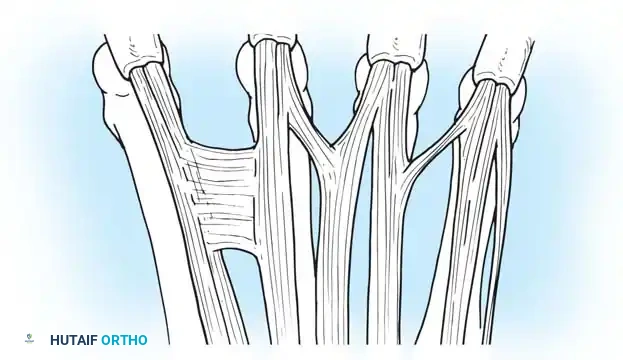
The most critical areas of sensory innervation required for pinch, grip, and spatial orientation include:
1. Ulnar side of the Thumb: Essential for all pinch mechanisms.
2. Radial side of the Index Finger: Critical for key pinch and fine manipulation.
3. Radial side of the Middle Finger: Supports the index finger in tripod pinch.
4. Ulnar side of the Little Finger: Crucial for ulnar border contact, resting the hand on surfaces, and preventing unrecognized burns or trauma.
Pitfall: Failing to prioritize the ulnar digital nerve of the thumb or the radial digital nerve of the index finger in a multiply-injured hand will result in profound, irreversible deficits in fine motor pinch and manipulation.
Step-by-Step Microsurgical Epineurial Repair
Primary repair is indicated for acute nerve transections where the nerve ends can be approximated without tension.
1. Positioning and Preparation:
* Position the patient supine with the arm extended on a radiolucent hand table.
* Apply a well-padded pneumatic tourniquet to the upper arm to ensure a bloodless field.
* Utilize operating loupes (minimum 3.5x to 4.5x magnification) or an operating microscope for the repair.
2. Exposure and Debridement:
* Extend the traumatic wound using Bruner (zigzag) or mid-lateral incisions to expose the proximal and distal nerve stumps.
* Identify the nerve ends. The proximal stump often exhibits a neuroma if the injury is not acute, while the distal stump may be shrunken.
* Using a fresh scalpel blade or specialized nerve scissors, sharply resect the contused or scarred nerve ends until healthy, "mushrooming" fascicles are visualized.
3. Alignment and Coaptation:
* Mobilize the nerve proximally and distally to gain length, taking care not to strip the segmental blood supply (vasa nervorum).
* Align the nerve ends by matching the longitudinal epineurial blood vessels and the cross-sectional fascicular topography.
* Perform an epineurial repair using 8-0 or 9-0 non-absorbable monofilament suture (e.g., Nylon).
* Place the first two sutures 180 degrees apart to act as stay sutures. Place subsequent sutures to accurately approximate the epineurium without causing fascicular buckling.
Surgical Warning: Tension is the enemy of nerve regeneration. If the nerve ends cannot be approximated with an 8-0 suture without the suture tearing through the epineurium, a primary repair is contraindicated. In such cases, a nerve graft (e.g., sural nerve, medial antebrachial cutaneous nerve) or a biological/synthetic nerve conduit must be utilized to bridge the gap.
Postoperative Protocol and Rehabilitation
The success of a meticulously performed nerve repair relies heavily on the postoperative rehabilitation protocol.
Phase 1: Immobilization and Protection (Weeks 0-3)
- Postoperatively, the hand and wrist are immobilized in a dorsal blocking splint. The wrist is typically placed in 20-30 degrees of flexion, and the metacarpophalangeal (MCP) joints in 70-80 degrees of flexion to minimize tension on the digital nerve repair.
- Active extension within the confines of the splint is permitted to prevent tendon adhesions, provided it does not place tension on the neurorrhaphy.
Phase 2: Mobilization and Nerve Gliding (Weeks 3-6)
- The splint is gradually discontinued.
- Gentle active and active-assisted range of motion exercises are initiated.
- Nerve gliding exercises are introduced to prevent the nerve from adhering to the surrounding scar bed, which can lead to traction neuropathy.
Phase 3: Sensory Re-education and Desensitization (Weeks 6 and Beyond)
- Desensitization: As the nerve regenerates and hyperesthesia develops, desensitization techniques (tapping, massage, immersion in varying textures like rice or fluidotherapy) are employed to raise the pain threshold.
- Sensory Re-education: Once protective sensation (ability to perceive the 4.31 Semmes-Weinstein monofilament) returns, formal sensory re-education begins. This involves training the brain to correctly interpret the altered afferent impulses. Patients practice identifying objects of different shapes, sizes, and textures with their eyes closed, relying on cognitive feedback to remap the sensory cortex.
By adhering to these rigorous biological principles, standardized evaluation metrics, and precise microsurgical techniques, the orthopaedic surgeon can maximize the potential for functional nerve regeneration and restore meaningful utility to the injured hand.
📚 Medical References
- Peripheral nerve regeneration in Gore-tex chambers, Scand J Plast Reconstr Surg 22:207, 1988.
- Dellon AL: Reinnervation of denervated Meissner corpuscles: a sequential histologic study in the monkey following fascicular nerve repair, J Hand Surg 1:98, 1976.
- Dellon AL, Witebsky FG, Terrill RE: The denervated Meissner: a sequential histologic study following nerve division in the Rhesus monkey, Plast Reconstr Surg 56:182, 1975.
- Ducker TB, Kempe LG, Hayes GJ: The metabolic background for peripheral nerve surgery, J Neurosurg 30:270, 1969.
- Flores AJ, Lavernia CJ, Owens PW: Anatomy and physiology of peripheral nerve injury and repair, Am J Orthop 29:167, 2000.
- Foerster O: The dermatomes in man, Brain 56:1, 1933.
- Grewal R, Xu J, Sotereanos DG, et al: Biomechanical properties of peripheral nerves, Hand Clin 12:195, 1996.
- Jabaley ME, Wallace W, Heckler FR: Internal topography of major nerves of the forearm and hand: a current view, J Hand Surg 5:82, 1980.
- Kristensson K: Retrograde signaling of nerve cell body response to trauma. In Gorio A, Millesi H, Mingrino S, eds: Posttraumatic peripheral nerve regeneration, New York, 1981, Raven. Lasek JR, Shelanske ML, Brinkely BR, et al: Cytoskeletons and the architecture of nervous systems, Neurosci Res Program Bull 19:1, 1981.
- Livingston WK: Evidence of active invasion of denervated areas by sensory fi bers from neighboring nerves in man, J Neurosurg 4:140, 1947.
- Lundborg G, Branemark PI: Microvascular structure and function of the peripheral nerves: vital microscopic studies of the tibial nerve in the rabbit, Adv Microcirc 1:66, 1968.
- Lundborg G, Dahlin LB: Anatomy, function, and pathophysiology of peripheral nerves and nerve compression, Hand Clin 12:185, 1996.
- Mackinnon SE, Dellon AL, Lundborg G, et al: A study of neurotropism in the primate model, J Hand Surg 11A:888, 1986.
- Omer G, Spinner M, eds: Management of peripheral nerve problems, Philadelphia, 1980, Saunders. Raji AM: An experimental study of the effects of pulsed electromagnetic fi eld (diapulse) on nerve repair, J Hand Surg 9B:105, 1984.
- Raji ARM, Bowden REM: Effects of high-peak pulsed electromagnetic fi eld on the degeneration and regeneration of the common peroneal nerve in rats, J Bone Joint Surg 65B:478, 1983.
- Seckel BR, Ryan SE, Gagne JR, et al: Target-specifi c nerve regeneration through a nerve guide in the rat, Plast Reconstr Surg 78:793, 1986.
- Seddon HJ, Medawar PB, Smith H: Rate of regeneration of peripheral nerves in man, J Physiol 102:191, 1943.
- Shizhen Z, Xiangluo T, Muzhil L: The microsurgical anatomy of peripheral nerves. In Shizhen Z, Yongjian M, Wencyun Y, eds: Microsurgical anatomy, Lancaster, UK, 1985, MTP Press. Smith BH, Kornblith PL: Axoplasmic transport in neurological surgery, Neurosurgery 10:268, 1982.
- Starkweather RJ, Neviaser RJ, Adams JP, et al: The effect of devascularization on the regeneration of lacerated peripheral nerves: an experimental study, J Hand Surg 3:163, 1978.
- Sunderland S: The intraneural topography of the radial, medial and ulnar nerves, Brain 68:243, 1945.
- Sunderland S, Bradley KC: Endoneurial tube shrinkage in the distal segment of a severed nerve, J Comp Neurol 93:411, 1950.
- Terzis JK, Feeker BL, Sismoue EN: A computerized study of the intraneural organization of the median nerve, J Hand Surg 9A:605, 1984.
- Williams HB, Jabaley ME: The importance of internal anatomy of the peripheral nerves to nerve repair in the forearm and hand, Hand Clin 2:689, 1986.
- Refl ex Sympathetic Dystrophy Cooper DE, DeLee JC, Ramamurthy S: Refl ex sympathetic dystrophy of the knee, J Bone Joint Surg 71A:365, 1989.
- Davidoff G, Werner R, Cremer S, et al: Predictive value of the three-phase technetium bone scan in diagnosis of refl ex sympathetic dystrophy syndrome, Arch Phys Med Rehabil 70:135, 1989.
- Forster RS, Fu FH: Refl ex sympathetic dystrophy in children: a case report and review of the literature, Orthopedics 8:475, 1985.
- Gerard RW: The physiology of pain: abnormal neuron states in causalgia and related phenomena, Anesthesiology 12:1, 1951.
- Kleinert HE, Cook NM, Wayne L, et al: Post-traumatic sympathetic dystrophy, Orthop Clin North Am 4:917, 1973.
- Kline SC, Holder LE: Segmental refl ex sympathetic dystrophy: clinical and scintigraphic criteria, J Hand Surg 18A:853, 1993.
- Kozin F: The refl ex sympathetic dystrophy syndrome, Am J Med 70:23, 1981.
- Kozin F, Soin JS, Ryan LM: Bone scintigraphy in the refl ex sympathetic dystrophy syndrome, Radiology 138:437, 1981.
- Lankford LL: Refl ex sympathetic dystrophy. In Omer G, Spinner M, eds: Management of peripheral nerve problems, Philadelphia, 1980, Saunders. Lankford LL: Refl ex sympathetic dystrophy. In Green DP, ed: Operative hand surgery, 2nd ed, New York, 1988, Churchill Livingstone. Lankford LL, Thompson JE: Refl ex sympathetic dystrophy, upper and lower extremity: diagnosis and management, Instr Course Lect 26:163, 1977.
- Lee GW, Weeks PM: The role of bone scintigraphy in diagnosing refl ex sympathetic dystrophy, J Hand Surg 20A:458, 1995.
- Linson MA, Leffert R, Todd DP: The treatment of upper extremity refl ex sympathetic dystrophy with prolonged continuous stellate ganglion blockade, J Hand Surg 8A:153, 1983.
- Mackinnon SE, Holder LE: The use of three-phase radionuclide bone scanning in the diagnosis of refl ex sympathetic dystrophy, J Hand Surg 9A:556, 1984.
- Mayfi eld FH, Devine JH: Causalgia, Surg Gynecol Obstet 80:631, 1945.
- McKain CW, Urban BJ, Goldner JL: The effects of intravenous regional guanethidine and reserpine, J Bone Joint Surg 65A:808, 1983.
- O’Donoghue JP, Powe JE, Mattar AG, et al: Three-phase bone scintigraphy: asymmetric patterns in the upper extremities of asymptomatic normals and refl ex sympathetic dystrophy patients, Clin Nucl Med 18:829, 1993.
- Poplawski ZJ, Wiley AM, Murray JF: Post-traumatic dystrophy of the extremities, J Bone Joint Surg 65A:642, 1983.
- Rasmussen TB, Freedman H: Treatment of causalgia: an analysis of 100 cases, J Neurosurg 3:165, 1946.
- Reuben SS, Rosenthal EA, Steinberg RB: Surgery on the affected upper extremity of patients with a history of
You Might Also Like