Krukenberg Reconstruction: Comprehensive Surgical Technique and Biomechanics
Key Takeaway
The Krukenberg reconstruction is a specialized surgical procedure designed to convert a below-elbow amputation stump into a sensate, pincer-like mechanism. By separating the radius and ulna, surgeons create functional "chopsticks" powered by native forearm musculature. This technique is particularly indicated for bilaterally amputated, visually impaired pediatric patients, offering unparalleled tactile feedback and prehension that mechanical prostheses cannot replicate.
Introduction to the Krukenberg Reconstruction
First described by Hermann Krukenberg in 1917, the Krukenberg reconstruction remains a highly specialized and functionally profound surgical intervention in the field of upper extremity amputation surgery. The procedure involves the longitudinal separation of the radius and ulna in a below-elbow amputation stump to create a sensate, "chopstick"-like prehensile forearm.
While modern myoelectric and targeted muscle reinnervation (TMR) prostheses have advanced significantly, they universally lack the high-fidelity proprioceptive and tactile feedback inherent to native biological tissue. The Krukenberg procedure capitalizes on the retained sensation of the forearm skin and the intact motor units of the proximal forearm musculature, converting a terminal stump into an active, sensate pincer.
This comprehensive guide details the indications, biomechanical principles, meticulous surgical technique, and postoperative rehabilitation protocols required to successfully execute a Krukenberg reconstruction.
Indications and Patient Selection
Patient selection is the most critical determinant of success in Krukenberg reconstruction. The procedure is inherently disfiguring, transforming a standard amputation stump into a bifurcated limb. Consequently, extensive preoperative counseling with the patient and their family is mandatory.
Primary Indications
- Bilateral Below-Elbow Amputations in Visually Impaired Patients: This is the absolute classic indication. For a blind patient, tactile feedback is their primary interface with the world. A mechanical or myoelectric prosthesis provides zero tactile sensation, rendering it virtually useless for fine spatial navigation or delicate object manipulation. The Krukenberg forearm provides direct cutaneous sensation and proprioception.
- Pediatric Bilateral Amputees: Younger children exhibit remarkable neuroplasticity and adapt rapidly to the bifurcated limb. Parents frequently observe their children advancing from a limited, one-handed activity pattern to a highly functional two-handed pattern.
- Patients in Developing Nations: In regions with limited access to advanced prosthetic resources, maintenance, or funding, the Krukenberg procedure offers a durable, biological solution that requires no external hardware.
Secondary Considerations
Swanson and Swanson extensively reported on the functional outcomes of this procedure, noting that individuals with bilateral amputations found the biological adaptation vastly superior to mechanical prostheses for activities of daily living (ADLs). Furthermore, when a patient utilizes a standard prosthesis on one limb and a Krukenberg reconstruction on the contralateral limb, dominance almost universally transfers to the Krukenberg limb due to the superior sensory feedback.
💡 Clinical Pearl: Prosthetic Compatibility
A common misconception is that a Krukenberg reconstruction precludes the use of a standard prosthesis. This is false. The bifurcated limb can be held in a closed (adducted) position and fitted with a standard cosmetic or functional socket when desired for social or aesthetic reasons.
Surgical Prerequisites
To achieve a functional pincer, the following anatomical prerequisites must be met:
1. Adequate Stump Length: A minimum stump length of 8 cm measured from the insertion of the biceps tendon is required. Shorter stumps lack the necessary lever arm and muscle bulk to generate sufficient pinch strength.
2. Viable Forearm Musculature: The proximal musculature must be relatively normal and well-innervated to power the radial and ulnar rays.
3. Psychological Readiness: The patient and family must be fully prepared for the aesthetic outcome and committed to the rigorous postoperative rehabilitation required to train the new motor patterns.
Biomechanics of the Krukenberg Forearm
The functional success of the Krukenberg procedure relies on reassigning the remaining forearm musculature to act as adductors (closing the pincer) and abductors (opening the pincer) of the newly separated radial and ulnar rays. Motion at the proximal ends of these rays occurs at the radiohumeral and proximal radioulnar joints.
The Radial Ray
The radial ray is the more mobile of the two and acts as the active "thumb" in the prehensile construct.
* Adductors (Pinch Closure): Pronator teres (the most critical adductor), supinator, flexor carpi radialis (FCR), the radial half of the flexor digitorum sublimis (FDS), and the palmaris longus.
* Abductors (Pinch Opening): Brachioradialis, extensor carpi radialis longus (ECRL), extensor carpi radialis brevis (ECRB), the radial half of the extensor digitorum communis (EDC), and the biceps brachii.
The Ulnar Ray
The ulnar ray acts as the stable post against which the radial ray compresses, analogous to the index/long fingers in a native hand.
* Adductors (Pinch Closure): Flexor carpi ulnaris (FCU), the ulnar half of the flexor digitorum sublimis (FDS), brachialis, and anconeus.
* Abductors (Pinch Opening): Extensor carpi ulnaris (ECU), the ulnar half of the extensor digitorum communis (EDC), and the triceps brachii.
⚠️ Surgical Warning: The Pronator Teres
The pronator teres is the primary motor unit responsible for the adduction (strong pinch) of the radial ray. Under no circumstances should the pronator teres be resected, denervated, or compromised during the debulking phase of the operation.
Surgical Technique
The procedure is performed under general anesthesia or regional brachial plexus block. A sterile tourniquet is applied high on the arm.
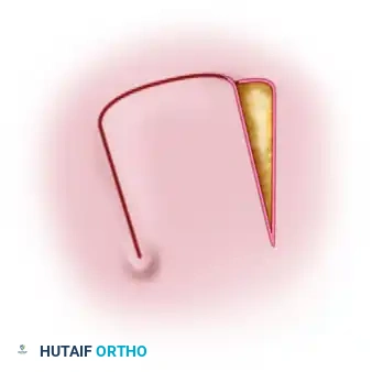
1. Incision Design and Flap Elevation
Careful soft tissue handling is paramount, as the skin must be rotated to cover the opposing surfaces of the newly created rays without placing suture lines directly in the web space or on the contact surfaces.
Standard Incision Approach:
* Volar Incision: Make a longitudinal incision on the flexor surface of the forearm, biased slightly toward the radial side.
* Dorsal Incision: Make a similar longitudinal incision on the dorsal surface, biased slightly toward the ulnar side.
* Web Flap: On the dorsal surface, elevate a V-shaped flap designed to drop into and form the new commissure (web space) at the proximal junction of the separated rays.
Alternative Approach (Nathan and Trung):
Modified S-shaped incisions can be utilized to optimize skin rotation and minimize the need for split-thickness skin grafting. The solid line typically indicates the volar incision, while the dotted line indicates the dorsal incision, creating interlocking flaps.
2. Muscle Separation and Grouping
Deepen the incisions through the fascia. The critical step is longitudinally dividing the forearm musculature into two distinct functional groups corresponding to the radial and ulnar rays.
- Radial Group Allocation: Carefully mobilize and carry the following structures to the radial side:
- Radial wrist flexors (FCR) and extensors (ECRL, ECRB)
- Radial half of the flexor digitorum sublimis (FDS)
- Radial half of the extensor digitorum communis (EDC)
- Brachioradialis and Palmaris longus
- Pronator teres (Protect meticulously)
- Ulnar Group Allocation: Mobilize and carry the following structures to the ulnar side:
- Ulnar wrist flexors (FCU) and extensors (ECU)
- Ulnar half of the flexor digitorum sublimis (FDS)
- Ulnar half of the extensor digitorum communis (EDC)
3. Muscular Debulking
A common pitfall in Krukenberg reconstruction is leaving excessive muscle bulk, which prevents tension-free skin closure and creates a clumsy, overly thick pincer. Muscles that originally crossed the wrist to act on the digits are largely non-functional in this new construct and serve only to add unnecessary volume.
- Resection: If the stump is too bulky, aggressively resect the pronator quadratus, the flexor digitorum profundus (FDP), the flexor pollicis longus (FPL), the abductor pollicis longus (APL), and the extensor pollicis brevis (EPB).
- Preservation: Re-verify the integrity of the pronator teres. Do not disturb its insertion on the radius.
4. Division of the Interosseous Membrane
To achieve independent motion of the rays, the radius and ulna must be physically separated.
* Identify the interosseous membrane.
* Incise the membrane longitudinally throughout its entire length, staying strictly along its ulnar attachment.
* Neurovascular Protection: Extreme care must be taken during this step to identify and protect the anterior and posterior interosseous vessels and nerves. Laceration of the interosseous artery can lead to devastating ischemic complications of the flaps.
5. Ray Separation and Osteotomy (If Required)
Once the interosseous membrane is divided, the radial and ulnar rays should separate easily.
* Depending on the overall size and length of the forearm, the tips of the rays should separate by 6 to 12 cm when actively abducted.
* Apposition Check: Manually adduct the rays. The opposing distal ends of the radius and ulna must touch to allow for fine pinch.
* If the natural bow of the radius prevents the tips from touching, a corrective closing-wedge osteotomy of the radius or ulna must be performed to ensure perfect distal apposition.
6. Hemostasis and Skin Coverage
- Tourniquet Deflation: Remove the tourniquet prior to closure. Obtain meticulous hemostasis. The highly vascular muscle bellies and divided interosseous membrane are prone to hematoma formation, which can compromise the skin flaps.
- Flap Assessment: Observe the capillary refill and overall circulation in the mobilized skin flaps. Excise any excess subcutaneous fat to thin the flaps and improve pliability.
- Skin Rotation: Rotate the skin around each respective ray. The primary goal is to close the skin over each bone so that the longitudinal suture lines do not lie on the opposing (contact) surfaces of the rays. Scars on the contact surfaces will become painful and hypertrophic due to the constant friction of the pincer mechanism.
💡 Clinical Pearl: Managing Bone Length vs. Skin Closure
If skin closure is excessively tight, you may need to shorten the bones. However, in pediatric patients, the bones must NEVER be shortened. Growth at the distal epiphyses is still incomplete, and resecting bone will arrest growth, resulting in an unacceptably short stump at skeletal maturity. In children, rely entirely on split-thickness skin grafting to cover any defects.
7. Final Closure and Dressing
- Rudimentary Digits: If the patient has a congenital amputation with rudimentary digits at the stump terminus, preserve them if possible, as they often contain highly concentrated sensory end-organs.
- Web Space: Suture the V-shaped dorsal flap into place at the proximal junction (commissure) of the rays to create a smooth, deep web space.
- Grafting: Apply split-thickness skin grafts (STSG) to any remaining raw muscle surfaces. Do not close skin under tension.
- Drains: Insert small rubber drains or closed-suction drains into the deep spaces of both rays.
- Dressing: Apply a bulky, non-adherent compression dressing. Crucially, the dressing must be applied with the tips of the rays separated by at least 6 cm to prevent contracture of the web space during the initial healing phase.
Postoperative Care and Rehabilitation
The surgical reconstruction is only the first half of the Krukenberg procedure; rigorous, structured rehabilitation is required to achieve functional prehension.
Immediate Postoperative Phase (Days 1-14)
- Elevation: The limb must be constantly elevated for the first 3 to 4 days to minimize edema, which is the primary enemy of flap survival and graft take.
- Immobilization: The limb remains in the bulky compression dressing with the rays abducted.
- Wound Care: Drains are typically removed at 48-72 hours. Sutures are removed at approximately 14 days, provided the flaps are fully healed.
Early Rehabilitation Phase (Weeks 2-4)
- At 2 to 3 weeks postoperatively, once the wounds are stable, active rehabilitation begins.
- Motor Retraining: The patient is instructed to begin active abduction and adduction of the rays. Because the brain is accustomed to firing these muscles for wrist flexion/extension or pronation/supination, significant cognitive retraining is required.
- Biofeedback: Occupational therapists may use mirror therapy or electromyographic (EMG) biofeedback to help the patient isolate the pronator teres (for closure) and the brachioradialis/extensors (for opening).
Late Rehabilitation Phase (Weeks 4+)
- Strengthening: Progressive resistance exercises are introduced to increase the pinch strength of the adductors.
- Desensitization: The newly formed contact surfaces may be hypersensitive. A structured desensitization program using various textures is implemented.
- Activities of Daily Living (ADLs): Therapy transitions to functional tasks—picking up blocks, holding utensils, and manipulating clothing fasteners. Children typically master these tasks within weeks, whereas adults may require several months of dedicated occupational therapy.
Conclusion
The Krukenberg reconstruction is a masterclass in functional anatomical reassignment. By respecting the neurovascular anatomy, meticulously balancing the adductor and abductor muscle groups, and executing precise soft-tissue coverage, the orthopaedic surgeon can provide a bilaterally amputated or visually impaired patient with a sensate, highly functional limb. While aesthetically unconventional, the profound improvement in independence and quality of life makes the Krukenberg procedure an indispensable technique in the armamentarium of the reconstructive surgeon.
You Might Also Like