Complications, Rehabilitation, and Salvage in Microvascular Replantation
Key Takeaway
Microvascular replantation demands meticulous postoperative monitoring to detect early circulatory compromise. This comprehensive guide details the management of early and late complications, including thrombosis, compartment syndrome, and nonunion. It outlines evidence-based rehabilitation protocols and provides a step-by-step surgical technique for the re-exploration and salvage of failing replants, emphasizing the critical role of timely intervention and vein grafting in preserving limb viability.
INTRODUCTION TO REPLANTATION COMPLICATIONS
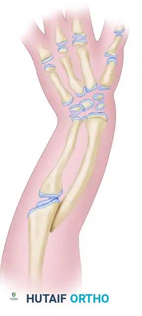
The success of microvascular replantation extends far beyond the survival of the amputated part; it is ultimately defined by the restoration of functional utility. While advancements in microsurgical techniques have significantly improved viability rates, the postoperative course remains fraught with potential pitfalls. Complications can be broadly categorized into early (within the first 1 to 3 weeks), which primarily threaten the survival of the replant, and late, which compromise the functional outcome. Mastery of these complications, coupled with a rigorous, evidence-based approach to postoperative monitoring, salvage re-exploration, and rehabilitation, is mandatory for the reconstructive microsurgeon.
EARLY POSTOPERATIVE COMPLICATIONS
Early complications typically manifest within the first 7 to 14 days and require immediate, decisive intervention to prevent catastrophic loss of the replanted part.
Circulatory Compromise
Circulatory failure is the most pressing and devastating early complication. It is typically secondary to arterial thrombosis, venous congestion, or severe vasospasm. Arterial insufficiency presents with a pale, cool digit lacking capillary refill and turgor. Venous congestion presents with a swollen, cyanotic, and rapidly engorging part with brisk but dark capillary bleeding.
🚨 SURGICAL WARNING: The "no-reflow" phenomenon, driven by ischemia-reperfusion injury, endothelial swelling, and microvascular thrombosis, can mimic anastomotic failure. Prompt differentiation between mechanical anastomotic failure and microvascular collapse is critical.
Hemorrhage and Hematoma Formation
Excessive bleeding may originate from unligated or uncauterized vessels that spasm during the initial surgery but dilate postoperatively. Furthermore, the aggressive systemic anticoagulant therapy (e.g., heparin, dextran, aspirin) routinely employed to maintain microvascular patency significantly elevates the risk of hematoma. A hematoma is not merely a source of blood loss; it acts as a mechanical compressive force on the fragile venous anastomoses, precipitating secondary venous thrombosis.
Skin Necrosis and Soft Tissue Loss
Significant skin necrosis frequently occurs after the primary closure of integument that initially appeared viable but subsequently succumbed to the sheer magnitude of the crush or avulsion injury sustained during the initial trauma.
* Management: Aggressive, serial débridement is required. Secondary closure utilizing local rotational flaps, regional flaps, or split-thickness skin grafts (STSG) must be performed once the wound bed is optimized.
Compartment Syndrome and Ischemia-Reperfusion Injury
Prolonged warm ischemia times, particularly in major limb replantations (macro-replantations of the arm, forearm, or distal leg), lead to severe ischemia-reperfusion injury. The influx of oxygenated blood generates reactive oxygen species (ROS), leading to massive intracellular edema and elevated intracompartmental pressures.
* Intervention: Prophylactic or early therapeutic fasciotomies of the arm, forearm, and hand are mandatory in major limb replantations. Failure to decompress the compartments will result in irreversible myonecrosis and secondary microvascular collapse.
Infection
While significant sepsis is relatively rare following replantation, the presence of necrotic tissue, hematoma, and foreign bodies (sutures, K-wires) creates an optimal environment for bacterial proliferation. Management relies on broad-spectrum intravenous antibiotics tailored to intraoperative cultures, aggressive wound débridement, and the establishment of adequate drainage.
LATE POSTOPERATIVE COMPLICATIONS
Late complications dictate the ultimate functional outcome and often necessitate secondary reconstructive procedures. These interventions are typically delayed until the soft tissue envelope has stabilized, usually 3 to 6 months post-replantation.
Osseous Complications: Nonunion and Malunion
Rigid skeletal fixation is the foundation of a successful replant. However, extensive periosteal stripping and devascularization can lead to delayed union or nonunion.
* Management: Symptomatic nonunions require takedown, meticulous preparation of the bone ends, autologous bone grafting (e.g., iliac crest or distal radius), and revision internal fixation.
Tendon Adhesions and Loss of Excursion
The biomechanics of tendon healing in a replanted digit are severely compromised by the surrounding scar tissue, leading to dense adhesions.
* Management: Tenolysis is frequently required to restore excursion. In cases of severe tendon substance loss or destruction of the flexor pulley system, staged tendon reconstruction utilizing a silicone Hunter rod followed by a free tendon graft (e.g., palmaris longus) is indicated.
Joint Stiffness
Articular damage, prolonged immobilization, and capsular contracture inevitably lead to joint stiffness.
* Management: Intensive hand therapy is the first line of defense. Refractory stiffness may require surgical capsulotomy. In selected patients with severe articular destruction, interposition arthroplasty or arthrodesis in a functional position may be necessary to salvage utility.
Neurological Deficits
Primary neurorrhaphy may fail due to extensive zone of injury, intraneural fibrosis, or inadequate resection of the damaged nerve ends.
* Management: If a primary repair fails to demonstrate advancing Tinel's sign or return of function within a reasonable timeframe (calculated at 1 mm/day of regeneration), re-exploration is warranted. Interpositional nerve grafting (e.g., sural nerve or medial antebrachial cutaneous nerve) is often required to bridge the resulting defect without tension.
POSTOPERATIVE MONITORING AND MEDICAL SALVAGE
A reliable, continuous monitoring system is the cornerstone of postoperative care. The goal is the immediate detection of vascular compromise, as the salvage rate drops precipitously with delayed intervention.
Monitoring Techniques
- Clinical Observation: Color, pulp turgor, and capillary refill remain the gold standard.
- Surface Temperature Monitoring: A drop of >2°C compared to the adjacent normal digit, or an absolute temperature <30°C, is highly indicative of vascular compromise.
- Instrumental Monitoring: Implantable venous Dopplers, pulse oximetry, and quantitative fluoroscopy can provide objective data, particularly in buried flaps or heavily bandaged digits.
Medical Salvage Strategies
When a replant exhibits signs of distress, immediate medical maneuvers should be instituted while preparing the operating room:
1. Positioning: For arterial insufficiency, lower the limb to a dependent position. For venous congestion, elevate the limb.
2. Environmental Control: Ensure the patient is warm, adequately hydrated, and free from pain or anxiety to minimize sympathetic tone.
3. Pharmacologic Intervention:
* Brachial Plexus Block: Highly effective for breaking severe vessel spasm by providing a profound sympathectomy.
* Heparin Bolus: An intravenous bolus of 3,000 to 5,000 Units of Heparin may be administered to halt propagating thrombus in a failing part.
💡 CLINICAL PEARL: The decision to return to the operating room must be made promptly. Reoperation is significantly more likely to be successful if performed within 4 to 6 hours of the onset of ischemic signs. "Time is tissue."
SURGICAL TECHNIQUE 63-11: RE-EXPLORATION OF THE FAILING REPLANT
When medical salvage fails, emergent surgical re-exploration is indicated. The surgeon must systematically evaluate all anastomoses, guided by the clinical presentation (arterial vs. venous failure).
1. Preparation and Exposure
- Positioning: Supine with the affected limb on a radiolucent hand table. Apply a pneumatic tourniquet, but do not inflate it unless catastrophic, uncontrollable hemorrhage is encountered, as tourniquet ischemia will exacerbate the existing microvascular compromise.
- Incision: Carefully remove all sutures and open the previous incisions. Evacuate any hematoma and irrigate the wound with warm, heparinized saline.
2. Arterial Evaluation
- Inspection: Even if clinical signs point to venous failure, inspect the arterial anastomoses first to ensure inflow. Assess for patency using the empty-and-refill (Acland) test.
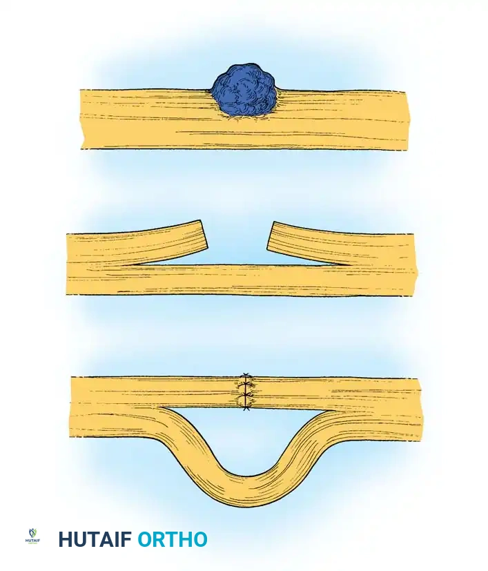
- Resection of Thrombosis: If an arterial anastomosis is thrombosed, do not attempt thrombectomy through the suture line. Excise the anastomosis entirely.
- Inflow Assessment: Release the micro-clamp proximally. There must be vigorous, pulsatile, "spurting" flow. If flow is weak, the vessel is in spasm or there is proximal injury. Dilate the vessel gently with vessel dilators or apply topical papaverine (30 mg/mL).
- Vein Grafting: If proximal flow remains inadequate, or if the vessel wall appears contused or delaminated, dissect further proximally until healthy intima is found. Never anastomose under tension. Interpose a reversed segment of autologous vein graft (e.g., from the volar forearm or dorsal foot).
- Distal Assessment: Evaluate the distal arterial tree. If the primary digital artery is unsalvageable, explore the contralateral digital artery for substitution.
3. Venous Evaluation
- Inspection: If arterial inflow is robust but the part remains congested, direct immediate attention to the venous system.
- Mechanical Obstruction: Inspect veins proximal and distal to the anastomoses to exclude extrinsic compression (tight skin closure, hematoma) or torsion (twisted pedicle).
- Revision: If venous thrombosis is identified, excise the thrombosed segment. Repair the vessel end-to-end only if completely tension-free; otherwise, utilize an interpositional vein graft (non-reversed).
4. Alternative Salvage Techniques for Digits
If all available veins have been exhausted, or if the distal venous tree is too severely crushed to accept an anastomosis, alternative methods to provide venous outflow must be employed to allow the digit to survive until neovascularization occurs (typically 5 to 8 days).
* Pulp Incisions and Nail Wedge Excision: Deep incisions into the pulp or excision of a wedge of the nail bed can promote continuous venous oozing.
* Medicinal Leeches (Hirudo medicinalis): The application of leeches provides active blood removal and secretes hirudin, a powerful local anticoagulant.
* Systemic Monitoring: When utilizing these techniques, the patient's hemoglobin and hematocrit must be monitored every 6 to 8 hours. Blood volume loss can be profound and must be corrected promptly with transfusions. Prophylactic antibiotics (e.g., Ciprofloxacin or Ceftriaxone) are mandatory to prevent Aeromonas hydrophila infections associated with leech therapy.
5. The Decision to Reamputate
If, after exhaustive exploration, resection of damaged segments, and vein grafting, satisfactory arterial inflow and venous outflow cannot be restored, the surgeon must proceed with reamputation. Prolonging the attachment of a non-viable part risks severe systemic toxicity, sepsis, and psychological trauma to the patient.
REHABILITATION PROTOCOLS
The rehabilitation of a replanted part is a complex, multidisciplinary endeavor that must be highly individualized. It requires a delicate balance between protecting the fragile microvascular anastomoses and preventing the devastating complications of tendon adhesions and joint contractures.
Phase I: Protection and Immobilization (Weeks 0 - 3)
- Immobilization: For the first 3 weeks, the primary goal is the protection of the vascular repairs. The limb is immobilized in a bulky, non-compressive dressing with a dorsal blocking splint (if flexor tendons were repaired) or a volar resting splint.
- Positioning: The limb is kept elevated to promote venous and lymphatic drainage.
- Movement: Absolutely no active or passive movement of the involved joints is permitted, as sheer stress can induce microvascular thrombosis.
Phase II: Early Controlled Motion (Weeks 3 - 6)
- Biomechanical Rationale: By week 3, the microvascular anastomoses have endothelialized, and the tendon repairs have achieved sufficient tensile strength to withstand gentle stress. Controlled stress promotes collagen realignment and prevents dense scar formation.
- Therapy: The patient is transitioned to a graduated program of active and active-assisted range-of-motion (ROM) exercises.
- Splinting: Dynamic splinting (e.g., rubber band traction for flexor tendon repairs) or static progressive splinting is introduced to safely guide motion while protecting the repairs.
Phase III: Strengthening and Integration (Weeks 6 and Beyond)
- Progression: Passive stretching is cautiously introduced. If bone healing is confirmed radiographically, progressive resistance exercises are initiated.
- Sensory Re-education: As nerve regeneration progresses, sensory re-education programs are vital to help the brain interpret altered afferent signals, maximizing the functional utility of the replanted part.
- Return to Function: The ultimate goal is the integration of the replanted part into activities of daily living (ADLs) and occupational tasks, acknowledging that maximal medical improvement may take 12 to 18 months.
You Might Also Like