Herniated Disc: Get Relief with Nonsurgical Treatment Options
Key Takeaway
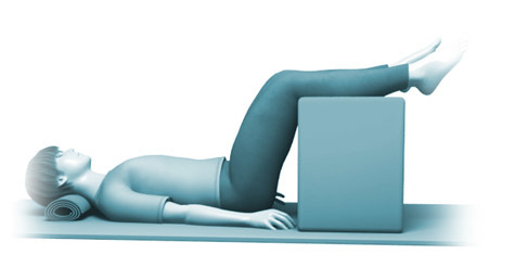
Learn more about Herniated Disc: Get Relief with Nonsurgical Treatment Options and how to manage it. Disc nonsurgical treatment for a slipped disc primarily involves conservative approaches. These include staying active with exercise, relaxation techniques, specific positioning like the psoas position, and medications such as NSAIDs, muscle relaxants, or anti-inflammatory drugs. Manual and physical therapy are also common to relieve pain and improve mobility, with an emphasis on avoiding prolonged bed rest.
As academic orthopedic surgeons and medical educators, our understanding of lumbar disc herniation (LDH) and its management must be grounded in both the natural history of the condition and the evidence-based indications for surgical intervention. While a significant proportion of patients experience resolution of symptoms with conservative measures, a detailed appreciation of surgical anatomy, meticulous technique, and judicious patient selection remains paramount for those who ultimately require operative management.
Introduction and Epidemiology
Lumbar disc herniation, often colloquially referred to as a "slipped disc," represents a common pathology within spinal medicine, characterized by displacement of intervertebral disc material beyond the confines of the annulus fibrosus. This can lead to compression of neural elements, most commonly the traversing nerve root or, in severe cases, the cauda equina. The epidemiology of LDH demonstrates a peak incidence in individuals aged 30 to 50 years, with a predilection for the L4-L5 and L5-S1 spinal segments due to their biomechanical demands. Lifetime prevalence ranges from 1% to 3%, contributing significantly to back pain and radiculopathy.
The natural history of LDH is characterized by a high rate of spontaneous resolution. Approximately 70-90% of patients with symptomatic LDH experience significant improvement or complete resolution of radicular pain within six to twelve weeks, irrespective of specific conservative interventions. This phenomenon is often attributed to the inflammatory cascade subsiding, dehydration and shrinkage of the herniated fragment, and enzymatic degradation. Consequently, initial management overwhelmingly prioritizes non-surgical approaches. As noted in the foundational understanding, the shift from historical recommendations of prolonged bed rest to advocating for continued activity is crucial. Prolonged immobility can contribute to muscular deconditioning, joint stiffness, and impaired physiological recovery, ultimately hindering functional restoration. Structured exercise programs, judicious use of analgesics, physical therapy, and various pain management techniques, including relaxation exercises, are cornerstones of initial treatment, aimed at mitigating symptoms and improving mobility while the natural healing process unfolds. This document, however, transcends the initial non-operative paradigm to provide a comprehensive academic review of surgical considerations for patients who fail to improve with extensive conservative care or present with emergent indications.
Surgical Anatomy and Biomechanics
A thorough understanding of the surgical anatomy of the lumbar spine and the biomechanics underlying disc herniation is indispensable for safe and effective operative intervention.
Vertebral Column and Intervertebral Discs
The lumbar spine typically comprises five vertebrae, each consisting of a robust vertebral body anteriorly and a posterior arch formed by pedicles, laminae, transverse processes, and a spinous process. The intervertebral disc, situated between adjacent vertebral bodies, acts as a primary shock absorber and permits spinal motion. It is composed of the annulus fibrosus, a concentric lamellar structure of collagen fibers, and the central nucleus pulposus, a gelatinous core rich in proteoglycans. Herniation occurs when the nucleus pulposus extrudes through a defect in the annulus fibrosus. Disc herniations are classified morphologically as protrusions (broad-based bulging without full annular rupture), extrusions (displaced nuclear material extending beyond the annular confines, but remaining continuous with the parent disc), and sequestrations (free fragments of nuclear material detached from the parent disc).
Neural Elements and Ligamentous Structures
Within the vertebral canal, the spinal cord typically terminates as the conus medullaris around the L1-L2 level. Below this, the cauda equina, a bundle of nerve roots, descends to exit at their respective neural foramina. Lumbar nerve roots are particularly vulnerable to compression by disc herniations. Understanding the relationship between the exiting and traversing nerve roots at each level is critical. For example, at L4-L5, a posterolateral herniation typically compresses the L5 (traversing) nerve root, while an extreme lateral or far lateral herniation would compress the L4 (exiting) nerve root. The dorsal root ganglion, often located within the neural foramen, is highly sensitive to compression.
Key ligamentous structures include the anterior and posterior longitudinal ligaments, which stabilize the vertebral bodies. The posterior longitudinal ligament (PLL) offers some containment to posterior disc herniations but is thinner and weaker in the posterolateral aspect, explaining the prevalence of posterolateral herniations. The ligamentum flavum, a thick elastic ligament connecting adjacent laminae, forms the posterior boundary of the spinal canal and must be resected during surgical decompression.
Myology and Vascularity
The paraspinal musculature (erector spinae group: iliocostalis, longissimus, spinalis; and deep intrinsic muscles: multifidus, rotatores) is meticulously dissected during posterior approaches. Minimizing muscle trauma is essential to reduce postoperative pain and accelerate recovery. Arterial supply to the lumbar spine is segmental, with branches from the aorta. Venous drainage occurs via epidural venous plexuses, which can be a significant source of bleeding during surgery if not carefully managed.

Indications and Contraindications
Patient selection for operative management of lumbar disc herniation is a critical determinant of surgical success. While the majority of patients respond to conservative care, specific indications necessitate surgical intervention.
Indications for Operative Intervention
- Cauda Equina Syndrome (CES): This is an absolute surgical emergency characterized by severe, progressive bilateral neurological deficits including saddle anesthesia, bowel or bladder dysfunction (retention or incontinence), and bilateral lower extremity weakness. Immediate surgical decompression is required to preserve neurological function.
- Progressive Neurological Deficit: Documented worsening motor weakness despite appropriate non-operative management. This typically involves a decline of at least one grade on the Medical Research Council (MRC) scale (0-5). Persistent or worsening foot drop is a common presentation.
- Intractable Radicular Pain: Severe, debilitating radicular pain that has failed to respond to a minimum of 6-12 weeks of comprehensive, supervised conservative management. This includes physical therapy, oral analgesics (NSAIDs, neuropathic agents), activity modification, and often epidural steroid injections. The pain must be severe enough to significantly impair quality of life and functional independence.
- Correlation of Clinical Symptoms and Imaging: Clear correlation between the patient's neurological symptoms and signs (e.g., L5 radiculopathy) and imaging findings (e.g., L4-L5 disc herniation compressing the L5 nerve root on MRI).
- Recurrent Disc Herniation: Symptomatic recurrence of disc herniation in a patient who previously experienced successful non-operative or operative treatment.
Contraindications for Operative Intervention
- Resolving Symptoms: Patients whose symptoms are actively improving or have completely resolved do not require surgery.
- Mild, Tolerable Symptoms: Surgical intervention is generally not indicated for mild radicular pain that can be managed with conservative measures, even if imaging reveals a herniation.
- Absence of Neurological Deficit: While severe pain alone can be an indication, the absence of objective neurological deficits (motor weakness, sensory loss, reflex changes) often suggests a higher likelihood of non-operative success or a greater need to exclude non-spinal pain generators.
- Significant Psychological Overlay or Secondary Gain Issues: Patients with unaddressed psychological comorbidities (e.g., depression, anxiety, somatization) or significant secondary gain factors often have suboptimal surgical outcomes. A thorough psychosocial assessment is advisable.
- Uncontrolled Systemic Infection: Active systemic infection must be treated prior to elective spinal surgery.
- Significant Medical Comorbidities: Severe cardiac, pulmonary, or other systemic conditions that significantly increase anesthetic and surgical risk, particularly for elective procedures.
Operative vs. Non-Operative Indications Summary
| Condition | Operative Indication | Non-Operative Indication |
|---|---|---|
| Cauda Equina Syndrome (CES) | Absolute Emergency: Acute onset of bowel/bladder dysfunction, saddle anesthesia, progressive bilateral motor weakness. | Not applicable; requires immediate surgery. |
| Progressive Neurological Deficit | Documented worsening motor weakness (e.g., >1 MRC grade decline), particularly with impending foot drop or significant functional impairment. | Stable or improving motor weakness, mild motor deficits that do not significantly impact function. |
| Radicular Pain | Severe, debilitating radicular pain refractory to 6-12 weeks of comprehensive conservative management, significantly impacting quality of life. | Mild to moderate radicular pain responsive to activity modification, analgesics, physical therapy, and/or epidural steroid injections; symptoms duration less than 6-12 weeks, or improving with conservative care. |
| Spinal Instability | Associated spinal instability (e.g., spondylolisthesis) contributing to radiculopathy, typically requiring fusion in addition to decompression. | No evidence of spinal instability or segmental hypermobility. |
| Imaging Correlation | Clear correlation between clinical radiculopathy (e.g., L5 radiculopathy) and MRI findings (e.g., L4-L5 disc extrusion compressing L5 nerve root). | Incidental disc herniation on imaging without correlating clinical symptoms, or non-specific back pain without radiculopathy. |
Pre Operative Planning and Patient Positioning
Meticulous preoperative planning and proper patient positioning are paramount for optimizing surgical outcomes and minimizing complications during lumbar discectomy.
Preoperative Assessment and Planning
- Clinical Evaluation: A comprehensive history, including symptom onset, duration, alleviating/aggravating factors, and prior treatments, is essential. A thorough neurological examination documenting motor strength, sensory deficits, deep tendon reflexes, and gait assessment confirms the neurological level and severity.
- Imaging Review: Magnetic Resonance Imaging (MRI) is the gold standard for diagnosing LDH, providing detailed visualization of the disc, nerve roots, and spinal cord. High-resolution axial and sagittal T2-weighted sequences are crucial. Computed Tomography (CT) may be used if MRI is contraindicated (e.g., pacemakers, certain metallic implants) or to assess bony anatomy. Dynamic flexion/extension radiographs may be considered if spinal instability is suspected.
- Medical Optimization: Patients should be medically optimized by their primary care physician or appropriate subspecialists. This includes managing comorbidities (e.g., diabetes, hypertension, coagulopathies), discontinuing anticoagulants as per institutional protocols, and smoking cessation counseling.
- Informed Consent: A detailed discussion with the patient regarding the proposed procedure, including its goals, anticipated benefits, potential risks (e.g., nerve injury, dural tear, infection, bleeding, persistent pain, recurrence), alternatives (e.g., continued conservative management, other surgical options), and expected postoperative course.
- Anesthesia Consultation: Evaluation by the anesthesia team to assess airway, cardiac, and pulmonary status, formulate an anesthetic plan, and discuss pain management strategies.
Patient Positioning
The patient is typically positioned prone on a specialized radiolucent spinal operating table (e.g., Jackson table, Andrews frame, Wilson frame) designed to allow the abdomen to hang freely. This is critical to decompress the inferior vena cava and epidural venous plexus, thereby reducing epidural bleeding and improving visualization of the surgical field.
- Padding: Meticulous padding of pressure points is essential to prevent iatrogenic injuries. This includes:
- Eyes: Protected from direct pressure to prevent retinal ischemia or corneal abrasion.
- Chest and Pelvis: Supported on bolsters or frames, ensuring the abdomen is free.
- Knees: Flexed slightly with padding.
- Ankles/Feet: Padded and free-floating to prevent pressure sores or peroneal nerve palsies.
- Arms: Positioned on arm boards, abducted less than 90 degrees, with elbows padded to prevent ulnar nerve compression.
- Fluoroscopy: Intraoperative fluoroscopy is used to accurately identify and confirm the operative level before incision. A metallic marker (e.g., K-wire, hemostat) is placed on the skin at the suspected level, and an AP and lateral fluoroscopic image are obtained.
- Preparation and Draping: The surgical site (typically from mid-thoracic to gluteal cleft) is prepped with an antiseptic solution and sterilely draped, ensuring adequate exposure for incision and potential extension if needed.
- Urinary Catheter: A Foley catheter is typically inserted for procedures of longer duration or in patients with suspected bladder dysfunction.
Detailed Surgical Approach and Technique
The gold standard for surgical management of symptomatic lumbar disc herniation refractory to conservative treatment is microdiscectomy, which can be performed as an open, minimally invasive (tubular), or endoscopic procedure. The principles of decompression remain consistent regardless of the approach.
Lumbar Microdiscectomy
This technique involves a small midline incision and microscopic or loupe magnification to perform nerve root decompression and disc fragment removal.
-
Incision and Dissection:
- A 3-4 cm midline skin incision is made, centered over the confirmed operative level.
- The subcutaneous tissue is incised, and the thoracolumbar fascia is identified.
- The fascia is incised longitudinally, typically just lateral to the spinous processes, on the side of the herniation.
- Subperiosteal dissection of the paraspinal muscles (multifidus and longissimus) is performed using Cobb elevators, elevating them off the lamina and facet capsule. Self-retaining retractors (e.g., McCulloch, Gelpi, or specialized tubular retractors for MIS) are then placed to maintain exposure.
- Tubular Microdiscectomy Variant: For minimally invasive approaches, a small skin incision is made, and sequential dilators are used to gently separate the muscle fibers down to the lamina, avoiding extensive subperiosteal dissection. A tubular retractor is then docked, and an endoscope or microscope is introduced.
-
Localization and Bony Resection (Laminotomy/Hemilaminotomy):
- The operative level is definitively confirmed with intraoperative fluoroscopy by placing an instrument (e.g., probe, rongeur tip) on the lamina.
- A limited laminotomy or hemilaminotomy is performed using a high-speed burr or Kerrison rongeurs. The aim is to remove enough bone from the inferior aspect of the superior lamina and/or the superior aspect of the inferior lamina to gain access to the underlying ligamentum flavum and epidural space. Facet joint integrity should be preserved as much as possible to avoid iatrogenic instability.
-
Ligamentum Flavum Excision:
- The ligamentum flavum is carefully resected using Kerrison rongeurs and a fine-tipped probe. This exposes the underlying dura and nerve root. Meticulous technique is critical to avoid dural tears or nerve root injury.
- The epidural fat and veins are often encountered beneath the ligamentum flavum. Bipolar cautery is used sparingly and carefully for hemostasis to avoid thermal injury to neural structures.
-
Neural Decompression and Disc Identification:
- Once the ligamentum flavum is resected, the dura mater and the traversing nerve root are identified. The nerve root is gently retracted medially or laterally using a nerve root retractor (e.g., Penfield #4) to expose the posterolateral aspect of the disc space and the herniated fragment. The integrity of the dura should be continuously monitored.
- The herniated disc fragment is often readily visible, pressing on the nerve root.
-
Discectomy:
- A small annulotomy (incision into the annulus fibrosus) is made directly over the herniation using a scalpel (e.g., #11 blade) or a fine pituitary rongeur.
- The extruded or sequestered disc fragment is carefully grasped and removed using pituitary rongeurs. Multiple passes are often required to ensure removal of all free fragments.
- The disc space is then gently explored to remove loose nuclear material. While aggressive curettage of the entire disc space was historically performed, current evidence suggests that a more limited discectomy (removing only symptomatic fragments) may reduce the risk of future disc space collapse or instability, without increasing recurrence rates significantly.
- Confirmation of adequate decompression is achieved by gently probing the nerve root to ensure it is freely mobile and no longer under tension or compression. The epidural space should be clear of residual disc fragments.
-
Closure:
- Once hemostasis is achieved, the retractors are removed.
- The thoracolumbar fascia is meticulously reapproximated with absorbable sutures (e.g., 0-Vicryl).
- Subcutaneous tissues are closed with absorbable sutures (e.g., 2-0 Vicryl), and the skin is closed with staples or subcuticular sutures (e.g., 3-0 Monocryl).
- A sterile dressing is applied.
Key Technical Considerations
- Magnification: Use of an operating microscope or high-power loupes enhances visualization and precision, reducing iatrogenic injury.
- Hemostasis: Meticulous hemostasis throughout the procedure is vital to prevent blood obscuring the field and to reduce postoperative hematoma formation.
- Nerve Root Protection: Constant awareness of the nerve root's location and gentle handling are paramount. Aggressive retraction can lead to neurological deficits.
- Facet Preservation: Minimizing resection of the facet joint capsule and articular processes is crucial to maintain spinal stability and prevent future issues like spondylolisthesis.
- Identification of Recurrent Herniation: In revision cases, scar tissue can obscure anatomical landmarks, necessitating careful dissection.
Complications and Management
Despite advancements in surgical techniques, complications can still arise during or after lumbar discectomy. Prompt recognition and appropriate management are crucial for optimal patient outcomes.
Common Complications
| Complication | Incidence | Salvage Strategy |
|---|---|---|
| Dural Tear | 1-10% | Intraoperative: Direct repair with non-absorbable sutures (e.g., 4-0 Nurolon) and/or patch graft (fat, fascia, allograft) if primary repair is difficult. Sealant (fibrin glue) application. Strict postoperative bed rest for 24-48 hours. Postoperative (CSF leak/fistula): Bed rest, lumbar drain, re-exploration and repair if persistent. |
| Nerve Root Injury | <1% | Intraoperative: Gentle handling, avoid aggressive retraction. If transected, microsurgical repair may be attempted. Postoperative: Symptomatic management, potential exploration if deficit is severe/progressive. |
| Recurrent Disc Herniation | 5-15% (2-year) | Symptomatic/Refractory: Re-exploration and revision discectomy. Consider fusion if associated with instability or severe degenerative changes. |
| Wound Infection | 1-3% (superficial), <1% (deep) | Superficial: Oral antibiotics, local wound care. Deep: Surgical debridement, intravenous antibiotics, wound vac, prolonged therapy. |
| Vascular Injury | <0.1% | Intraoperative (major vessel): Immediate recognition, direct compression, vascular surgery consultation, potentially open laparotomy for repair. |
| Persistent Radiculopathy | 5-10% | Postoperative: Rule out residual compression, new herniation, or nerve root inflammation/fibrosis. Consider epidural steroid injections, physical therapy, neuropathic pain medications. Further imaging or neurophysiological studies. |
| Cauda Equina Syndrome (Iatrogenic) | Extremely rare | Intraoperative: Immediate decompression, ensure no retained fragments or hematoma. Postoperative: Emergent re-exploration, hematoma evacuation, ensure complete decompression. |
| Wrong Level Surgery | <0.5% | Intraoperative: Verification with fluoroscopy. If identified before closure, extend incision or perform new incision at correct level. Postoperative: Revision surgery at correct level. |
Management Strategies for Complications
- Dural Tear: Small dural tears identified intraoperatively are typically repaired primarily with fine, non-absorbable sutures. If primary repair is difficult due to tissue fragility or large defect size, an onlay graft of fat, muscle, or fascia, along with fibrin glue, can be utilized. Postoperatively, strict bed rest and sometimes a lumbar drain are employed to reduce cerebrospinal fluid (CSF) leak. Persistent CSF leaks or pseudomeningoceles may require surgical re-exploration.
- Nerve Root Injury: Direct nerve root injury is rare with careful microdiscectomy. If it occurs, the extent of injury (stretch, partial, complete laceration) dictates management. Direct microsurgical repair is considered for transections. Postoperatively, neurological deficits are closely monitored, and symptomatic treatment is provided.
- Recurrent Disc Herniation: Recurrence typically presents with return of radicular symptoms. Diagnostic MRI confirms the diagnosis. Management depends on symptom severity and progression. Re-operation (revision microdiscectomy) is often effective. If there is associated instability or significant degenerative changes, fusion may be considered.
- Infection: Superficial wound infections are treated with oral antibiotics and local wound care. Deep infections (discitis, epidural abscess, osteomyelitis) are serious and require urgent surgical debridement, culture-directed intravenous antibiotics, and sometimes prolonged treatment.
- Vascular Injury: Major vascular injury (e.g., iliac artery or vein) is a rare but life-threatening complication, typically occurring anteriorly to the vertebral body during over-penetration with instruments. Immediate recognition, direct compression, and emergent consultation with a vascular surgeon for open repair are critical.
- Persistent Radiculopathy (Failed Back Surgery Syndrome - FBSS): This complex multifactorial condition refers to persistent or new radicular pain after spinal surgery. Causes include inadequate decompression, recurrent herniation, epidural fibrosis, segmental instability, nerve root inflammation, psychological factors, or incorrect diagnosis. Management is individualized and may involve physical therapy, pain management strategies (e.g., neuromodulation), further diagnostic imaging, and occasionally revision surgery.
Post Operative Rehabilitation Protocols
Postoperative rehabilitation after lumbar microdiscectomy is crucial for functional recovery, pain management, and preventing recurrence. Protocols typically follow a phased approach, balancing early mobilization with protection of the healing surgical site.
Phase I: Immediate Postoperative (Days 0-14)
- Goals: Pain control, wound healing, prevention of complications, independent functional mobility, patient education.
- Activity Restrictions:
- Lifting: Strict avoidance of lifting anything heavier than 5-10 lbs (e.g., a gallon of milk).
- Bending: Avoidance of forward bending at the waist.
- Twisting: Avoidance of lumbar rotation.
- Sitting: Limit prolonged sitting (e.g., 20-30 minutes max) to reduce intradiscal pressure.
- Early Mobilization:
- Ambulation initiated on the day of surgery or postoperative day 1. Gradual increase in walking distance as tolerated.
- Emphasis on proper body mechanics for getting in/out of bed, sitting, and standing.
- Pain Management: Multimodal analgesia (acetaminophen, NSAIDs if appropriate, short-term opioids, muscle relaxants) as needed.
- Wound Care: Instruction on incision site care, monitoring for signs of infection.
Phase II: Early Rehabilitation (Weeks 2-6)
- Goals: Restore basic lumbar motion, initiate core stabilization, improve endurance.
- Activity: Gradually increase duration of sitting and standing. Continue to adhere to lifting, bending, and twisting precautions.
- Physical Therapy: Referral to outpatient physical therapy.
- Core Stabilization: Initiation of gentle exercises for transverse abdominis and multifidus activation. Focus on neutral spine position.
- Flexibility: Gentle hamstring, hip flexor, and piriformis stretches if tolerated. Avoid aggressive lumbar flexion.
- Aerobic Conditioning: Low-impact activities such as walking, stationary cycling, or elliptical trainer (as tolerated).
- Patient Education: Reinforce ergonomic principles for daily activities and work.
Phase III: Intermediate Rehabilitation (Weeks 6-12)
- Goals: Progressive strengthening of core and lower extremities, improve balance and proprioception, increase functional capacity.
- Activity: Gradual easing of lifting restrictions (e.g., up to 20-25 lbs). Increased tolerance for prolonged sitting/standing.
- Physical Therapy:
- Progressive Strengthening: Advance core exercises to include planks, side planks, bird-dogs. Incorporate gluteal and back extensor strengthening.
- Functional Exercises: Introduce activities simulating daily tasks or work-related movements.
- Cardiovascular: Increase intensity and duration of aerobic exercise. Swimming may be introduced.
- Sport-Specific Training: For athletes, gradual return to sport-specific drills, ensuring proper form and biomechanics.
- Work Hardening/Conditioning: For patients returning to physically demanding occupations, a work hardening program may be beneficial.
Phase IV: Advanced Rehabilitation and Long-Term Management (Beyond 12 Weeks)
- Goals: Maintain strength and flexibility, prevent recurrence, ensure long-term functional independence.
- Activity: Most restrictions lifted, with continued emphasis on proper body mechanics and lifting techniques. Gradual return to full activities, including recreational sports, as tolerated.
- Maintenance:
- Continued home exercise program focused on core strength, flexibility, and aerobic fitness.
- Importance of lifestyle modifications, including maintaining a healthy weight and smoking cessation.
- Ergonomic assessment of work and home environments.
- Follow-up: Regular follow-up with the surgeon and physical therapist to monitor progress and address any concerns.
Patient compliance with the rehabilitation protocol is crucial for maximizing outcomes and reducing the risk of re-herniation. Individualized protocols, tailored to patient-specific factors, activity levels, and recovery progression, are often employed.
Summary of Key Literature and Guidelines
The management of lumbar disc herniation has evolved significantly, guided by landmark clinical trials and the development of evidence-based guidelines.
Key Clinical Trials and Meta-Analyses
The Spine Patient Outcomes Research Trial (SPORT) is arguably the most influential study comparing surgical and non-surgical treatments for LDH. The initial results (2006-2007) and long-term follow-up (up to 8 years, 2014) consistently demonstrated that patients randomized to surgical discectomy reported greater improvement in pain, function, and satisfaction compared to non-operative treatment, particularly in those with persistent symptoms beyond 6 weeks. However, it also highlighted a significant crossover rate, indicating that many patients initially assigned to non-operative care eventually opted for surgery, and vice-versa, making an "intention-to-treat" analysis challenging. When comparing "as-treated" groups, surgery consistently showed superior outcomes for radiculopathy.
Subsequent meta-analyses and systematic reviews have largely supported these findings, affirming the effectiveness of microdiscectomy for carefully selected patients with persistent, debilitating radiculopathy refractory to conservative care. These reviews often emphasize that while surgery provides faster and more complete relief of radicular pain in the short to medium term, the long-term outcomes (beyond 2-4 years) between surgical and non-surgical groups tend to converge, especially for neurological outcomes, due to the high rate of spontaneous resolution or eventual crossover to surgery.
Evidence-Based Guidelines
Professional organizations such as the American Academy of Orthopaedic Surgeons (AAOS) and the North American Spine Society (NASS) have developed comprehensive guidelines for the management of LDH. These guidelines consistently advocate for:
- Initial Conservative Management: A trial of non-operative care (typically 6-12 weeks) as the first-line treatment for nearly all patients with symptomatic LDH, except in cases of cauda equina syndrome or progressive neurological deficit.
- Surgical Indications: Microdiscectomy is recommended for patients with persistent, functionally limiting radicular pain despite adequate conservative management, provided there is clear clinical and radiographic correlation.
- Specific Scenarios:
- Cauda Equina Syndrome: Recognized as a surgical emergency requiring urgent decompression.
- Progressive Neurological Deficit: Also warrants urgent surgical intervention.
- Minimally Invasive Approaches: Increasingly favored due to reduced muscle dissection, shorter hospital stays, and potentially faster recovery, with comparable efficacy to open techniques.
- Limited vs. Aggressive Discectomy: Current evidence supports limited removal of only the herniated fragment, rather than aggressive curettage of the entire disc space, to potentially reduce recurrence rates and preserve disc height.
- Postoperative Rehabilitation: Emphasized as a critical component of recovery to optimize functional outcomes and prevent recurrence.
Future Directions
Ongoing research in LDH management focuses on several areas:
- Predictive Biomarkers: Identifying genetic or imaging biomarkers to better predict which patients will respond to conservative care versus those requiring early surgical intervention.
- Biologics and Disc Regeneration: Exploring novel biological therapies (e.g., cell-based therapies, growth factors) to promote disc repair and prevent degeneration.
- Improved Minimally Invasive Techniques: Further refinement of endoscopic and ultra-minimally invasive approaches to minimize tissue trauma and enhance recovery.
- Artificial Disc Replacement: While primarily indicated for degenerative disc disease, its role in specific cases of herniation without significant facet pathology or instability is being explored.
In conclusion, the management of lumbar disc herniation demands a nuanced, evidence-based approach. While conservative management remains the initial cornerstone, a profound understanding of surgical indications, meticulous technique, and diligent postoperative care are indispensable for patients who require operative intervention to achieve lasting relief and functional restoration.
You Might Also Like

