Achilles Tendon Rupture: Comprehensive Guide to Epidemiology, Anatomy, Biomechanics, and the Critical Watershed Zone
Key Takeaway
An Achilles tendon rupture is a common injury in middle-aged 'weekend warriors.' Most ruptures occur in the hypovascular 'watershed zone,' 2-6 cm proximal to the calcaneal insertion. This critical zone's limited blood supply contributes to degenerative changes and reduced healing, making its understanding paramount for effective diagnosis and management.
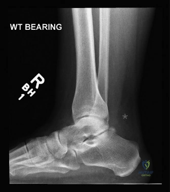
A 42-year-old amateur squash player presents to the emergency department after feeling a "pop" in his calf during a match. He describes a sensation of being kicked in the back of the leg. On examination, there is a palpable defect 4cm proximal to the calcaneal insertion and a positive Thompson test. Please interpret the provided radiograph and discuss the primary clinical concern regarding the anatomy of this region.
Candidate: The radiograph demonstrates a disruption in the Kager’s fat pad silhouette, which is consistent with an acute Achilles tendon rupture. The primary concern is the "watershed zone," located 2-6cm proximal to the calcaneal insertion. This area is relatively hypovascular, receiving its primary blood supply from the paratenon rather than the musculotendinous or osseotendinous junctions, which predisposes it to both degenerative change and rupture.
Candidates often focus solely on the "rupture" diagnosis without discussing the specific anatomical vulnerability. Failing to mention the blood supply mechanism (the paratenon vs. the intrinsic vascularity) or omitting the specific distance (2-6 cm) from the insertion demonstrates a lack of deep anatomical knowledge expected of an FRCS candidate.
A perfect answer identifies the obliteration of Kager's triangle on the lateral view as a classic radiographic sign. It then links this to the "watershed zone" (2-6cm proximal to the insertion), explaining that this region is the intersection of three vascular supplies—the musculotendinous junction, the paratenon, and the calcaneal arcade—where the supply from the paratenon is insufficient, creating a relative zone of avascularity that drives both the pathophysiology of chronic tendinopathy and the high incidence of acute failure.
The patient is a 45-year-old active male who wants to return to squash. Based on the clinical evidence, would you recommend operative or non-operative management? Structure your answer by comparing the outcomes and risks of both approaches.
Candidate: For an active, young-to-middle-aged patient, there is a strong case for operative management. Operative repair typically carries a lower rerupture rate (approx. 2-5%) compared to non-operative treatment. However, I must balance this against the risk of surgical complications, such as wound dehiscence and sural nerve injury, which are higher in open surgery. If he chooses non-operative management, it must be accompanied by an aggressive functional rehabilitation protocol to maintain strength and reduce the risk of stiffness.
Giving a dogmatic, one-sided answer ("I always operate" or "Surgery is never necessary") is a major red flag. Examiners want to see a balanced discussion of the "Operative/Non-operative" spectrum based on current evidence (e.g., Soroceanu/Bhandari meta-analyses), acknowledging that both are valid but carry different risk profiles.
The candidate should categorize the decision by: 1. Patient factors (Age, activity demands); 2. Injury factors (Size of gap, delay in presentation); 3. Risk profile (Wound healing, DM/PVD status). Mentioning the shift towards "accelerated functional rehabilitation" regardless of the chosen path shows the candidate is up-to-date with current literature, noting that the choice of surgery is now about reducing rerupture risk in high-demand individuals while accepting a higher procedural risk profile.
During your open repair, you are about to incise the paratenon. What are the key anatomical structures you must protect, and how do you minimize the risk of iatrogenic complications?
Candidate: The primary structure at risk is the sural nerve. It typically runs along the lateral border of the tendon in the distal third. To minimize risk, I would use a longitudinal incision slightly medial or lateral to the midline—ideally lateral if carefully retracted, but ensuring I don't compromise the skin bridge. I would identify and protect the nerve early in the dissection, avoid excessive retractors, and preserve as much paratenon as possible to protect the vascular supply and facilitate healing.
Failing to mention the paratenon's role in vascularity or assuming the sural nerve is only at risk in percutaneous repairs. The examiner wants to hear about meticulous soft-tissue handling during the open approach, specifically avoiding the use of blunt, aggressive retraction.
The gold standard answer addresses the "Sural Nerve" risk specifically, noting it crosses the lateral border 10-15cm proximal to the calcaneus. The candidate should demonstrate "surgical maturity" by discussing the skin bridge management (avoiding midline incisions that put the skin at risk of necrosis) and the "paratenon preservation" technique, which is critical for revascularization of the watershed zone post-operatively.
