Masterclass in Hand Surgical Incisions & Approaches
Key Takeaway
Mastering hand surgical incisions requires a profound understanding of cutaneous biomechanics and underlying neurovascular anatomy. This guide details evidence-based principles for volar, dorsal, and digital approaches. By avoiding deep creases, preventing perpendicular joint crossings, and utilizing offset fascial dissections, orthopedic surgeons can optimize exposure while minimizing ischemic necrosis and debilitating scar contractures in the hand and wrist.
INTRODUCTION TO HAND CUTANEOUS BIOMECHANICS
The hand is a highly specialized, dynamic organ where the integumentary system is inextricably linked to underlying musculoskeletal function. Unlike other anatomical regions where skin serves primarily as a passive envelope, the skin of the hand—particularly the glabrous volar surface—is a critical component of the gripping mechanism. Consequently, surgical incisions in the hand and wrist demand meticulous planning.
As long as fundamental biomechanical and anatomical principles are strictly observed, skin incisions can be safely executed anywhere on the hand. The primary objective of any hand incision is to provide extensile exposure to deep structures without compromising the vascularity of the skin flaps, while simultaneously preventing the formation of restrictive scar contractures that could tether gliding tendons or limit joint excursion.
FUNDAMENTAL PRINCIPLES OF INCISION PLACEMENT
The execution of a flawless surgical approach in the hand relies on several non-negotiable principles. Failure to adhere to these tenets frequently results in delayed wound healing, ischemic necrosis, or debilitating functional impairment.
Avoidance of Deep Creases
Incisions placed directly within deep flexion creases must be universally avoided. In these specific anatomical zones, the subcutaneous fat layer is exceptionally sparse, and the dermis is densely tethered to the underlying deep fascia.
Surgical Warning: Moisture naturally accumulates within deep palmar and digital creases. Incisions placed directly within these folds are highly susceptible to maceration of the skin edges, leading to wound dehiscence and secondary bacterial colonization.
Tension and Extensile Exposure
An incision must be of adequate length to expose the deep structures without necessitating excessive, traumatic retraction of the skin edges. Vigorous retraction causes microvascular thrombosis within the dermal plexus, leading to marginal necrosis.
To achieve greater exposure without extending the skin incision unnecessarily, the skin and subcutaneous fat should be sharply dissected as a full-thickness flap from the underlying investing fascia. This technique preserves the subdermal vascular plexus and allows for safe, dynamic retraction.
The Offset Incision Technique
A critical concept in hand surgery is that the placement of the skin incision applies only to the cutaneous surface. Entries into deeper structures (fascia, tendon sheaths, joint capsules) are dictated by their specific anatomy and should frequently be offset or even opposite in direction to the skin incision.
For example, when performing a release for de Quervain stenosing tenosynovitis, the optimal skin incision over the radial styloid is transverse (parallel to Langer’s lines). However, the subsequent incision through the extensor retinaculum to decompress the first dorsal compartment must be longitudinal.
Clinical Pearl: The offset incision prevents the skin scar from adhering directly to the underlying healing tendon or bone. By undermining a full-thickness flap on one side, the deep fascial approach is made parallel to, but physically offset from, the cutaneous wound, creating a barrier of healthy subcutaneous tissue between the two healing layers.
Vascular Preservation and Parallel Incisions
Parallel or nearly parallel incisions that are placed too closely together must be strictly avoided. The vascular supply to the skin of the hand relies on a delicate network of perforating vessels. Narrow bipedicled flaps created by closely spaced parallel incisions are at an exceptionally high risk of ischemic necrosis. If multiple incisions are required, they must be separated by a wide bridge of skin to ensure adequate perfusion.
Joint Motion and Scar Contracture
Joint motions in the hand are approximately perpendicular to the long axis of the skin creases. Therefore, a cardinal rule of hand surgery is that incisions must never cross a crease at or near a right angle.
When a linear scar crosses a flexion crease perpendicularly, the natural process of wound contraction (mediated by myofibroblasts) creates a longitudinal tether. In the hand and fingers, this bowstringing effect results in severe flexion contractures. If an incision must cross a crease, it should be designed to cross obliquely (e.g., using a Bruner zig-zag or a lazy-S configuration) so that the forces of scar contraction do not align with the axis of joint motion.
DORSAL HAND AND WRIST APPROACHES
The skin on the dorsum of the hand is thin, highly mobile, and possesses a robust subdermal venous and lymphatic network. Because of this mobility, shorter incisions frequently suffice on the dorsum, as the skin can be easily retracted to expose adjacent structures.
Dorsal Wrist Incisions
A straight longitudinal or a lazy-S incision on the mid-dorsum of the wrist provides excellent, extensile exposure. By elevating full-thickness dermo-fat flaps off the extensor retinaculum, structures can be accessed from the extreme radial aspect (first dorsal compartment) to the extreme ulnar aspect (sixth dorsal compartment) through a single incision.
As illustrated above, dorsal approaches include the longitudinal incision to expose the metacarpal shaft (F) and the versatile dorsal wrist incision (O), which can be modified into a lazy-S to prevent linear contracture across the radiocarpal joint.
When exposing the metacarpal shafts for fracture fixation, longitudinal incisions are preferred. These should be placed slightly off-center from the extensor tendons to prevent direct adherence of the tendon to the healing skin scar.
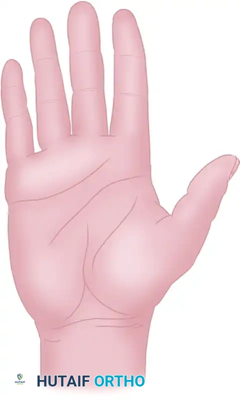
VOLAR HAND AND WRIST APPROACHES
The volar skin is thick, glabrous, and anchored to the palmar aponeurosis by dense vertical fibrous septa. This unique anatomy prevents the skin from sliding during gripping but makes surgical elevation more challenging.
Palmar Incisions
Incisions in the palm must carefully navigate the complex neurovascular anatomy and the superficial palmar arch.
The volar surface accommodates a variety of specialized incisions: (G) Distal palmar fascia exposure; (H) Mid-palmar structures; (I & K) L-shaped and S-shaped incisions at the base of the finger to avoid perpendicular crease crossing; (J) Short transverse incision for A1 pulley release; (L) Proximal flexor sheath of the thumb; (M) Thenar eminence approach; (N) Extensive palmar-wrist incision (e.g., for severe trauma or Dupuytren's); (P) Transverse volar wrist incision; (Q) Base of the thumb approach.
When extending a palmar incision into the wrist (such as for an extended carpal tunnel release or volar compartment syndrome), the incision must cross the wrist flexion creases obliquely or in a zig-zag fashion (as seen in incision N) to prevent a debilitating volar flexion contracture.
DIGITAL INCISIONS AND APPROACHES
Digital incisions require the highest level of precision. The development of a contracture in a finger results in profound functional impairment.
The Midlateral Finger Incision
The midlateral approach is the workhorse incision for exposing the flexor tendon sheath, phalangeal shafts, and digital neurovascular bundles. It is placed along the neutral line of the finger—the axis that neither stretches during flexion nor buckles during extension.
To identify the midlateral line, connect the apices of the digital flexion creases when the finger is fully flexed.
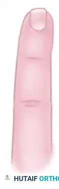
The midlateral incision in the finger (A) and the thumb (C). This approach provides unparalleled access to the volar structures while keeping the scar completely off the tactile palmar surface.
Surgical Technique for the Midlateral Approach:
1. Incision: The skin is incised along the exact midlateral line.
2. Neurovascular Bundle Management: The surgeon has two options regarding the digital neurovascular (NV) bundle:
* Volar Retraction (Preferred for flexor sheath access): The dissection is carried dorsal to the NV bundle. Cleland’s ligaments (which lie dorsal to the bundle) are divided. The NV bundle is then mobilized and carried volarward within the volar skin flap. This protects the bundle and provides excellent exposure of the flexor apparatus.
* Superficial Dissection (Preferred for dorsal/lateral access): The dissection is carried superficial to the NV bundle.
3. Flap Thickness: If dissecting superficial to the NV bundle, extreme care must be taken to avoid making the skin flaps too thin, which will inevitably lead to flap necrosis.

A closer view reinforcing the midlateral digital approach. Notice how the incision remains strictly lateral, avoiding the volar tactile pads and the dorsal extensor apparatus.
Dorsal Digital Incisions
Dorsal digital incisions are primarily utilized for exposing the extensor mechanism, performing arthrodesis, or addressing dorsal phalangeal fractures.
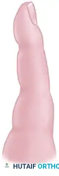
Dorsal digital approaches include the curved or lazy-S incision to expose the central slip of the extensor tendon (D), and the inverted-V (or Y-shaped) incision (E) specifically designed for arthrodesis or extensive exposure of the distal interphalangeal (DIP) joint.
When exposing the central slip (D), a straight longitudinal incision should be avoided over the proximal interphalangeal (PIP) joint to prevent scar contracture that could limit PIP flexion. The inverted-V incision (E) over the DIP joint creates a robust distally based flap that provides excellent exposure of the joint articular surfaces while preserving the germinal matrix of the nail.
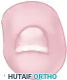
Fingertip and Felon Incisions
Infections of the distal pulp space (felons) require prompt surgical drainage to prevent ischemic necrosis of the pulp and osteomyelitis of the distal phalanx. The pulp space is divided into multiple closed compartments by dense fibrous septa radiating from the periosteum to the skin.
Incisions for draining a felon must decompress the septal compartments without compromising the tactile surface. (B) represents a unilateral longitudinal incision, while (R) represents a J-shaped or hockey-stick alternative.
Pitfall: The classic "fish-mouth" incision around the entire fingertip is historically obsolete and should be strictly avoided. It frequently results in an unstable, painful, and insensate fingertip scar.
For felon drainage, a unilateral longitudinal incision (B) is preferred. It should be placed on the ulnar aspect of digits II, III, and IV, and the radial aspect of the thumb and small finger (to avoid the primary pinch contact surfaces). The incision must be placed dorsal to the tactile pad, and the knife should be swept across the pulp space to divide the vertical fibrous septa, ensuring complete decompression.
POSTOPERATIVE PROTOCOLS AND SCAR MANAGEMENT
The success of a meticulously planned hand incision is heavily dependent on postoperative care.
- Hemostasis and Closure: Prior to closure, the tourniquet should be deflated to ensure meticulous hemostasis, preventing postoperative hematoma formation which can lead to fibrosis and stiffness. Skin is typically closed with non-absorbable monofilament sutures (e.g., 4-0 or 5-0 nylon) using a tension-free technique.
- Immobilization: The hand is immobilized in a bulky, non-compressive dressing. The wrist is typically placed in 20-30 degrees of extension, the MCP joints in 70-90 degrees of flexion, and the IP joints in full extension (the "intrinsic-plus" or "safe" position) to prevent collateral ligament contracture.
- Elevation and Edema Control: Strict elevation above the level of the heart is mandatory for the first 48-72 hours to minimize interstitial edema, which is the primary precursor to joint stiffness.
- Rehabilitation: Early active range of motion (ROM) is initiated as soon as the specific surgical procedure allows. Once sutures are removed (typically at 10-14 days) and the wound is fully epithelialized, aggressive scar massage and silicone gel sheeting are employed to soften the scar and promote the gliding of underlying structures.
You Might Also Like
