Wrist Biomechanics and Kinematics: A Comprehensive Surgical Guide
Key Takeaway
Understanding wrist biomechanics is essential for diagnosing and treating complex carpal instabilities. The wrist functions through a highly coordinated interaction of capsuloligamentous structures and articular contours, with the proximal capitate serving as the primary center of rotation. This guide explores fundamental kinematic theories, force transmission pathways, and pathomechanical injury patterns, providing orthopedic surgeons with the foundational knowledge required for advanced operative interventions.
INTRODUCTION TO WRIST BIOMECHANICS
The human wrist is an evolutionary marvel of biomechanical engineering, designed to provide a vast global range of motion while maintaining the rigid stability necessary for powerful grip and precise fine motor function. The stability of the wrist during both isolated and interrelated motions depends entirely on the delicate interplay between capsuloligamentous integrity and the highly specific contact surface contours of the carpal bones.
Unlike other major joints, the proximal carpal row lacks direct tendinous insertions, functioning instead as an intercalated segment. Its movement is entirely dictated by mechanical forces exerted by surrounding articulations, ligaments, and the spanning tendons of the forearm. For the operative orthopedic surgeon, a profound understanding of wrist kinematics, force transmission, and pathomechanics is not merely academic—it is the foundational prerequisite for diagnosing carpal instability, planning surgical reconstructions, and executing precise salvage procedures.
FUNDAMENTAL KINEMATICS OF THE WRIST
The Center of Rotation
The global center of rotation for most multi-planar wrist motions is generally considered to be located within the proximal pole of the capitate. This central axis dictates the arc of motion for both the radiocarpal and midcarpal joints.
Flexion and Extension Kinematics
Wrist flexion and extension are complex, coupled motions distributed across multiple articulations.
* Flexion: In vivo kinematic studies utilizing ultrafast computed tomography (CT) demonstrate that the radiocarpal and midcarpal joints contribute almost equally to total wrist flexion.
* Extension: Conversely, the midcarpal joint contributes a significantly greater proportion to wrist extension than the radiocarpal joint.
Clinical Pearl: When performing partial wrist fusions (e.g., radiocarpal arthrodesis), preserving the midcarpal joint is critical, as it allows the patient to retain a functional arc of extension, which is biomechanically more vital for grip strength than terminal flexion.
Radial and Ulnar Deviation
Coronal plane motion at the wrist requires a highly synchronized, three-dimensional rotation of the carpal bones.
* Radial Deviation: During radial-to-ulnar deviation, the proximal carpal row undergoes obligatory palmar flexion. Simultaneously, the proximal row translocates (shifts) ulnarly at both the midcarpal and radiocarpal joints. The scaphoid flexes to avoid impingement against the radial styloid.
* Ulnar Deviation: During ulnar-to-radial deviation, the proximal carpal row extends (dorsal rotation), with the majority of this motion occurring within the intercarpal joints.
The Intercalated Segment Concept
The proximal carpal row (scaphoid, lunate, triquetrum) functions as a mechanical intercalated segment linking the forearm to the hand. Because no tendons insert directly onto the lunate, its position is dictated by the scaphoid (which pulls it into flexion via the scapholunate interosseous ligament) and the triquetrum (which pulls it into extension via the lunotriquetral ligament). The scaphoid acts as the primary stabilizing strut bridging the proximal and distal rows.
CONCEPTUAL MODELS OF WRIST BIOMECHANICS
To conceptualize how forces are transmitted and how carpal positions are controlled by ligaments and articular contours, several biomechanical models have been popularized in orthopedic literature.
Novarro’s Three-Column Theory
Novarro popularized the concept of the wrist functioning as three distinct longitudinal columns:
1. The Central (Force-Bearing) Column: Comprises the distal articular surface of the radius, the lunate, and the capitate. Some expanded definitions include the proximal two-thirds of the scaphoid, the trapezoid, and the articulations with the second and third metacarpal bases.
2. The Radial Column: Includes the radius, the scaphoid, the trapezium, the trapezoid, and the thumb carpometacarpal (CMC) joint.
3. The Ulnar (Control) Column: Comprises the triangular fibrocartilage complex (TFCC), the hamate, the triquetrum, and the CMC joints of the ring and little fingers.
Taleisnik’s Columnar Modification
Taleisnik refined the columnar theory, proposing that:
* The Central Column includes the entire distal carpal row and the lunate.
* The Lateral Column consists solely of the scaphoid, acting as a stabilizing outrigger.
* The Medial Column consists of the triquetrum, functioning as a rotary pivot point for the carpus.
Lichtman’s Ring Concept
Lichtman proposed a ring concept of wrist kinematics, which is highly applicable to understanding carpal instability.
* The wrist is viewed as an oval ring formed by the semirigid proximal and distal carpal rows, stabilized by interosseous ligaments.
* Limited, physiologic mobility occurs at the mobile links: the scaphotrapezial joints radially and the triquetrohamate joints ulnarly.
* Pathologic Instability: Disruption of the bone or ligaments within this ring creates predictable instability deformities.
* DISI (Dorsal Intercalated Segmental Instability): Occurs with scapholunate ligament disruption. The lunate, freed from the flexing force of the scaphoid, extends dorsally under the influence of the intact triquetrum.
* VISI (Volar Intercalated Segmental Instability): Occurs with lunotriquetral ligament disruption. The lunate, freed from the extending force of the triquetrum, flexes palmarly under the influence of the intact scaphoid.
Surgical Warning: Failure to recognize and anatomically reduce a DISI or VISI deformity prior to ligamentous reconstruction or partial arthrodesis will result in altered kinematics, restricted motion, and accelerated radiocarpal arthrosis.
FORCE TRANSMISSION AND LOAD BEARING
The wrist is subjected to immense physiological loads. Biomechanical studies of force transmission reveal that the distal carpal row may bear more than 10 times the force applied to the fingertips during a power grip.
Load Distribution Across the Carpus
Approximately 55% to 60% of the total axial load on the distal row is transmitted centrally through the capitate, scaphoid, and lunate.
At the radiocarpal level, the distribution of axial load in a wrist with neutral ulnar variance is highly specific:
* Radioscaphoid Joint: Bears 50% to 56% of the load.
* Radiolunate Joint: Bears 29% to 30% of the load.
* Ulnolunate Joint (via TFCC): Bears 10% to 21% of the load.
Pitfall: Alterations in ulnar variance drastically shift these load-bearing mechanics. A positive ulnar variance of just 2.5 mm can increase the load transmitted through the ulnocarpal joint to over 40%, leading to ulnar impaction syndrome and degenerative tears of the TFCC. Conversely, negative ulnar variance concentrates stress on the radiolunate joint, predisposing the patient to Kienböck's disease (avascular necrosis of the lunate).
PATHOMECHANICS AND INJURY PATTERNS
The mechanical properties of the ligaments and bones dictate specific, reproducible patterns of injury based on the vector of the applied force.
Scaphoid Fractures
Scaphoid fractures typically result from a fall on an outstretched hand (FOOSH) with the wrist in forced extension and radial deviation. In this position, the scaphoid is locked within the scaphoid fossa, and the dorsal articular margin of the radius serves as a rigid fulcrum. The proximal pole is held tightly by the radioscaphocapitate ligament, causing the scaphoid to bend and fracture at its waist.
Perilunate Dislocations
Carpal dislocations, particularly perilunate dislocations, follow a predictable pathomechanical cascade (Mayfield's stages) resulting from extreme wrist extension, ulnar deviation, and intercarpal supination. The energy propagates from radial to ulnar, sequentially stripping the lunate of its ligamentous attachments.
Ulnar-Sided Ligament Injuries
Conversely, injuries involving forced wrist flexion and pronation contribute heavily to ligamentous trauma on the ulnar side of the wrist. This mechanism places maximum tension on the lunotriquetral interosseous ligament and the dorsal radiocarpal ligaments, leading to VISI deformities or ulnocarpal instability.
CLINICAL EVALUATION OF WRIST INSTABILITY
A thorough clinical evaluation must correlate the patient's symptoms with the underlying biomechanical derangement.
History and Symptomatology
For long-standing problems, the surgeon must correlate the chief complaint with exacerbating or alleviating factors.
* Assess the relationship of pain to specific work or recreational activities.
* Document the presence and exact location of swelling, bruising, and aching.
* Mechanical Symptoms: Sensations of clicking, popping, snapping, grating, and crunching are hallmark signs of dynamic or static carpal instability.
* Consider systemic factors, including a personal or family history of inflammatory arthritides, which can cause generalized capsuloligamentous attenuation.
Provocative Physical Examination
- Watson Scaphoid Shift Test: Evaluates scapholunate integrity. Pressure is applied to the palmar tuberosity of the scaphoid as the wrist is moved from ulnar to radial deviation. A palpable "clunk" indicates scaphoid subluxation over the dorsal rim of the radius.
- Lunotriquetral Ballottement (Shuck) Test: Evaluates lunotriquetral integrity. The lunate is stabilized while the triquetrum is translated dorsally and palmarly. Pain or excessive laxity indicates a positive test.
- Midcarpal Pivot Shift Test: Evaluates for midcarpal instability. The wrist is brought from radial to ulnar deviation under axial load. A sudden "catch" or "clunk" indicates a sudden catch-up clunk of the proximal row transitioning from flexion to extension.
SURGICAL IMPLICATIONS AND OPERATIVE PRINCIPLES
Understanding wrist biomechanics directly informs surgical positioning, approaches, and reconstructive techniques.
Indications for Operative Intervention
- Acute, repairable ligamentous disruptions (e.g., acute scapholunate tears).
- Static carpal instability (DISI/VISI) with reducible carpal alignment.
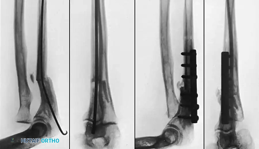
- Displaced scaphoid fractures or nonunions altering carpal kinematics.
- Perilunate fracture-dislocations.
- Symptomatic ulnar impaction syndrome requiring joint leveling.
Patient Positioning and Setup
- Anesthesia: Regional block (supraclavicular or axillary) or general anesthesia.
- Positioning: Supine with the operative arm extended on a radiolucent hand table.
- Tourniquet: A well-padded pneumatic upper arm tourniquet is applied and inflated to 250 mmHg (or 100 mmHg above systolic pressure) after exsanguination.
- Traction: For complex intra-articular fractures or arthroscopy, utilize a wrist traction tower with sterile finger traps (index and middle fingers) applying 10-15 lbs of longitudinal traction to distract the radiocarpal and midcarpal joints.
Surgical Approaches
The Dorsal Approach (Berger’s Capsular Flap)
The dorsal approach is the workhorse for addressing carpal instability, proximal row carpectomy, and partial wrist fusions.
1. Incision: A longitudinal incision is made over the dorsal wrist, centered over Lister's tubercle.
2. Retinacular Flaps: The extensor retinaculum is incised over the third extensor compartment. The extensor pollicis longus (EPL) is mobilized and retracted radially.
3. Compartment Elevation: The second and fourth extensor compartments are elevated subperiosteally.
4. Capsulotomy: A ligament-sparing dorsal capsulotomy (Berger flap) is performed. An inverted "V" or "U" shaped incision is made, preserving the dorsal radiocarpal and dorsal intercarpal ligaments. This flap is elevated distally to expose the radiocarpal and midcarpal joints.
5. Closure: The capsular flap is meticulously repaired with non-absorbable sutures to restore dorsal stability and prevent postoperative palmar subluxation of the carpus. The EPL is often left transposed subcutaneously.
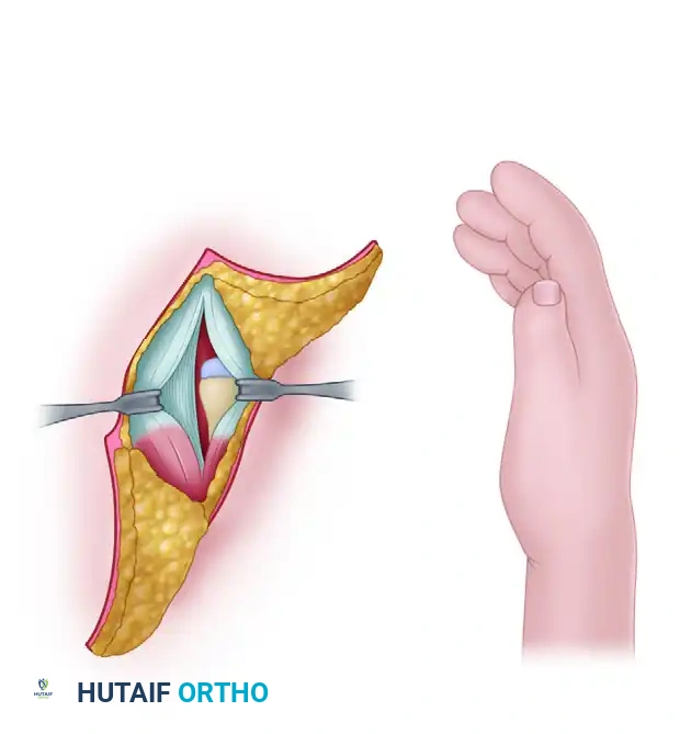
The Volar Approach (Modified Henry)
Utilized for volar scaphoid fixation, distal radius fractures, and volar capsular repairs.
1. Incision: A longitudinal incision is made over the flexor carpi radialis (FCR) tendon.
2. Superficial Dissection: The FCR sheath is incised, and the tendon is retracted ulnarly (protecting the median nerve).
3. Deep Dissection: The floor of the FCR sheath is incised to expose the pronator quadratus and the volar wrist capsule.
4. Capsulotomy: A longitudinal or T-shaped volar capsulotomy exposes the radioscaphocapitate and long radiolunate ligaments. Extreme care must be taken to repair these ligaments during closure to prevent postoperative ulnar translation of the carpus.
POSTOPERATIVE PROTOCOLS
Postoperative rehabilitation is heavily dictated by the biomechanical demands of the reconstructed structures.
- Phase I: Immobilization (0-4 Weeks): Following ligamentous reconstruction or fracture fixation, the wrist is immobilized in a short-arm cast or rigid orthosis. The position of immobilization depends on the pathology (e.g., slight extension and radial deviation for SL repairs to offload the ligament).
- Phase II: Protected Motion (4-8 Weeks): Transition to a removable splint. Initiate active and active-assisted range of motion. Emphasize the "dart-thrower's motion" (extension/radial deviation to flexion/ulnar deviation), which minimizes stress on the scapholunate interval while maximizing functional midcarpal motion.
- Phase III: Strengthening and Proprioception (8-12+ Weeks): Progressive resistance exercises are introduced. Proprioceptive training (e.g., using a gyroscope or balance board) is critical to retrain the dynamic neuromuscular stabilizers of the wrist, compensating for the loss of static ligamentous integrity. Return to heavy manual labor or contact sports is typically restricted until 4 to 6 months postoperatively.
You Might Also Like