Anatomical Double-Bundle Anterior Cruciate Ligament Reconstruction: A Comprehensive Surgical Guide
Key Takeaway
Anatomical double-bundle anterior cruciate ligament (ACL) reconstruction aims to restore native knee kinematics by independently reconstructing the anteromedial and posterolateral bundles. Utilizing a three-portal technique, surgeons can precisely place grafts within the native femoral and tibial footprints. This advanced procedure requires meticulous tunnel placement, specific tensioning protocols, and careful consideration of notch morphology to optimize rotational stability and postoperative outcomes.
Comprehensive Introduction and Patho-Epidemiology
Evolution of Anterior Cruciate Ligament Reconstruction
The historical trajectory of anterior cruciate ligament (ACL) reconstruction has been characterized by a continuous pursuit of optimal kinematic restoration. For decades, the gold standard involved non-anatomical, isometric single-bundle techniques, frequently relying on a transtibial drilling approach. While this methodology successfully eliminated the anterior drawer sign and restored basic sagittal plane stability, it inadvertently resulted in excessively vertical graft placements. These vertical grafts, positioned high and deep within the intercondylar notch, failed to replicate the native oblique orientation of the ACL. Consequently, a significant subset of patients continued to experience residual rotatory laxity, clinically manifested as a persistent pivot-shift phenomenon, which ultimately compromised their ability to return to high-demand, pivoting sports.
The paradigm shifted significantly with the advent of anatomical ACL reconstruction, spearheaded by pioneers who recognized the profound limitations of isometric positioning. The transition to anatomical double-bundle reconstruction represents the zenith of this evolution, aiming to place the femoral and tibial grafts directly into the native, anatomically defined footprints of the ACL. Extensive biomechanical and clinical investigations have corroborated that this technique yields knee joint kinematics that more accurately replicate the intact, native knee. By abandoning the "clock-face" method in favor of footprint-driven tunnel placement, surgeons can now address the complex, multi-planar instability that characterizes the ACL-deficient knee.
Kinematic Rationale for the Double-Bundle Approach
The native ACL is not a simple, homogenous cylindrical structure; rather, it is a complex continuum of fibers that can be functionally divided into two distinct bundles, named according to their tibial insertion sites: the anteromedial (AM) bundle and the posterolateral (PL) bundle. These bundles exhibit a dynamic, reciprocal tensioning pattern throughout the knee's range of motion. The AM bundle maintains tension primarily at higher degrees of knee flexion, serving as the dominant restraint to anterior tibial translation in the flexed position. Conversely, the PL bundle is taut in extension and serves as the primary restraint to both anterior translation and rotatory loads as the knee approaches full extension.
By reconstructing both the AM and PL bundles independently, the surgeon can meticulously restore this reciprocal tensioning relationship, thereby reinstating the complex, multi-planar stability of the knee joint. The primary biomechanical advantage of the double-bundle technique lies in its superior restoration of rotational stability. While a well-placed anatomical single-bundle reconstruction can provide excellent outcomes, double-bundle reconstructions have been shown in in vitro robotic testing to more reliably eliminate the pivot-shift phenomenon under combined valgus and internal rotation torques. This kinematic superiority is highly correlated with enhanced patient satisfaction, improved subjective knee scores, and a more confident return to high-level sports.
Epidemiology of Rotatory Laxity
Epidemiological data suggests that up to 20% to 30% of patients undergoing traditional single-bundle ACL reconstruction may exhibit a residual, low-grade pivot shift postoperatively. This residual rotatory laxity is not merely a benign physical examination finding; it is a critical precursor to early-onset osteoarthritis and subsequent graft failure. The incidence of ACL injuries continues to rise, particularly among young, highly active, and skeletally immature populations. As athletic demands increase, the tolerance for residual micro-instability decreases proportionally.
The double-bundle technique directly addresses this epidemiological challenge by providing a more robust biomechanical construct. Patients with generalized ligamentous laxity or those presenting with an explosive, high-grade pivot shift preoperatively are particularly susceptible to residual instability if treated with a single-bundle construct. In these high-risk cohorts, the epidemiological evidence strongly supports the utilization of a double-bundle approach to mitigate the risk of revision surgery and to preserve the long-term integrity of the menisci and articular cartilage.
Detailed Surgical Anatomy and Biomechanics
Osteology and the Femoral Footprint
A profound, three-dimensional understanding of the lateral wall of the intercondylar notch is the absolute prerequisite for successful anatomical double-bundle reconstruction. The surgeon must transition away from relying on the traditional, two-dimensional "clock-face" reference and instead locate the ideal ACL insertion sites utilizing distinct bony landmarks and soft tissue remnants. When viewing the lateral wall of the notch with the knee flexed to 90 degrees, the native femoral insertion site encompasses the lower 30% to 35% of the notch wall, extending significantly lower than historically appreciated.
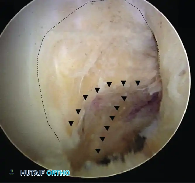
Two critical osseous landmarks must be meticulously identified during notch preparation. The first is the Lateral Intercondylar Ridge, historically referred to as "Resident's Ridge." This prominent ridge runs anterior to the ACL footprint when the knee is in extension, but it marks the superior boundary of the ACL origin when the knee is flexed at 90 degrees. It is a fundamental anatomical rule that no native ACL fibers attach superior (anterior in extension) to this ridge. Failure to identify the lateral intercondylar ridge is the most common cause of non-anatomical, excessively vertical graft placement.
The second critical landmark is the Lateral Bifurcate Ridge. This smaller, often less distinct osseous prominence runs perpendicular to the lateral intercondylar ridge and physically separates the AM bundle footprint from the PL bundle footprint. The AM bundle originates proximal and posterior to this ridge, while the PL bundle originates distal and anterior to it (relative to the axis of the femur). The surgeon must clear the overlying synovial tissue meticulously until these bony ridges and the posterior articular cartilage border are unequivocally visualized.
Tibial Insertion Site Morphology
The tibial footprint of the ACL is considerably larger and more robust than its femoral counterpart, presenting a distinct set of anatomical challenges and opportunities. The native tibial insertion spans a wide area between the medial and lateral tibial eminences, intimately associated with the anterior horn of the lateral meniscus. The AM bundle inserts anteromedially, adjacent to the medial tibial spine, while the PL bundle inserts posterolaterally, extending towards the posterior aspect of the anterior horn of the lateral meniscus.
Understanding the morphology of the tibial footprint is critical for preventing anterior graft impingement against the intercondylar roof during full extension. The center of the entire ACL footprint is typically located approximately 15 mm posterior to the anterior margin of the tibia, aligning with the posterior margin of the anterior horn of the lateral meniscus. When separating the footprint into its two constituent bundles, the center of the AM bundle is located more anteriorly and medially, while the center of the PL bundle is positioned more posteriorly and laterally.
During surgical preparation, preserving the tibial remnants is highly advantageous. These remnants not only serve as precise, patient-specific guides for anatomical tunnel placement but also contain mechanoreceptors that may facilitate postoperative proprioceptive recovery. The surgeon must carefully debride only the central, non-functional scar tissue while leaving the peripheral footprint architecture intact to guide the placement of the tibial aiming guides.
Biomechanical Contributions of the AM and PL Bundles
The biomechanical synergy between the AM and PL bundles is a marvel of evolutionary engineering, providing dynamic stability across the entire arc of motion. The AM bundle, owing to its more isometric nature, acts as the primary restraint to anterior tibial translation when the knee is flexed beyond 30 degrees. Its fibers are oriented to maximally resist the anterior shear forces generated by the quadriceps mechanism during deep flexion activities, such as squatting or landing from a jump.
Conversely, the PL bundle plays a paramount role as the knee approaches full extension. In this position, the PL bundle becomes maximally taut, providing the primary restraint not only to anterior tibial translation but also to internal rotation and valgus stress. This specific biomechanical profile makes the PL bundle the critical anatomical structure responsible for controlling the pivot-shift phenomenon. When the knee is flexed, the PL bundle relaxes and its femoral origin rotates horizontally, allowing the AM bundle to assume the primary stabilizing role.
Reconstructing both bundles independently allows the surgeon to replicate this complex, dynamic interplay. A single-bundle reconstruction, even when placed anatomically in the center of the footprint, often fails to provide the broad, flat, ribbon-like construct necessary to adequately tension the knee in both flexion and extension. The double-bundle construct, by utilizing two distinct grafts with separate tensioning vectors, effectively restores the native footprint's surface area and biomechanical function, thereby optimizing the knee's kinematic profile.
Exhaustive Indications and Contraindications
Patient Selection Criteria
The anatomical double-bundle reconstruction, while offering superior kinematic restoration, is a highly technically demanding procedure that necessitates meticulous patient selection. The ideal candidates for this advanced technique are high-demand, pivoting-sport athletes (e.g., soccer, basketball, rugby players) who require absolute rotational stability to perform at their peak. Furthermore, patients presenting with a high-grade (Grade III) pivot shift on preoperative clinical examination are prime candidates, as this indicates a profound deficiency in the PL bundle that a single-bundle reconstruction may fail to adequately address.
Another critical demographic for the double-bundle technique includes patients with generalized ligamentous laxity or hypermobility syndromes. These individuals inherently possess a higher baseline risk for graft failure and recurrent instability due to the inherent compliance of their secondary soft-tissue restraints. The robust, multi-planar stability provided by the double-bundle construct offers a vital mechanical advantage in this hyperlax population. Additionally, revision ACL reconstruction scenarios, where the primary single-bundle graft has failed due to unrecognized rotatory instability, often warrant a double-bundle approach, provided the anatomical constraints allow for it.
Anatomical Feasibility and Constraints
Despite the clinical advantages, the patient's native anatomy serves as the ultimate arbiter regarding the feasibility of a double-bundle reconstruction. Preoperative magnetic resonance imaging (MRI) and precise intraoperative measurements are absolutely critical. The intercondylar notch width is the first major constraint. The width of the notch entrance and its overall morphology (e.g., A-shaped vs. U-shaped) dictate whether two separate femoral tunnels can be safely drilled and two grafts accommodated without causing catastrophic graft impingement against the lateral wall or the PCL. Generally, a notch width no smaller than 12 mm is considered the absolute minimum required for a double-bundle procedure.
The size of the native insertion sites is the second critical constraint. Both the tibial and femoral footprints must be meticulously measured intraoperatively using an arthroscopic ruler. If the insertion site is smaller than 14 mm in its maximum diameter, accommodating two separate tunnels while maintaining an adequate, structurally sound bony bridge (minimum 2 mm) becomes highly challenging, if not impossible. Attempting a double-bundle technique in a diminutive footprint significantly increases the risk of tunnel convergence, which effectively converts the procedure into a massive single-bundle reconstruction and severely compromises graft fixation.
Absolute and Relative Contraindications
| Category | Condition | Rationale / Clinical Implication |
|---|---|---|
| Absolute Contraindication | Intercondylar Notch Width < 12 mm | High risk of severe graft impingement, restricted range of motion, and early graft attrition. |
| Absolute Contraindication | Femoral/Tibial Footprint < 14 mm | Inability to maintain a safe 2 mm bony bridge between tunnels; high risk of tunnel confluence and fixation failure. |
| Absolute Contraindication | Open Physes (Tanner Stage I/II) | Drilling four transphyseal tunnels poses an unacceptable risk of iatrogenic growth arrest and angular deformity. |
| Absolute Contraindication | Severe Osteoarthritis (Kellgren-Lawrence III/IV) | Altered joint kinematics and bone quality make the complex procedure unwarranted; osteotomy or arthroplasty may be indicated. |
| Relative Contraindication | Multiple Ligament Knee Injury (MLKI) | Prolonged surgical time and fluid extravasation risks; single-bundle often preferred to expedite complex reconstructions. |
| Relative Contraindication | Severe Bone Bruising / Osteopenia | Poor bone stock compromises the aperture fixation required for the dual tibial tunnels. |
Pre-Operative Planning, Templating, and Patient Positioning
Advanced Imaging and Templating
Preoperative planning for an anatomical double-bundle ACL reconstruction demands a more rigorous imaging protocol than standard single-bundle procedures. High-resolution, multi-planar Magnetic Resonance Imaging (MRI) is the cornerstone of this planning phase. The surgeon must carefully evaluate the oblique coronal and sagittal sequences to assess the integrity of the secondary stabilizers, the presence of meniscal pathology, and, crucially, the dimensions of the intercondylar notch and the native ACL footprints.
In cases of revision surgery or suspected anatomical anomalies, a three-dimensional computed tomography (3D CT) reconstruction is highly recommended. 3D CT provides unparalleled visualization of the osseous anatomy, allowing the surgeon to precisely map the lateral intercondylar ridge and the lateral bifurcate ridge preoperatively. Furthermore, it enables accurate measurement of the notch width and footprint dimensions, allowing the surgeon to definitively determine the feasibility of the double-bundle technique before the patient enters the operating room, thereby avoiding intraoperative surprises and forced alterations to the surgical plan.
Operating Room Setup and Patient Positioning
The patient is placed in the supine position on a standard operating table. The choice of leg stabilization is critical and depends on the surgeon's preference and the specific portal technique utilized. A lateral post positioned at the level of the mid-thigh, combined with a foot roll, is frequently utilized to allow for the application of a valgus stress, thereby opening the medial compartment for meniscal work. Crucially, the setup must permit the knee to be hyper-flexed to at least 120 degrees, which is an absolute requirement for drilling the femoral tunnels through the accessory anteromedial portal without risking posterior cortical blowout.
Alternatively, a dedicated leg holder can be employed. If a leg holder is used, it must be positioned proximally enough to avoid obstructing the creation of the accessory anteromedial portal and to allow for unobstructed hyperflexion. A well-padded tourniquet is applied to the proximal thigh but is typically inflated only if visualization becomes compromised by bleeding. Standard sterile draping is performed, ensuring that the contralateral leg is meticulously padded and protected from compression injuries throughout the duration of the procedure.
Examination Under Anesthesia
Prior to the initial incision, a comprehensive Examination Under Anesthesia (EUA) is mandatory. The complete relaxation afforded by anesthesia allows the surgeon to accurately quantify the degree of sagittal and rotatory instability without the confounding factor of patient guarding. The Lachman test is performed to assess anterior tibial translation, but the pivot-shift test is the paramount maneuver in this context.
The pivot-shift must be carefully graded (Grade I to III) to establish a baseline for postoperative comparison. A highly explosive, Grade III pivot-shift strongly reinforces the indication for a double-bundle reconstruction. Furthermore, the surgeon must meticulously evaluate the knee for concurrent collateral ligament laxity or posterolateral corner (PLC) deficiency. Failure to recognize and concomitantly address a PLC injury will inevitably lead to excessive forces being transmitted to the newly reconstructed ACL grafts, ultimately resulting in premature graft failure, regardless of how perfectly the double-bundle technique is executed.
Step-by-Step Surgical Approach and Fixation Technique
Portal Placement and Diagnostic Arthroscopy
The foundation of a successful anatomical double-bundle reconstruction is the establishment of a meticulous three-portal approach. This configuration provides a comprehensive, unobstructed view of the entire ACL footprint and facilitates the independent, precise drilling of the femoral tunnels. The standard Anterolateral (AL) portal is established first, adjacent to the lateral border of the patellar tendon, slightly superior to the joint line. This portal serves primarily as the viewing portal, providing a critical top-down perspective of the lateral wall of the intercondylar notch.
The Central Medial portal is then created under direct intra-articular visualization using a spinal needle. While viewing through the AL portal, the spinal needle is inserted directly through the center of the patellar tendon or just adjacent to its medial border, directed in a proximal-to-distal trajectory. This portal is essential for clearing the intercondylar notch, performing meniscal interventions, and managing the preparation of the tibial footprint.
The Accessory Anteromedial (AAM) portal is the critical working portal for drilling the femoral tunnels. It must be created precisely. Using a spinal needle for localization, the AAM portal is established superior to the medial joint meniscus, approximately 2 cm medial to the medial border of the patellar tendon. The surgeon must ensure that specialized instruments, such as flexible or rigid reamers, passed through this portal can comfortably reach the femoral footprint without causing iatrogenic scuffing or damage to the articular cartilage of the medial femoral condyle.
Notch Preparation and Footprint Identification
Once the portals are established and diagnostic arthroscopy is complete, attention turns to notch preparation. The surgeon must carefully debride the ruptured ACL using a motorized shaver and a radiofrequency ablation wand. It is a critical surgical principle to preserve the tibial and femoral remnants of the native ACL whenever possible. These remnants serve as invaluable, patient-specific anatomical landmarks that guide the precise placement of the tunnels.
Using the radiofrequency wand or a specialized arthroscopic awl, the surgeon marks the exact centers of the AM and PL bundles on both the tibia and the femur. On the femoral side, the lateral intercondylar ridge and the lateral bifurcate ridge must be clearly delineated. On the tibial side, the relationship to the anterior horn of the lateral meniscus and the medial tibial spine dictates the bundle centers. At this juncture, the surgeon must utilize an arthroscopic ruler to measure the footprints. If the anatomical constraints (footprint < 14 mm, notch < 12 mm) are violated, the surgeon must exercise clinical judgment, abort the double-bundle plan, and proceed with a robust single-bundle reconstruction.
Tunnel Drilling Sequence and Trajectory
The sequence of tunnel drilling is a critical logistical component of the procedure, designed to maintain optimal visibility and prevent complications related to fluid extravasation and bone debris. The sequence generally proceeds as follows:
- Femoral Posterolateral (PL) Tunnel: This tunnel is drilled first through the accessory anteromedial (AAM) portal. The knee is hyper-flexed to at least 110-120 degrees. The PL footprint is located distal and anterior to the bifurcate ridge. Placing the PL tunnel first is crucial because if the AM tunnel were drilled first, the resulting bleeding and bone debris would obscure the inferiorly located PL footprint.
- Tibial Anteromedial (AM) and Posterolateral (PL) Tunnels: Standard tibial aiming guides are introduced through the central medial portal. The guides are set at approximately 45 to 50 degrees. The AM and PL tunnels are drilled to intersect the exact center of their respective native insertion sites.
- Femoral Anteromedial (AM) Tunnel: Finally, the femoral AM tunnel is drilled. This can be executed through the AAM portal with the knee in hyperflexion. Alternatively, it can be drilled through the previously established tibial AM or PL tunnel, but only if this transtibial trajectory allows the reamer to reach the precise, native femoral AM insertion site without compromising the tunnel aperture or resulting in a vertical graft.
Surgical Pitfall: Tunnel Convergence. When determining the diameter of the reamers, the surgeon must aim to restore as much of the native insertion site as possible. However, it is an absolute biomechanical imperative to maintain an approximately 2-mm bony bridge between the AM and PL bundles on both the femur and the tibia. Failure to maintain this bridge will result in tunnel confluence, effectively converting the meticulously planned double-bundle procedure into a massive single-bundle reconstruction, severely compromising aperture fixation and graft incorporation.
Graft Preparation and Passage
While the tunnels are being drilled, the surgical assistant prepares the grafts on the back table. The graft sizes must be meticulously matched to their respective tunnel diameters. Typically, the AM bundle, which bears higher loads in flexion, is reconstructed with a larger diameter graft (e.g., 7-8 mm). The PL bundle is reconstructed with a slightly smaller graft (e.g., 5-6 mm), accurately reflecting the native anatomical proportions. Hamstring autografts (semitendinosus and gracilis) are the most common choice, but the quadriceps tendon is an excellent alternative, particularly when a large volume of robust tissue is required.
Once prepared, the grafts are passed into the joint. The PL graft is typically passed first to ensure it does not become entangled with the AM graft. Passing sutures are retrieved through the respective tibial tunnels, passed through the joint, and out the corresponding femoral tunnels. The grafts are then advanced into the femoral sockets under direct arthroscopic visualization, ensuring that they are fully seated and that no soft tissue interposition occurs.
Tensioning Protocol and Fixation Strategy
The fixation strategy and, more importantly, the tensioning angles are paramount to the success of the double-bundle construct. The goal is to replicate the distinct biomechanical roles of the two bundles.
- Femoral Fixation: Suspensory fixation (e.g., adjustable or fixed-loop cortical buttons) is highly recommended on the femoral side. Suspensory fixation avoids the disruption of the delicate intra-articular insertion site architecture, which can easily occur with aperture interference screw fixation in a crowded double-bundle setup.
- Tibial Fixation: Interference screw fixation is typically utilized on the cortical tibial side to provide rigid, aperture-level fixation. This minimizes the "bungee cord" effect and promotes rapid graft incorporation at the joint line.
- Tensioning Protocol: The bundles must be tensioned separately. The Anteromedial (AM) graft is tensioned and fixed while the knee is held in approximately 45 to 60 degrees of flexion, the position where the native AM bundle is most taut. Subsequently, the Posterolateral (PL) graft is tensioned and fixed with the knee in full extension (0 degrees), securing the joint against rotatory loads.
Complications, Incidence Rates, and Salvage Management
Intraoperative Complications and Tunnel Convergence
The technical complexity of the double-bundle procedure inherently carries a higher risk of intraoperative complications compared to single-bundle techniques. The most dreaded intraoperative complication is tunnel convergence or confluence. This occurs when the bony bridge separating the AM and PL tunnels fractures or is inadvertently reamed away. This complication effectively destroys the independent mechanical integrity of the two bundles, leading to a massive, unstable single tunnel. If this occurs, the surgeon must immediately pivot to a salvage strategy, typically involving the use of a large, single-bundle graft (such as a bone-patellar tendon-bone or a robust quadriceps tendon) fixed with a large interference screw or specialized hybrid fixation techniques.
Another significant intraoperative risk is posterior cortical blowout during femoral tunnel drilling. Because the AAM portal trajectory requires hyperflexion, the reamer is directed towards the posterior cortex of the lateral femoral condyle. If the starting point is too posterior or the angle is incorrect, the reamer can breach the thin posterior cortex, compromising suspensory fixation. Surgeons must meticulously measure the femoral depth and ensure a minimum of 5-7 mm of intact posterior cortex remains.
Postoperative Failures and Stiffness
Postoperative complications can broadly be categorized into biological failures and mechanical failures. Arthrofibrosis and postoperative stiffness are more common following double-bundle reconstructions due to the increased surgical trauma, the larger volume of graft tissue within the notch, and the potential for graft impingement if the tunnels were not perfectly placed. Meticulous notchplasty is occasionally required, but precise anatomical placement is the best prevention. If stiffness persists despite aggressive rehabilitation, arthroscopic lysis of adhesions and manipulation under anesthesia may be necessary.
Graft failure, while statistically lower in terms of rotatory instability compared to single-bundle constructs, can still occur due to traumatic re-injury or insidious stretching. The PL bundle is particularly susceptible to stretching if it is over-tensioned during fixation or if the patient is mobilized too aggressively in the early postoperative phase.
Complications and Salvage Strategies
| Complication | Estimated Incidence | Etiology / Risk Factors | Salvage / Management Strategy |
|---|---|---|---|
| Tunnel Convergence | 3% - 8% | Footprint < 14mm, oversized reamers, failure to maintain 2mm bridge. | Convert to single-bundle reconstruction; utilize large interference screw or backup cortical fixation. |
| Posterior Cortical Blowout | 1% - 4% | Incorrect AAM portal trajectory, inadequate hyperflexion during drilling. | Abort suspensory button; use aperture interference screw or over-the-top routing technique. |
| Graft Impingement | 2% - 5% | Notch < 12mm, anteriorly placed tibial tunnels, inadequate notchplasty. | Arthroscopic debridement, targeted notchplasty, aggressive ROM physiotherapy. |
| Arthrofibrosis / Stiffness | 4% - 10% | Prolonged surgical time, excessive tissue volume in notch, delayed rehab. | Aggressive physical therapy; arthroscopic lysis of adhesions if no progress by 12 weeks. |
| Iatrogenic Chondral Injury | 1% - 3% | Instrument crowding, scuffing medial condyle via AAM portal. | Careful portal placement; use of flexible reamers; chondroplasty for minor scuffs. |
Phased Post-Operative Rehabilitation Protocols
Immediate Postoperative Phase and Tissue Protection
Postoperative rehabilitation following an anatomical double-bundle ACL reconstruction must follow a highly structured, phased approach. The primary objective in the immediate postoperative phase (Phase 1: 0-2 weeks) is to protect the healing grafts while mitigating the inflammatory response. Because the PL bundle is tensioned and fixed in full extension, achieving and maintaining full, passive terminal extension immediately after surgery is absolutely critical. Failure to achieve full extension early can lead to permanent flexion contractures and PL bundle dysfunction.
During this initial phase, the focus is on reducing effusion through cryotherapy and elevation. Quadriceps activation is initiated immediately using isometric sets and straight leg raises. We generally proceed more cautiously with hamstring strengthening when a hamstring autograft has been utilized, due to the initial profound weakness of the flexor mechanism and the biological time required for soft-tissue-to-bone healing within the tunnels. Weight-bearing status is typically dictated by concurrent meniscal or chondral procedures, but isolated double-bundle reconstructions can often bear weight as tolerated with crutches and a brace locked in extension.
Intermediate Phase and Neuromuscular Control
As the patient progresses into Phase 2 (2-6 weeks), the emphasis shifts towards the progressive restoration of full knee flexion and the initiation of closed kinetic chain exercises. The grafts are undergoing the vulnerable phase of ligamentization, where the tissue is weakest. Therefore, open kinetic chain quadriceps exercises, particularly those applying high shear forces to the tibia (e.g., leg extensions from 0 to 30 degrees), are strictly avoided. Closed kinetic chain exercises, such as mini-squats and leg presses, are preferred as they promote co-contraction of the hamstrings and quadriceps, thereby protecting the grafts.
Phase 3 (6-12 weeks) marks the introduction of advanced proprioceptive training and the gradual normalization of gait mechanics. Balance boards, single-leg stance exercises, and perturbation training are incorporated to restore the neuromuscular control that is vital for dynamic joint stability. Light jogging may be introduced towards the end of this phase, but strictly contingent upon the patient meeting specific strength criteria (e.g., quadriceps strength reaching at least 70% of the contralateral limb) and demonstrating an absence of effusion.
Return to Sport and Advanced Functional Testing
The final phase of rehabilitation, Phase 4 (3-9 months), is dedicated to sport-specific agility training, plyometrics, and cutting maneuvers. The double-bundle construct, with its superior rotational stability, theoretically allows for a more confident progression through these high-demand activities. However, the biological timeline of graft maturation cannot be accelerated.
The patient is generally not permitted to return to full, unrestricted athletic activity until approximately 9 to 12 months postoperatively. This return is not dictated merely by time, but by the patient passing rigorous, objective functional testing. Criteria for return to sport include symmetrical quadriceps and hamstring strength (isokinetic testing demonstrating >90% symmetry), excellent performance on hop testing batteries (e.g., single hop, triple hop, crossover hop), and a psychological readiness to return. Premature return to sport, prior to the complete neuromuscular and biological integration of the grafts, significantly increases the risk of catastrophic graft rupture.
Summary of Landmark Literature and Clinical Guidelines
Biomechanical Evidence
The biomechanical superiority of the anatomical double-bundle reconstruction is well-documented in the seminal literature. In vitro robotic studies by pioneers such as Freddie Fu, Kuroda, and Yasuda have consistently demonstrated that while single-bundle reconstructions effectively restore anterior-posterior translation to intact knee levels, they frequently fail to control the combined internal rotation and valgus torques that simulate the pivot-shift mechanism. In contrast, the double-bundle construct, by explicitly recreating the PL bundle, significantly reduces this rotatory laxity, bringing the knee's kinematic profile much closer to that of the native, uninjured state.
These biomechanical investigations have also highlighted the critical importance of anatomical tunnel placement. Studies have shown that non-anatomical double-bundle reconstructions (e.g., placing the tunnels outside the native footprints) yield kinematics that are inferior even to an anatomically placed single bundle. Therefore, the literature underscores that the "anatomical" aspect of the procedure is just as critical, if not more so, than the "double-bundle" aspect.
Long-Term Clinical Outcomes
While the biomechanical data overwhelmingly favors the double-bundle technique, the clinical literature presents a more
