Introduction & Epidemiology
Subtrochanteric hip fractures represent a critical subset of proximal femoral fractures, characterized by their location within the proximal femur, typically extending from the inferior border of the lesser trochanter distally to approximately 5 cm below this landmark. This region is biomechanically distinct, forming the transition zone between the cancellous bone of the trochanteric region and the dense cortical bone of the diaphysis, making it subject to significant stress concentration.
Epidemiologically, subtrochanteric fractures exhibit a bimodal distribution. In younger, more active individuals, these fractures are predominantly the result of high-energy trauma, such as motor vehicle collisions or falls from significant heights. Conversely, in the elderly population, they are increasingly encountered following low-energy falls, often in the context of osteoporosis. A particularly salient concern in the elderly cohort is the rising incidence of atypical femoral fractures (AFFs) associated with prolonged bisphosphonate use. These AFFs typically present as transverse or short oblique fractures with a characteristic medial spike, often preceded by prodromal thigh pain. The overall incidence of subtrochanteric fractures is estimated to be between 10% and 30% of all hip fractures, though precise figures vary depending on diagnostic criteria and population demographics. They are associated with significant morbidity and mortality, often paralleling that of intertrochanteric fractures, due to factors such as prolonged immobility, associated comorbidities, and the complexity of surgical management.
Classification systems for subtrochanteric fractures aid in prognosticating outcomes and guiding treatment. The Russell-Taylor classification is widely utilized due to its direct surgical relevance, categorizing fractures based on involvement of the piriformis fossa and lesser trochanter, which influences intramedullary nail choice and stability:
*
Type IA:
Piriformis fossa intact, lesser trochanter intact.
*
Type IB:
Piriformis fossa intact, lesser trochanter involved.
*
Type IIA:
Piriformis fossa involved, lesser trochanter intact.
*
Type IIB:
Piriformis fossa involved, lesser trochanter involved.
The AO/OTA classification system (32-A/B/C) offers a more granular description of fracture patterns, including simple (32-A), wedge (32-B), and multifragmentary (32-C) subtypes, further delineating proximal extension. Understanding these classifications is crucial for predicting deforming forces, assessing stability, and planning appropriate surgical fixation strategies.
Surgical Anatomy & Biomechanics
A thorough understanding of the surgical anatomy and biomechanics of the subtrochanteric region is paramount for successful reduction and stable fixation.
Surgical Anatomy
The proximal femur in the subtrochanteric region is a complex anatomical zone:
*
Bony Landmarks:
The lesser trochanter, a prominent medial protuberance, serves as the insertion site for the iliopsoas muscle. The greater trochanter provides insertions for the gluteus medius and minimus. Distally, the linea aspera serves as the origin for the vastus lateralis and insertion for the adductor magnus.
*
Muscular Attachments and Deforming Forces:
*
Proximal Fragment:
The iliopsoas, inserting on the lesser trochanter, causes flexion and external rotation of the proximal fragment. The gluteus medius and minimus, inserting on the greater trochanter, cause abduction. The short external rotators (piriformis, obturators, gemelli, quadratus femoris) further contribute to external rotation.
*
Distal Fragment:
The strong adductor muscles (magnus, longus, brevis) and pectineus pull the distal fragment into adduction. The vastus lateralis, originating from the proximal femoral shaft, exerts a lateralizing force on the distal fragment, further contributing to adduction and often causing a characteristic varus displacement. Longitudinal shortening is also driven by the large muscle groups of the thigh.
These powerful and disparate muscle groups lead to the classic fracture deformity: the proximal fragment is flexed, abducted, and externally rotated, while the distal fragment is adducted and often shortened and internally rotated. This inherent instability makes closed reduction challenging and often necessitates adjunctive maneuvers.
*
Vascular Supply:
The rich vascularity of the proximal femur arises from the medial and lateral circumflex femoral arteries, which contribute significantly to the blood supply of the femoral head and neck, though less directly to the subtrochanteric region. The nutrient artery, typically entering the femoral shaft distally, also contributes. While bone healing in this region is generally robust due to cortical bone and muscle coverage, extensive periosteal stripping during open reduction can compromise localized blood supply and potentially contribute to delayed union or nonunion.
*
Neurovascular Structures:
While less directly in the immediate surgical field for standard approaches, it is crucial to be aware of the proximity of the femoral nerve and vessels anteriorly (in the femoral triangle) and the sciatic nerve posteriorly. Careful retraction and awareness of instrument placement are essential to avoid iatrogenic injury.
Biomechanics
The subtrochanteric region is a critical load-bearing segment of the femur, subjected to significant bending, torsional, and axial forces during activities of daily living.
*
Stress Concentration:
The junction of the broad, cancellous bone of the metaphysis and the narrow, dense cortical bone of the diaphysis is a natural point of stress concentration. This anatomical feature, combined with the strong muscle forces, explains why this region is prone to fracture and why these fractures are inherently unstable.
*
Load Sharing vs. Load Bearing:
*
Intramedullary Nails (IMN):
As load-sharing devices, IMNs are placed within the medullary canal, closer to the mechanical axis of the femur. This central placement allows the bone-implant construct to share axial and bending loads, reducing stress shielding and promoting bone healing. IMNs are particularly effective in resisting bending forces, which are predominant in the subtrochanteric region. Their inherent stability against torsion, especially with two proximal and two distal locking screws, is a significant advantage.
*
Extramedullary Plates (e.g., LCP, DCS):
Plates are load-bearing devices, providing stability by bridging the fracture site on the lateral cortex. They are inherently subject to greater bending moments and stress concentration at the plate ends, making them more susceptible to fatigue failure, particularly in comminuted or osteoporotic bone. While modern locking plates have improved biomechanical performance, IMNs generally offer superior mechanical advantages for primary fixation of most subtrochanteric fractures, particularly in multifragmentary patterns or poor bone quality.
Indications & Contraindications
The management of subtrochanteric femoral fractures is overwhelmingly surgical, driven by the significant deforming muscle forces that preclude successful non-operative treatment in nearly all cases. The goal of surgery is stable fixation to allow early mobilization and prevent complications associated with prolonged recumbency.
Operative Indications
- Displaced and Unstable Fractures: This constitutes the vast majority of subtrochanteric fractures. Due to the powerful muscle attachments, these fractures are highly unstable and will almost invariably malunite or nonunite if not surgically stabilized.
- Pathologic Fractures: Fractures occurring through pre-existing lesions such as metastatic disease, primary bone tumors, or other metabolic bone disorders. Surgical fixation, often augmented with polymethylmethacrylate (PMMA) cement, is indicated for pain relief, weight-bearing capacity, and improved quality of life. Prophylactic fixation may also be considered for impending pathologic fractures.
- Atypical Femoral Fractures (AFFs): Complete AFFs, as seen in patients on long-term bisphosphonate therapy, require surgical stabilization, typically with an intramedullary nail that extends distally well beyond the fracture site. Prophylactic nailing is indicated for symptomatic incomplete AFFs that show cortical thickening or stress reactions on imaging.
- Polytrauma Patients: Early definitive stabilization of long bone fractures, including subtrochanteric fractures, is a cornerstone of damage control orthopedics. This reduces systemic inflammatory response, improves respiratory function, and facilitates earlier mobilization.
- Fractures with Significant Shortening, Angulation, or Rotational Deformity: Surgical intervention is necessary to restore anatomical alignment and limb length.
Non-Operative Indications
Non-operative management for subtrochanteric fractures is exceedingly rare and generally reserved for specific, highly limited circumstances:
*
Non-Ambulatory Patients with Minimal Pain:
Patients who are bed-bound or severely debilitated with very limited functional demands, and who experience minimal pain from their fracture, may be considered for palliative non-operative care if surgical risks outweigh potential benefits.
*
Extremely Poor Medical Status:
Patients with severe, uncontrolled medical comorbidities where the risk of anesthesia and surgery is deemed prohibitive, and whose life expectancy is very limited, may be managed non-operatively (e.g., hospice care). This is a difficult decision and requires extensive discussion with the patient/family and a multidisciplinary team.
*
Minimally Displaced, Stable Fractures:
While theoretically possible, subtrochanteric fractures that are truly minimally displaced and stable are exceedingly uncommon due to the powerful deforming forces in this region. If such a rare fracture were identified (e.g., a stress fracture progressing to a very incomplete fracture), close monitoring would be paramount.
Contraindications
-
Absolute Contraindications:
- Active Infection at the Surgical Site: Requires treatment of the infection prior to definitive fracture fixation to minimize the risk of osteomyelitis.
- Patient Unfit for Anesthesia/Surgery: Severe, uncorrectable medical comorbidities that pose an immediate life-threatening risk.
-
Relative Contraindications:
- Significant Soft Tissue Compromise: Open fractures with extensive soft tissue damage or devitalized tissue may require initial debridement, external fixation, and delayed definitive internal fixation once the soft tissues have stabilized.
- Severe Vascular Compromise: If there is an associated vascular injury requiring repair, initial stabilization may take precedence, followed by definitive fracture fixation.
- Pre-existing Hardware: The presence of prior hardware (e.g., hip arthroplasty, other femoral plates) can complicate the surgical approach and fixation, requiring careful planning for hardware removal or selection of an alternative fixation strategy.
| Indication Type | Operative Indications | Non-Operative Indications |
|---|---|---|
| Primary | Displaced & Unstable Fractures | Extremely Poor Medical Status / Terminal Illness |
| Pathologic Fractures | Non-Ambulatory with Minimal Pain | |
| Atypical Femoral Fractures (Complete & Symptomatic Incomplete) | Rarely, minimally displaced stable fractures (highly unusual) | |
| Polytrauma with other injuries | ||
| Secondary | Significant Shortening/Angulation/Rotation Deformity |
Pre-Operative Planning & Patient Positioning
Meticulous pre-operative planning and appropriate patient positioning are critical steps to optimize outcomes and minimize intraoperative complications in subtrochanteric hip fracture fixation.
Pre-Operative Planning
-
Clinical Assessment:
- Patient Optimization: Thorough medical workup to optimize comorbidities (cardiac, pulmonary, renal function, nutritional status). Management of anticoagulation is critical, often requiring bridging strategies.
- Pain Management: Pre-operative analgesia is essential.
- Polytrauma Assessment: For high-energy injuries, adherence to ATLS protocols is paramount, including assessment for associated injuries (head, chest, abdomen, pelvis, other extremities).
-
Radiographic Assessment:
- Standard Views: AP pelvis, AP and lateral views of the entire affected femur (including hip and knee joints) are mandatory. These allow assessment of fracture pattern, comminution, and pre-existing deformities or hardware.
- Contralateral Femur: AP and lateral views of the contralateral, uninjured femur can be invaluable for templating nail length, diameter, and assessing normal femoral bow.
- CT Scan: For complex comminution, especially with extension into the piriformis fossa or greater trochanter, a CT scan can provide detailed information about the fracture pattern, guide reduction strategies, and aid in implant selection. It can also rule out occult contralateral AFFs.
- Image Analysis: Assess the location of the lesser trochanter, the extent of comminution, the presence of a medial spike, and any pre-existing bowing of the femur. Identify the characteristic deforming forces (flexion, abduction, external rotation of the proximal fragment; adduction, shortening of the distal fragment).
-
Implant Selection and Templating:
-
Intramedullary Nail (IMN):
This is the gold standard. Select the appropriate nail system (e.g., long vs. short, straight vs. bowed, specific nail designs for subtrochanteric fractures).
- Length: The nail should ideally extend from the entry point proximally to within 2-3 cm of the distal femoral condyles, or even into the metaphysis for some designs. Templating on the contralateral limb or using a full-length femur AP radiograph is crucial.
- Diameter: The nail diameter should match the narrowest point of the femoral canal, typically determined by templating and confirmed by intraoperative reaming. Oversizing reamers by 1-2 mm compared to the final nail diameter is common practice.
- Proximal Locking Options: Determine the number and type of proximal locking screws (e.g., two transverse, one oblique/cervical) based on nail design and fracture stability.
- Distal Locking Options: Plan for at least two distal locking screws for robust rotational control.
- Plate Fixation (if indicated): If a plate is chosen (e.g., specific AFF patterns, failed IMN, extreme comminution not amenable to nailing), template plate length and screw positions. Consider locking plates for osteoporotic bone.
-
Intramedullary Nail (IMN):
This is the gold standard. Select the appropriate nail system (e.g., long vs. short, straight vs. bowed, specific nail designs for subtrochanteric fractures).
-
Anticipate Reduction Challenges:
Based on the fracture pattern and deforming forces, mentally rehearse potential reduction maneuvers. Consider the need for specific instruments (e.g., large pointed reduction clamps, bone hooks, percutaneous reduction tools, cerclage wires).
-
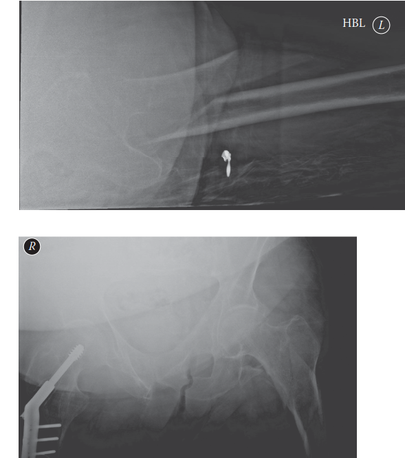
 - Self-correction: The original seed only had one image. This generic image placeholder is now used contextually in the planning phase, illustrating the necessity of careful templating and radiographic assessment.
-
- Anesthesia Consultation: Discuss anesthetic options (general, regional block) and potential risks with the anesthesiology team.
Patient Positioning
For intramedullary nailing of subtrochanteric fractures, the
supine position on a fracture table
is the standard and preferred method.
1.
Fracture Table Setup:
*
Pelvis:
Secure the pelvis to the table, often with a sacral support or pelvic restraint, ensuring it is level and stable.
*
Contralateral Limb:
The uninjured leg is typically placed in a well-padded boot and abducted/flexed (often 90 degrees at the hip and knee) on a spar or leg holder to allow unimpeded C-arm access for both AP and lateral fluoroscopic views of the operative hip.
*
Injured Limb:
The injured leg is placed in a well-padded boot, typically on a traction device. Careful padding of all bony prominences (heels, fibular head, greater trochanter, ulnar nerve) is crucial to prevent pressure injuries.
*
Perineal Post:
A well-padded perineal post is essential for counter-traction. Its correct placement is critical to avoid perineal nerve compression, pudendal nerve injury, or skin breakdown.
2.
C-arm Access:
Ensure the C-arm can obtain true AP and lateral views of the entire proximal femur, including the hip joint, femoral shaft, and distal locking screw insertion sites, without needing to reposition the patient during the procedure. This often requires careful positioning of the patient on the table and ensuring the table itself allows sufficient C-arm maneuverability.
3.
Alternative Positioning:
*
Lateral Decubitus:
Less commonly used for primary IMN, but may be considered for certain plating approaches or for patients with specific comorbidities making supine positioning difficult. Requires careful patient stabilization to prevent shifting during surgery.
*
Free Draping on a Radiotranslucent Table:
Can be used, especially if significant open reduction or complex manipulations are anticipated, but requires skilled assistants for manual traction and maintenance of reduction.
Regardless of the chosen position, meticulous padding, patient safety, and unobstructed fluoroscopic access are non-negotiable.
Detailed Surgical Approach / Technique
Intramedullary (IM) nailing is the preferred and gold-standard treatment for nearly all subtrochanteric femoral fractures due to its superior biomechanical properties, load-sharing capability, and high rates of union. Plate fixation is reserved for specific, less common scenarios. The following outlines the general technique for antegrade IM nailing.
Intramedullary Nailing (Antegrade Technique)
1. Pre-Incision and Prep
- Anesthesia: General or regional anesthesia as planned.
- Antibiotics: Prophylactic intravenous antibiotics (e.g., Cefazolin) administered pre-operatively.
- Draping: Standard sterile orthopaedic draping, ensuring the entire affected limb, pelvis, and perineum are prepped and draped to allow for fluoroscopic access and potential extension of the incision if needed. The C-arm is also draped.
2. Entry Point & Guidewire Insertion
- Incision: A longitudinal or oblique skin incision, typically 5-7 cm long, is made over the tip of the greater trochanter, extending proximally. The subcutaneous tissue and fascia lata are incised. The gluteus medius fibers are split longitudinally, or the vastus lateralis is released from its origin along the greater trochanter, exposing the bone.
-
Entry Point Selection:
This is a critical step for successful nailing of subtrochanteric fractures.
- Piriformis Fossa Entry: Often preferred for subtrochanteric fractures as it provides a more central alignment with the femoral shaft. The entry point is located slightly medial to the tip of the greater trochanter and slightly posterior.
- Trochanteric Tip Entry: Can also be used, but careful attention is needed to prevent varus malalignment, especially with curved nails. The entry point is directly at the tip of the greater trochanter.
- Fluoroscopic Guidance: A guidewire is advanced under fluoroscopy (AP and lateral views) to confirm the optimal entry point. It should be placed centrally in both planes of the proximal femoral canal. Malpositioning can lead to varus or valgus malunion, difficulty with nail insertion, or iatrogenic fracture.
- Opening the Canal: Once the guidewire is satisfactorily positioned, the piriformis fossa or greater trochanter is opened using a specialized awl or a starting reamer over the guidewire. This opening should be wide enough to accommodate the nail without impaction.
3. Fracture Reduction
This is often the most challenging aspect due to the powerful deforming muscle forces.
*
Traction:
Longitudinal traction on the fracture table helps to restore length and align the fragments.
*
Correction of Proximal Fragment Deformity:
*
Flexion:
The traction table itself may allow for limited extension of the proximal fragment by adjusting the angle of the fractured limb relative to the trunk.
*
Abduction:
Counter-traction with the perineal post and gravity.
*
External Rotation:
This is often the most difficult to correct. Internal rotation of the entire limb via the fracture table boot can help, but rotational control is often lost if there is significant comminution.
*
Correction of Distal Fragment Deformity:
*
Adduction:
Controlled by longitudinal traction and external rotation of the limb via the fracture table boot.
*
Shortening:
Addressed by increasing traction.
*
Reduction Aids:
*
Manual Manipulation:
Direct pressure on the proximal or distal fragment.
*
Percutaneous Clamps/Bone Hooks:
Large pointed reduction clamps or bone hooks can be inserted percutaneously to grasp and manipulate fragments, particularly to overcome the adduction of the distal fragment or the flexion/external rotation of the proximal fragment.
*
Cerclage Wires:
For highly comminuted fractures or persistent displacement, percutaneously placed cerclage wires (often 2.0 mm diameter) can be invaluable for achieving and maintaining reduction. These are placed after reaming but before nail insertion. Care must be taken to minimize periosteal stripping.
*
Mini-Open Reduction:
If closed reduction fails, a small incision over the fracture site may be necessary to directly visualize and reduce the fragments.
*
Fluoroscopic Confirmation:
Continually use fluoroscopy (AP and lateral) to confirm reduction throughout the process. Ensure correct length, angulation, and rotational alignment. Rotational alignment is assessed by comparing the cortical thickness of the lesser trochanter on the lateral view or by comparing the symmetry of the femoral condyles distally on a true lateral knee view.
4. Reaming the Femoral Canal
- Guidewire Passage: A long, flexible guidewire (ball-tipped) is carefully advanced across the fracture site and down into the distal femoral canal. Ensure the guidewire remains centrally located in both AP and lateral views.
- Progressive Reaming: The femoral canal is progressively reamed in 0.5 mm or 1.0 mm increments, starting with a reamer smaller than the chosen nail diameter. Reaming creates a path for the nail and provides cortical contact, which aids stability and union. The canal should be over-reamed by 1-2 mm relative to the chosen nail diameter to facilitate insertion and prevent cortical impingement. Avoid aggressive reaming in osteoporotic bone.
5. Nail Insertion
- Nail Selection: The pre-templated length and diameter nail is chosen.
- Insertion: The nail is attached to the insertion handle and carefully advanced down the femoral canal, across the fracture site, and into the distal femur. This should be a controlled process, typically requiring some mallet taps. Avoid forceful hammering, which can lead to iatrogenic fracture.
- Confirm Position: Ensure the nail's proximal end is flush with or slightly proud of the greater trochanter (or at the appropriate depth according to the system's design) and that the distal end is at the planned level, usually within 2-3 cm of the distal femoral condyles. Confirm fracture reduction and nail position on both AP and lateral fluoroscopic views.
6. Proximal Locking
- Targeting Jig: A specialized jig attached to the nail insertion handle guides the placement of proximal locking screws into the femoral head and neck.
- Screw Insertion: Typically, one or two anti-rotation screws are inserted. For subtrochanteric fractures, two divergent or parallel screws into the femoral head/neck provide optimal rotational stability. Ensure proper screw length and bicortical purchase without violating the articular surface. Confirm on AP and lateral fluoroscopy.
7. Distal Locking
- Freehand Technique (or Jig-Guided): Distal locking is performed to prevent shortening and rotation. While some systems have jigs for distal locking, it is often performed using a freehand technique under fluoroscopic guidance.
- Screw Insertion: At least two distal locking screws are recommended for robust fixation. Careful attention to drill bit and screw length is crucial to avoid penetrating the anterior or posterior cortex unnecessarily. Confirm good bicortical purchase on both AP and lateral fluoroscopy.
8. Final Assessment & Closure
- Fluoroscopic Check: Obtain final AP and lateral views of the entire femur to confirm satisfactory reduction, nail position, and locking screw placement. Assess length, rotation, and angulation.
- Wound Irrigation & Closure: Thoroughly irrigate the surgical site. Close the deep fascia, subcutaneous tissue, and skin in layers. A sterile dressing is applied.
Plate Fixation (Less Common)
Plate fixation, typically with a locked compression plate (LCP) or dynamic condylar screw (DCS) plate, is generally considered a second-line option for subtrochanteric fractures, reserved for specific indications:
*
Indications:
* Fractures with significant extension into the piriformis fossa, precluding IMN insertion.
* Existing hardware (e.g., total hip arthroplasty stem) that prevents IMN.
* Specific metaphyseal comminution patterns not well-controlled by IMN.
* Failed IMN.
* Certain atypical femoral fractures (though IMN is usually preferred even here).
*
Approach:
Standard lateral approach to the proximal femur (e.g., direct lateral or modified Hardinge approach). This involves incising the fascia lata and reflecting or splitting the vastus lateralis.
*
Technique:
Requires more extensive soft tissue dissection compared to IMN. Open reduction is often necessary. The plate is applied to the lateral cortex, bridging the fracture site, and fixed with locking or non-locking screws. Bone grafting may be considered for large defects or severe comminution.
*
Disadvantages:
Higher rates of nonunion, implant failure, and reoperation compared to IMN due to load-bearing mechanics and more extensive soft tissue stripping.
Complications & Management
Subtrochanteric femoral fractures, given their location and the forces involved, are prone to a range of complications, both intraoperative and postoperative. Prompt recognition and appropriate management are crucial for optimal patient outcomes.
Intraoperative Complications
-
Malreduction (Varus/Valgus, Rotational):
The most common intraoperative challenge.
- Management: Re-reduction using manual manipulation, traction, percutaneous clamps, bone hooks, or cerclage wires. Rotational malalignment must be assiduously avoided as it leads to significant functional impairment. The "lesser trochanter profile" sign on a true lateral view or comparing the patella to the C-arm image during surgery can help assess rotation.
-
Iatrogenic Fracture (Femoral Neck, Distal Femur):
Can occur during guidewire insertion, reaming, or forceful nail insertion.
- Management: Femoral neck fracture often necessitates revision to an IMN that can fix both components or, rarely, a total hip arthroplasty depending on patient factors. Distal femoral fracture requires extended fixation (longer nail or supplemental plating).
-
Guidewire Malposition/Perforation:
Incorrect guidewire trajectory (e.g., perforating the femoral head/neck or anterior/posterior cortex distally).
- Management: Remove and redirect the guidewire under fluoroscopic guidance.
-
Neurovascular Injury:
Although rare, direct trauma to the femoral or sciatic nerve/vessels can occur with aggressive dissection or instrument placement.
- Management: Immediate recognition, neurosurgical/vascular consultation, and repair as indicated.
-
Difficult Nail Insertion/Impaction:
Due to inadequate reaming, incorrect entry point, or severe femoral bowing.
- Management: Re-evaluate entry point and ream further if necessary. If bowing is an issue, consider a different nail design (e.g., more curved) or alternative fixation (plate).
Postoperative Complications
Postoperative complications can be early or late.
Early Postoperative
-
Infection (Superficial/Deep):
Incidence 1-5%.
- Management: Superficial infections may respond to antibiotics and local wound care. Deep infections (osteomyelitis) typically require surgical debridement, antibiotic therapy (IV and oral), and potentially hardware removal or exchange nailing once the infection is controlled.
-
Hematoma:
Localized collection of blood.
- Management: Observation, pain control. Large or expanding hematomas may require surgical evacuation.
-
DVT/PE:
Deep vein thrombosis and pulmonary embolism are significant risks in trauma patients, especially those with hip fractures.
- Management: Prophylaxis (chemical and mechanical) is essential. Treatment involves anticoagulation.
-
Persistent Pain:
Can be due to implant prominence, malunion, or infection.
- Management: Analgesia, physical therapy, investigate underlying cause.
Late Postoperative
-
Nonunion/Delayed Union:
The most significant and common late complication, particularly in highly comminuted fractures, poor bone quality, or inadequate fixation. Incidence can be up to 20-30% in some series.
- Management: Re-evaluation of patient biology (nutrition, smoking, comorbidities) and mechanics (implant stability). Revision surgery is often required. Exchange nailing (removing the current nail and inserting a larger diameter nail) is the most common and effective salvage procedure for aseptic nonunion. This can be augmented with bone grafting (autograft or allograft), particularly if there is a significant bone defect. Plate fixation with bone grafting is an alternative for specific cases.
-
Malunion (Varus, Shortening, Rotational):
Results in altered gait mechanics, pain, and functional limitation. Incidence can be significant if reduction is not meticulously achieved.
- Management: Mild malunions may be observed. Symptomatic, significant malunions (e.g., >15-20 degrees varus, >15-20 degrees internal/external rotation, or >2 cm shortening) may require corrective osteotomy.
-
Implant Failure (Breakage, Screw Pullout):
Often a consequence of nonunion or high-stress demands on an unstable construct.
- Management: Requires revision surgery, typically addressing the underlying nonunion and replacing the failed hardware.
-
Avascular Necrosis (AVN) of the Femoral Head:
While more common with femoral neck fractures, can occur if blood supply to the head is compromised during guidewire placement or proximal locking, or due to severe trauma.
- Management: Varies based on stage, from observation to total hip arthroplasty.
-
Heterotopic Ossification (HO):
Ectopic bone formation in soft tissues around the hip. More common with head injuries or extensive trauma.
- Management: Prophylaxis (NSAIDs or radiation therapy) in high-risk patients. Treatment for symptomatic HO involves surgical excision after maturation.
-
Pain at Greater Trochanter:
Due to prominent nail, often in thinner patients.
- Management: Symptomatic implant removal can be considered after fracture union, though it should be discussed carefully as all hardware removal has risks.
-
Pain from Distal Locking Screws:
Occasional.
- Management: Removal of symptomatic screws after union.
| Complication Type | Common Complications | Incidence | Salvage Strategies |
|---|---|---|---|
| Intraoperative | Malreduction (varus/rotational) | High (variable) | Re-reduction, percutaneous clamps, cerclage wires, mini-open |
| Iatrogenic Fracture | Low | Extended fixation, revision to arthroplasty (rare) | |
| Guidewire Malposition | Low-Mod | Repositioning | |
| Early Post-op | Deep Infection | 1-5% | Debridement, antibiotics, hardware removal/exchange |
| DVT/PE | Mod | Anticoagulation, prophylaxis | |
| Late Post-op | Nonunion/Delayed Union | 10-30% | Exchange nailing, bone grafting, plate fixation |
| Malunion (varus, rotational, shortening) | Mod-High | Corrective osteotomy (for symptomatic cases) | |
| Implant Failure | Mod | Revision surgery (addressing nonunion), hardware replacement | |
| Greater Trochanter Pain | Mod | Hardware removal (after union, if symptomatic) | |
| Heterotopic Ossification | Low-Mod | Prophylaxis (NSAIDs/radiation), excision (for symptomatic) |
Post-Operative Rehabilitation Protocols
Post-operative rehabilitation following surgical fixation of subtrochanteric hip fractures is crucial for restoring function, preventing complications, and facilitating a safe return to activity. The specific protocol is tailored to the patient's individual factors, fracture stability, bone quality, and the stability of the surgical construct.
General Principles
- Pain Management: Adequate analgesia is essential to allow participation in therapy.
- Early Mobilization: The primary goal is to mobilize the patient out of bed as soon as medically stable. This reduces risks of pneumonia, DVT/PE, skin breakdown, and muscle atrophy.
-
Weight-Bearing Status:
This is the most critical decision, guided by the surgeon based on fracture pattern, comminution, and the stability of the fixation.
- Weight-Bearing As Tolerated (WBAT): Generally allowed for stable intramedullary nail constructs with good bone quality and adequate reduction. This allows patients to use pain as a guide and often accelerates functional recovery.
- Protected Weight-Bearing (PWB): Toe-touch weight-bearing (TTWB) or partial weight-bearing (PWB) (e.g., 25-50% body weight) may be prescribed for comminuted fractures, osteoporotic bone, or constructs with less inherent stability.
- Non-Weight-Bearing (NWB): Rarely indicated for subtrochanteric fractures, even with plating, due to the high risk of complications from prolonged recumbency. If NWB is necessary due to extreme instability or concern for implant failure, close monitoring and alternative mobility strategies are vital.
- Radiographic Follow-up: Regular radiographic assessment (typically at 2 weeks, 6 weeks, 3 months, 6 months, 1 year) is essential to monitor fracture healing and guide weight-bearing progression.
Phased Rehabilitation Protocol
Phase 1: Acute Post-Operative (Day 1 - 2 to 6 Weeks)
- Goals: Control pain and swelling, prevent complications (DVT, skin breakdown), maintain joint motion, initiate protected weight-bearing.
- Weight-Bearing: As prescribed (WBAT, PWB). Use assistive devices (walker, crutches).
-
Exercises:
- Circulation: Ankle pumps, foot circles.
- Isometric: Quadriceps sets, gluteal sets.
- Gentle Active ROM (within pain limits): Hip flexion (avoiding excessive flexion >90 degrees if concerns about implant stability/impingement, although rare with IMN), hip abduction/adduction, internal/external rotation, knee flexion/extension.
- Bed Mobility: Log rolling, bridging, moving to edge of bed.
- Transfers: Sit-to-stand transfers with assistance.
- Gait Training: Initial ambulation with appropriate weight-bearing and assistive device. Focus on proper gait mechanics.
- Precautions: Avoid twisting motions of the hip. Adhere strictly to prescribed weight-bearing.
Phase 2: Intermediate (6 Weeks - 3 Months)
- Goals: Increase strength, improve range of motion, progress weight-bearing, normalize gait.
- Weight-Bearing: Progress as tolerated and as guided by radiographic union. Transition from walker to crutches, then to a single crutch or cane.
-
Exercises:
- Progressive Strengthening: Focus on hip abductors (side-lying abduction), hip extensors (prone hip extension), hip flexors (straight leg raises), quadriceps (knee extension, wall slides), hamstrings (heel slides).
- Balance and Proprioception: Single leg stance with support, tandem stance.
- Aerobic Conditioning: Stationary cycling, swimming (if wound healed).
- Continue ROM exercises.
- Gait Training: Focus on reducing assistive device use and improving gait pattern.
- Precautions: Continue to avoid high-impact activities or sudden twisting motions.
Phase 3: Advanced / Return to Activity (3 Months - 6+ Months)
- Goals: Achieve full functional range of motion and strength, return to pre-injury activities, manage long-term complications.
- Weight-Bearing: Full weight-bearing as tolerated.
-
Exercises:
- Advanced Strengthening: Lunges, squats, step-ups, deadlifts (with caution), resistive band exercises for hip abductors/adductors.
- Functional Training: Agility drills, sport-specific exercises (for younger, athletic patients).
- Cardiovascular Fitness: Jogging, running, hiking, cycling.
- Balance Training: Dynamic balance activities.
- Return to Sport/High-Impact Activities: Gradual return, typically not before 6 months post-op, and only after confirmed radiographic union, full strength, and balance have been restored. A formal functional assessment may be warranted.
- Long-Term Follow-up: Continued radiographic follow-up until solid union is confirmed. Address any issues like implant prominence or pain with implant. Hardware removal is generally not routine but can be considered for symptomatic patients after full union (typically 12-18 months post-op).
Throughout all phases, patient education regarding expected recovery, pain management, and activity modifications is paramount. Close communication between the surgeon, physical therapist, and patient optimizes adherence and outcomes.
Summary of Key Literature / Guidelines
The management of subtrochanteric hip fractures has evolved significantly, guided by robust clinical research and biomechanical studies. Current guidelines strongly favor intramedullary nailing, with key insights emerging from various publications.
Intramedullary Nailing vs. Plate Fixation
-
Superiority of IMN:
Numerous Level I and II studies, systematic reviews, and meta-analyses consistently demonstrate the superiority of intramedullary nailing (IMN) over extramedullary plate fixation for the treatment of most subtrochanteric femoral fractures.
- Bhandari et al. (2009) and Parker et al. (2005): These seminal works and subsequent meta-analyses have shown that IMN is associated with significantly lower rates of nonunion, implant failure, and reoperation compared to plate fixation (e.g., dynamic condylar screw or fixed-angle blade plates). This is largely attributed to the biomechanical advantages of IMN, which acts as a load-sharing device, resisting bending and torsional forces more effectively in the highly stressed subtrochanteric region. Plates, being load-bearing, are more susceptible to fatigue failure, particularly in comminuted fractures or osteoporotic bone.
- Modern Locking Plates: While modern locking compression plates (LCPs) offer improved angular stability, their use for primary fixation of subtrochanteric fractures remains generally limited to specific scenarios (e.g., extensive piriformis fossa comminution, specific atypical fracture patterns, or cases where IMN insertion is impossible due to existing hardware).
Atypical Femoral Fractures (AFFs)
- Bisphosphonate Association: The literature has firmly established the link between prolonged bisphosphonate use (and other anti-resorptive agents) and AFFs. These fractures have distinct radiographic features (transverse or short oblique, non-comminuted, often with a medial spike) and often involve prodromal thigh pain.
- Treatment Recommendations: Complete AFFs are best treated with long IMNs that extend well beyond the fracture site distally, ideally into the metaphyseal bone, to address the stress risers throughout the diaphysis. For incomplete, symptomatic AFFs with radiographic evidence of a stress reaction or cortical thickening, prophylactic nailing is strongly recommended to prevent complete fracture and avoid more complex revision surgery.
- Bisphosphonate Holiday: Cessation of bisphosphonate therapy (a "drug holiday") after AFF diagnosis and fixation is often recommended, though the optimal duration and necessity depend on individual patient risk factors for osteoporotic fractures. Adjuvant therapies to promote bone healing (e.g., teriparatide) are under investigation.
Role of Adjunctive Cerclage Wires
- Reduction Aid: For highly comminuted or unstable subtrochanteric fractures, particularly those with a large medial buttress fragment that cannot be reduced with traction alone, adjunctive cerclage wires have gained acceptance.
- Evidence: Studies (e.g., by Hak and other authors) suggest that judicious use of cerclage wires does not significantly increase the risk of infection or nonunion and can facilitate reduction and temporary stability, thereby improving IMN insertion and stability. Careful, minimally invasive placement to preserve periosteal blood supply is paramount.
Entry Point Considerations for IM Nailing
-
Piriformis Fossa vs. Trochanteric Entry:
While historically piriformis fossa entry was considered standard, modern nail designs and techniques have broadened the acceptable entry points.
- Piriformis Fossa: Offers a more central path for a straight nail into the femoral shaft, reducing the risk of varus malalignment, especially for subtrochanteric fractures. Concerns exist regarding potential damage to the medial femoral circumflex artery or gluteal tendons.
- Trochanteric Tip: Increasingly used with modern trochanteric entry nails, which are designed with a more lateral bend to accommodate the entry point. Proper reaming and guidewire placement are crucial to avoid varus malalignment.
- Guidance: Regardless of the chosen entry point, precise placement under fluoroscopic guidance (true AP and lateral) is essential to ensure central alignment within the femoral canal and prevent iatrogenic fracture or malreduction.
Polytrauma Management
- Early Definitive Fixation: Guidelines for polytrauma patients, such as those from the Orthopaedic Trauma Association (OTA), emphasize early definitive stabilization of major long bone fractures, including subtrochanteric fractures. This strategy aligns with damage control orthopedics principles, aiming to reduce systemic inflammatory response, minimize respiratory complications, and facilitate early mobilization, ultimately improving patient outcomes.
In summary, current orthopaedic trauma literature overwhelmingly supports intramedullary nailing as the primary and most effective treatment for subtrochanteric femoral fractures. Emphasis is placed on meticulous pre-operative planning, precise surgical technique, judicious use of reduction aids, and robust post-operative rehabilitation, all aimed at achieving stable fixation, promoting union, and restoring functional independence. Emerging evidence continues to refine strategies for specific challenges such as AFFs and complex comminution.
