Patient Presentation & History
A 34-year-old right-hand dominant male presented to the emergency department approximately 3 hours after sustaining a laceration to the dorsal aspect of his right hand. The mechanism of injury involved a fall onto a broken glass window while attempting to secure it during a storm. He immediately noticed pain and bleeding from the wound, followed by a subjective inability to fully extend his right ring finger.
His past medical history was unremarkable, with no known chronic conditions such as diabetes, rheumatoid arthritis, or peripheral vascular disease. He was a non-smoker and consumed alcohol socially. He denied any allergies. His occupation involved desk-based computer work, requiring fine motor control and dexterity in his dominant hand. There was no prior history of hand trauma or surgery. Tetanus status was current.
Upon presentation, the patient complained of sharp pain localized to the dorsal aspect of the right hand and an inability to actively extend the right ring finger at the metacarpophalangeal (MCP) joint, though he could flex it without difficulty. He also reported a localized area of numbness on the dorsal aspect of the ring finger, which he attributed to the immediate trauma.
Clinical Examination
Systemic Survey: The patient was hemodynamically stable and afebrile. No signs of systemic compromise were noted.
Local Examination (Right Hand):
-
Inspection: A transversely oriented, approximately 3 cm laceration was present on the dorsal aspect of the right hand, overlying the metacarpal shaft of the ring finger, just proximal to the MCP joint. The wound edges were relatively clean, but some glass debris was visible superficially. The hand exhibited a normal resting cascade, with the exception of the right ring finger, which displayed a slight flexed posture at the MCP joint, suggestive of an extensor lag. There was mild localized swelling and no obvious signs of active bleeding or gross contamination beyond the immediate glass particles. Skin turgor and color appeared normal.
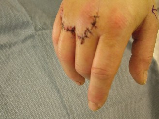
Figure 1: Intraoperative view showing the lacerated extensor digitorum communis tendon to the ring finger (Zone V injury) with visible retraction of the proximal stump. -
Palpation: Tenderness was elicited directly over the wound. There was no palpable crepitus or bony instability. The carpal bones and metacarpal shafts were stable to palpation. A gap was discernible on deep palpation within the extensor mechanism corresponding to the laceration site. Capillary refill in all digits was brisk (<2 seconds).
-
Range of Motion (ROM):
- Wrist: Full active and passive ROM in flexion, extension, radial, and ulnar deviation.
- Thumb: Full active and passive ROM.
- Index, Middle, Small Fingers: Full active and passive ROM at all joints (MCP, PIP, DIP). No extensor lag noted.
-
Right Ring Finger:
- Active Extension: Significant extensor lag at the MCP joint (approximately 40 degrees) was observed. The patient could not actively extend the ring finger from a flexed position beyond approximately 90 degrees of MCP flexion to 50 degrees of MCP flexion against gravity. Full active extension at the PIP and DIP joints was preserved with the MCP joint passively held in extension. This indicated a probable injury proximal to the juncture with the central slip but distal to the musculotendinous junction.
- Passive Extension: Full passive extension was achievable at the MCP, PIP, and DIP joints, confirming the absence of a fixed contracture or significant joint pathology.
- Individual Extensor Tests: With the wrist and MCP joints passively held in extension, the patient was unable to actively extend the ring finger at the MCP joint. The independent extensor digiti minimi and indicis tendons were intact.
-
Neurological Assessment:
- Motor: Intrinsic hand muscles (interossei, lumbricals) and extrinsic flexors were intact and strong (MRC Grade 5/5). While the primary complaint was extensor dysfunction of the ring finger, the clinical presentation was inconsistent with a complete radial nerve or posterior interosseous nerve (PIN) palsy, which would affect multiple extensor tendons (wrist extensors, MCP extensors of multiple digits). The isolated nature pointed toward a local tendon injury.
- Sensory: Light touch and two-point discrimination were intact in the median and ulnar nerve distributions. However, a small area of altered sensation (hypoesthesia) was noted on the dorsal-radial aspect of the proximal phalanx of the ring finger, suggestive of a superficial sensory nerve injury, likely a branch of the dorsal digital nerves.
-
Vascular Assessment: Radial and ulnar pulses were strong and symmetrical bilaterally. Allen's test was negative.
Based on clinical examination, a complete laceration of the extensor digitorum communis (EDC) tendon to the right ring finger, classified as a Verdan Zone V injury (dorsal hand, over metacarpals), was highly suspected, with a probable partial injury to a dorsal digital sensory nerve.
Imaging & Diagnostics
Plain Radiographs:
Standard anteroposterior, lateral, and oblique views of the right hand and wrist were obtained.
*
Findings:
The radiographs demonstrated no evidence of acute fracture, dislocation, or subluxation involving the carpal bones, metacarpals, or phalanges. Importantly, careful scrutiny of the soft tissues showed no radiopaque foreign bodies such as glass or metal within the wound track or surrounding tissues. This was crucial, as glass can often be radiolucent, but larger fragments can sometimes be visible. The joint spaces appeared preserved.
Ultrasonography:
Although not routinely indicated for acute, obvious open tendon lacerations, a point-of-care ultrasound was briefly considered to evaluate for retained foreign bodies, given the mechanism of injury (glass). However, given the clear clinical picture of a complete extensor tendon laceration and the need for immediate surgical exploration, further extensive pre-operative imaging was deemed unnecessary. In cases of equivocal clinical findings or suspected partial injury, dynamic ultrasound can be valuable for assessing tendon continuity and excursion.
CT/MRI Indications (Not indicated in this specific case):
*
CT Scan:
Would be indicated if there was suspicion of comminuted fractures, occult fractures, or deeply embedded radiolucent foreign bodies (e.g., wood, plastic, or very small glass fragments not seen on X-ray) that required precise localization for removal. For a clear-cut tendon laceration without bony involvement, CT offers little additional value.
*
MRI Scan:
Rarely indicated for acute open tendon lacerations, as the diagnosis is clinical and often confirmed intraoperatively. MRI is most useful for chronic tendon pathologies, differentiating between partial and complete tears, assessing tendon quality, or identifying associated soft tissue injuries (e.g., ligamentous, capsular) in closed injuries where the diagnosis is ambiguous. It can also differentiate between scar tissue and viable tendon in revision cases. For this patient, the open wound and clear extensor lag precluded the need for MRI.
Pre-operative Templating:
Templating is primarily relevant for reconstructive procedures involving bone (e.g., plate and screw fixation for complex fractures, arthroplasty). For an isolated extensor tendon repair, templating is not applicable. Surgical planning focused on selecting appropriate suture materials and understanding the anticipated tension required for repair.
Differential Diagnosis
The presentation of hand dysfunction, particularly an inability to extend a digit, necessitates considering a range of differential diagnoses beyond a simple extensor tendon laceration. A thorough clinical assessment is paramount in distinguishing these conditions.
| Feature | Extensor Tendon Laceration (Complete) | Posterior Interosseous Nerve (PIN) Palsy | Boutonnière Deformity (Central Slip Rupture) | Complex Fracture with Extensor Impairment |
|---|---|---|---|---|
| Mechanism | Direct sharp trauma (knife, glass, machinery). | Compression (e.g., tumor, lipoma, fracture), iatrogenic (surgery), direct trauma to forearm. | Direct blunt trauma to dorsum of PIP joint (e.g., sports injury), rheumatoid arthritis. | High-energy trauma (fall, crush), direct impact causing fracture and soft tissue injury. |
| Onset | Acute, immediately post-trauma. | Can be acute or insidious, depending on etiology. | Acute initially, but characteristic deformity often develops over 1-3 weeks. | Acute, immediately post-trauma. |
| Clinical Presentation | Inability to actively extend affected digit(s) at MCP/IP joint. Often associated with an open wound. Extensor lag. Passive ROM is full. | Weakness/paralysis of wrist and finger extensors (excluding ECRL). Wrist drop may be subtle (radial deviation with extension). No sensory deficit. | PIP flexion deformity, inability to actively extend PIP joint. Compensatory DIP hyperextension. No open wound typically. | Pain, swelling, deformity. Restricted active and passive ROM due to mechanical block/pain. Tendon injury may be secondary. |
| Wound | Usually open, visible laceration. | No open wound associated with the nerve injury itself. | No open wound associated with the central slip rupture. | May or may not have an open wound, depending on fracture type (open vs. closed). |
| Sensory Deficit | May have associated dorsal sensory nerve injury (e.g., digital nerve branch). | Pure motor nerve, no sensory deficits. | No sensory deficits. | May have associated nerve injury depending on fracture location/displacement. |
| Tenderness/Swelling | Localized tenderness at injury site. Variable swelling. | Diffuse tenderness in forearm extensor compartment in some cases, but not primary feature. | Tenderness over dorsum of PIP joint. Localized swelling. | Significant tenderness, swelling, ecchymosis over fracture site. |
| Active ROM | Inability to extend finger(s) against gravity. | Weak or absent active extension of MCP joints (digits 2-5) and wrist (ECU, ED, EDM, EIP). | Inability to actively extend PIP joint. | Severely limited active ROM due to pain, instability, or mechanical obstruction. |
| Passive ROM | Full passive ROM. | Full passive ROM. | Full passive ROM (initially). Fixed contracture if chronic. | May be restricted due to pain, edema, or mechanical block from displaced fragments. |
| Imaging | X-ray to rule out bony injury/foreign body. Ultrasound/MRI rarely for acute open. | X-ray (rule out bone pathology), EMG/NCS for confirmation, MRI for nerve compression. | X-ray (rule out avulsion fracture), MRI for soft tissue detail (central slip). | X-ray (definitive for fracture), CT for complex fractures, MRI for associated soft tissue. |
| Treatment | Surgical repair. | Conservative (splinting, observation) if neuropraxia; surgical exploration/release if compression/laceration. | Splinting (PIP extension) for 6-8 weeks; surgery for chronic/failed conservative. | Surgical fixation (ORIF) for unstable/displaced fractures; associated tendon repair. |
In our patient's case, the presence of a sharp open wound, the isolated nature of the extensor lag to a single digit (ring finger), and full passive ROM strongly supported a complete extensor tendon laceration over other differentials. The absence of wrist drop and preserved extension of other digits ruled out a broad radial nerve or PIN palsy. The location of the injury (dorsal hand, proximal to MCP) also excluded a central slip rupture (Boutonnière) which typically occurs at the PIP joint.
Surgical Decision Making & Classification
Rationale for Operative Intervention:
The presence of an open wound with a documented extensor lag and a palpable tendon gap unequivocally indicated a complete extensor digitorum communis (EDC) tendon laceration. This mandates surgical intervention for several critical reasons:
1.
Restoration of Function:
Primary repair is essential to restore active extension of the digit and prevent long-term functional deficits such as chronic extensor lag, stiffness, and subsequent grip weakness.
2.
Prevention of Scar Adhesion:
Surgical repair minimizes the risk of tendon ends retracting and forming non-functional scar tissue, which can make delayed repair significantly more challenging and less successful.
3.
Wound Management:
The open wound, despite being relatively clean, required thorough debridement, irrigation, and formal closure to minimize the risk of infection and facilitate primary healing.
4.
Foreign Body Removal:
Given the mechanism of injury (broken glass), surgical exploration provided the opportunity for meticulous identification and removal of any retained foreign bodies not visualized on radiographs, thereby preventing chronic pain, infection, or granuloma formation.
Timing of Surgery:
The decision was made for acute surgical exploration and primary repair within 6 hours of presentation. Early repair (within 24-72 hours) of extensor tendon lacerations is associated with better outcomes, as it minimizes tendon retraction, reduces edema, and allows for easier identification and repair of the tendon ends.
Relevant Classifications:
-
Verdan's Zones of Extensor Tendon Injuries: This classification is crucial for guiding surgical approach, repair technique, and post-operative rehabilitation protocols.
- Zone I: Distal to the DIP joint (Mallet finger).
- Zone II: Over the middle phalanx.
- Zone III: Over the PIP joint (Central slip injury, Boutonnière deformity).
- Zone IV: Over the proximal phalanx.
- Zone V: Over the MCP joint and dorsal hand (Metacarpal area). This was the zone of injury in our patient.
- Zone VI: Over the dorsal hand/carpals.
- Zone VII: Over the wrist joint (Retinaculum level).
- Zone VIII: Distal forearm.
- Our patient's injury was located proximal to the MCP joint, over the metacarpal shaft of the ring finger, thus classifying it as a Verdan Zone V injury . Injuries in this zone often involve the EDC tendons. The repair often involves careful identification of the retracted proximal stump and appropriate tensioning. Sagittal band injuries can also be seen in this zone.
-
Gustilo-Anderson Classification (Not applicable in this case): This classification system for open fractures is used to assess the severity of soft tissue damage and contamination. While the wound was open, there was no associated fracture, so this classification was not directly applied. However, principles of open wound management (debridement, irrigation) were still followed.
Surgical Technique / Intervention
Pre-operative Preparation:
*
Consent:
Informed consent was obtained, detailing the procedure, potential risks (infection, re-rupture, stiffness, nerve injury, persistent extensor lag, anesthesia risks), and expected outcomes.
*
Anesthesia:
The patient underwent regional anesthesia (axillary block) supplemented with conscious sedation, providing excellent intraoperative analgesia and post-operative pain control.
*
Patient Positioning:
The patient was positioned supine on the operating table, with the right upper extremity abducted and placed on a dedicated hand table.
*
Tourniquet:
A pneumatic tourniquet was applied to the upper arm and inflated to 250 mmHg after exsanguination with an Esmarch bandage, ensuring a bloodless field for meticulous dissection and repair.
*
Sterile Prep and Drape:
The entire right upper extremity was prepped with chlorhexidine solution and draped in a sterile fashion.
Surgical Approach and Exploration:
1.
Wound Extension:
The existing 3 cm transverse laceration was extended proximally and distally in a curvilinear fashion, allowing for adequate exposure of the extensor mechanism. A 'lazy-S' incision was utilized to avoid crossing skin creases perpendicularly and to facilitate broad access while optimizing cosmetic outcome and minimizing scar contracture.
2.
Debridement and Irrigation:
The wound edges were sharply debrided of any devitalized tissue. The wound was copiously irrigated with several liters of sterile normal saline to remove any superficial contaminants and potential microscopic glass particles.
3.
Tendon Identification:
The extensor digitorum communis (EDC) tendon to the ring finger was identified. The distal stump was present at the wound margin, attached to the junctional fibers distal to the MCP joint. The proximal stump, however, was retracted significantly, approximately 4 cm proximally, consistent with the elastic recoil of the musculotendinous unit. Careful dissection was performed proximally, following the line of the tendon sheath, to retrieve the retracted proximal end. The lumbrical and interosseous muscles were protected.
4.
Assessment of Associated Injuries:
The surrounding tissues were meticulously inspected. A small, superficial dorsal digital nerve branch to the radial side of the ring finger was found to be partially lacerated, consistent with the pre-operative sensory findings. There was no evidence of significant neurovascular injury to the digital arteries or other deep structures. The MCP joint capsule was found to be intact.
Tendon Repair:
1.
Preparation of Tendon Ends:
Both the proximal and distal tendon ends were débrided of any frayed or contused tissue, ensuring healthy, well-vascularized tissue for repair. Minimal shortening was necessary.
2.
Suture Material:
A 3-0 non-absorbable, monofilament suture (e.g., Prolene or Nylon) was selected for the core repair, providing excellent tensile strength and minimal tissue reaction. A 5-0 absorbable monofilament suture (e.g., PDS II) was chosen for the peripheral epitendinous repair.
3.
Core Suture Technique:
A modified Kessler locking-loop technique was employed for the core repair. This technique provides a strong, reliable repair with good opposition of the tendon ends, distributing stress across the repair site and allowing for early protected motion. Two separate strands of 3-0 Prolene were passed through each tendon stump, approximately 1 cm from the cut edge, creating two locking loops on each side, and then tied centrally.
4.
Tensioning:
The wrist was positioned in approximately 30 degrees of extension and the MCP joints in 10-15 degrees of flexion to reduce tension on the repair site. The tendon was repaired with appropriate tension, ensuring smooth glide and full passive excursion without gapping at the repair site. Over-tightening can restrict motion, while insufficient tension can lead to an extensor lag.
5.
Epitendinous Repair:
Following the core suture, a running epitendinous suture (5-0 PDS II) was performed around the circumference of the repair site. This peripheral repair contributes approximately 10-25% of the overall strength, flattens the repair site, minimizes gapping, and helps to prevent adhesions by creating a smooth surface.
6.
Nerve Repair:
The partially lacerated dorsal digital nerve branch was repaired with 8-0 Nylon using an epineurial repair technique under microscopic magnification, given its small caliber.
Wound Closure:
* The integrity of the repair was checked through gentle passive range of motion, confirming smooth tendon glide and absence of gapping.
* The deep tissues were irrigated again.
* The subcutaneous layer was closed with interrupted 4-0 absorbable sutures.
* The skin was closed with interrupted 5-0 non-absorbable monofilament sutures (Nylon), ensuring everted edges for optimal healing.
* A sterile dressing was applied.
Splinting:
A dorsal blocking splint was fabricated and applied immediately post-operatively. The wrist was held in 30 degrees of extension, the MCP joints in 0-10 degrees of flexion, and the interphalangeal joints in neutral extension. This position protects the repair by minimizing tension on the extensor tendon.
Post-Operative Protocol & Rehabilitation
Post-operative management of extensor tendon repairs is critical for successful outcomes, balancing protection of the repair with early, controlled motion to prevent adhesions and restore functional ROM.
Phase 1: Immobilization and Early Protected Motion (Weeks 0-3/4)
*
Immobilization:
A static dorsal blocking splint was applied immediately post-operatively, maintaining the wrist in 30 degrees of extension, MCP joints in 0-10 degrees of flexion, and IP joints in neutral extension. This position is crucial for reducing tension on the repaired extensor tendon. The patient was instructed to wear the splint continuously, removing it only for prescribed exercises.
*
Hand Therapy Referral:
Immediate referral to a certified hand therapist (CHT) was made.
*
Exercises:
*
Passive Flexion:
Under the guidance of the hand therapist, the patient commenced gentle, controlled passive flexion of the involved ring finger (MCP, PIP, DIP joints) within the confines of the splint or using specific splint designs that allow for controlled flexion against elastic traction (dynamic splinting). The goal is to achieve 30-45 degrees of MCP flexion, ensuring the extensor repair is not stressed.
*
Active Extension:
No active extension against resistance or gravity was permitted. The patient was instructed to gently relax the finger into the splint or assist with passive extension if a dynamic splint was used.
*
Tendon Gliding Exercises:
For uninvolved digits, to maintain their ROM and prevent stiffness.
*
Wound Care:
Daily wound inspections and dressing changes. Sutures were typically removed at 10-14 days.
Phase 2: Progressive Mobilization (Weeks 3/4 - 6/8)
*
Splint Modification:
The dorsal blocking splint was adjusted or replaced with a dynamic extension splint (e.g., using outrigger systems) that allowed for active flexion and passive extension, or a serial static splint with gradually decreasing wrist extension.
*
Active ROM:
Gradual introduction of active MCP extension, initially gravity-assisted, then against light resistance. The therapist guided progression based on the strength of the repair and the absence of extensor lag.
*
Differential Tendon Gliding:
Specific exercises to promote differential gliding of the EDC and intrinsic tendons.
*
Scar Management:
Initiation of scar massage, silicone gel sheets, or compression garments to minimize hypertrophic scarring and adhesions.
*
Strengthening:
Light isometric exercises or very gentle resistance exercises may be introduced towards the latter part of this phase, focusing on intrinsic muscles initially.
Phase 3: Strengthening and Functional Return (Weeks 6/8 - 12+)
*
Splint Weaning:
The splint was gradually weaned, initially for daily activities, then completely removed, depending on the patient's progress and the absence of extensor lag. Night-time splinting might continue for an additional few weeks.
*
Progressive Strengthening:
A comprehensive strengthening program was initiated, targeting extrinsic and intrinsic hand muscles. This included grip strengthening, pinch strength, and fine motor dexterity exercises.
*
Advanced Tendon Gliding:
Continued emphasis on full active and passive range of motion.
*
Return to Activity:
Gradual return to light functional activities. Avoidance of heavy lifting, gripping, or contact sports was recommended until at least 12 weeks post-surgery, or as advised by the surgeon and hand therapist. Full unrestricted activity clearance was often not until 4-6 months.
*
Sensory Re-education:
For the patient's dorsal digital nerve repair, sensory re-education exercises were initiated to help the brain interpret regenerating nerve signals.
Complications and Monitoring:
The patient was closely monitored for potential complications:
*
Infection:
Managed with antibiotics and local wound care.
*
Re-rupture:
Indicated by sudden loss of active extension. Requires re-exploration and re-repair or tendon grafting.
*
Adhesions:
Limiting tendon glide and range of motion. Managed with aggressive hand therapy, sometimes requiring tenolysis.
*
Extensor Lag:
Persistent inability to achieve full active extension. May require further therapy or revision surgery.
*
Stiffness:
Common sequela, mitigated by early motion.
*
Nerve Neuroma:
At the site of the digital nerve repair, causing persistent pain or hypersensitivity.
Our patient demonstrated excellent compliance with the post-operative protocol. At 3 months, he had regained full active extension of his ring finger with only a minor, non-functional extensor lag of less than 5 degrees at the MCP joint. His grip strength was returning to baseline, and sensory return in the dorsal ring finger was progressing positively, albeit not yet fully recovered.
Pearls & Pitfalls (Crucial for FRCS/Board Exams)
Pearls for Extensor Tendon Lacerations:
- Meticulous Clinical Examination: A systematic approach to hand examination is paramount. Assess wound characteristics, active vs. passive ROM, and neurovascular status. Isolated extensor lag with full passive ROM is diagnostic.
- Verdan's Zones: Thorough understanding of Verdan's zones is crucial as each zone dictates specific surgical considerations (e.g., proximity to joints, retinaculum, juncturae tendinum) and post-operative protocols. A Zone V injury, as in our case, requires careful attention to the juncturae and potential sagittal band involvement.
- Thorough Wound Exploration: Never underestimate the potential for retained foreign bodies, especially with glass or wood mechanisms. Meticulous debridement and copious irrigation are non-negotiable.
- Retrieval of Retracted Ends: Proximal extensor tendon stumps, particularly in Zones V-VIII, can retract significantly due to muscle belly pull. Often, a combination of proximal extension of the incision, gentle milking of the forearm musculature, and hyperflexion of the wrist can help deliver the retracted end. Avoid aggressive blind probing.
-
Appropriate Suture Technique & Material:
- Core Suture: Use a strong, non-absorbable monofilament suture (e.g., 3-0/4-0 Prolene, Nylon). The modified Kessler or similar locking-loop techniques provide robust repairs.
- Epitendinous Suture: A running peripheral suture (e.g., 5-0/6-0 PDS II, Nylon) is essential for increasing repair strength (up to 25%), minimizing gap formation, and creating a smooth gliding surface to reduce adhesions.
- Correct Tendon Tensioning: This is critical. The repaired tendon should have physiological tension, allowing for full passive range of motion without gapping, and maintaining a natural resting cascade. Over-tensioning restricts motion; under-tensioning leads to extensor lag.
- Associated Injuries: Always look for concomitant injuries: nerve lacerations (especially dorsal digital nerves), associated fractures (e.g., metacarpal fractures), or joint capsule involvement.
- Early Protected Motion: Modern protocols favor early controlled active and/or passive motion (e.g., Duran, Kleinert, or specific extensor tendon protocols) to prevent adhesions and promote tendon healing and glide. This requires close collaboration with a skilled hand therapist.
- Dorsal Blocking Splint: The initial splint position (wrist extension, MCP slight flexion, IP neutral/extension) is key to protect the extensor repair by minimizing tension.
- Patient Compliance: Rehabilitation success heavily relies on patient adherence to the therapy program and splinting regimen. Education is vital.
Pitfalls to Avoid in Extensor Tendon Lacerations:
- Inadequate Debridement and Irrigation: Leaving contaminated tissue or foreign bodies can lead to infection, non-healing wounds, or chronic inflammation.
- Missed Secondary Injuries: Failing to identify associated nerve, vascular, or joint capsule injuries can lead to persistent deficits or complications despite a successful tendon repair.
- Poor Tendon End Preparation: Frayed or devitalized tendon ends will not hold sutures securely and will heal poorly, increasing the risk of re-rupture or gapping.
-
Improper Suture Placement/Tension:
- Too close to cut edge: Suture pull-out.
- Insufficient number of passes/loops: Weak repair.
- Overly tight repair: Restricts motion, causes gapping in other tendon segments, or suture breakage.
- Loose repair: Extensor lag and re-rupture.
- Excessive Tendon Shortening or Lengthening: Altering the critical tendon length during repair will lead to either restricted flexion or a persistent extensor lag.
- Inadequate Immobilization/Protection: Premature or unprotected active motion can lead to repair gapping or rupture, especially in the early phases of healing.
- Over-Immobilization: Prolonged static immobilization beyond the recommended period can result in severe joint stiffness, capsular contractures, and adhesions, ultimately compromising functional recovery.
- Failure to Involve a Hand Therapist: Hand therapists are indispensable for guiding safe and effective post-operative rehabilitation. Their expertise in splint fabrication, specific exercise progression, and scar management is critical.
- Ignoring Scar Management: Neglecting scar care can lead to painful, hypertrophic scars that limit tendon glide and cosmesis.
- Underestimating Psychological Impact: Hand injuries can significantly impact a patient's daily life and occupation. Addressing concerns and managing expectations are important aspects of holistic care.
