Controversies
1. We prefer the posterior approach, irrespective of the previous approach(s).
1. In revision of a previous lateral approach, it avoids further damage to the gluteus medius and minimus, which have often not healed (see Fig. 2).
2. It allows extensile exposure of the acetabulum. The excellent femoral exposure often allows implant removal without the need for femoral/trochanteric osteotomy and its related morbidity.
3. In revising failed internal fixation of hip fractures, the approach can be performed and the hip dislocated prior to removal of metalwork, which requires minimal further dissection.
Femoral Stem Revision: Posterior Approach
Indications
1. Revision of failed primary hip replacement
2. Second-stage revision of infected hip replacement
3. Revision of failed proximal femoral fracture fixation
Examination/Imaging
4. Anteroposterior and lateral radiographs of the pelvis and femur
5. Preoperative templating and planning
1. Is the stem to be revised cemented or uncemented?
2. Is the stem or cement loose? If the stem or cement is loose, it can be removed straightforwardly.
3. Has a varus femoral deformity occurred?
4. If distal cement and/or a sclerotic bone pedestal is present, it can be removed with the aid of a bone window.
5. If removing a well-fixed stem, an extended trochanteric osteotomy may be required.
6. Ensure familiarity with the revision system.
Treatment Options
1. Lateral approach
2. Anterolateral approach
7. Ensure that inventory allows for revision of all components with compatible head sizes if the acetabulum is to be retained.
8. Be prepared for intraoperative complications such as fracture.
Surgical Anatomy
6. See posterior approach for primary hip replacement in Procedure 5.
7. To increase exposure, the quadratus femoris and gluteus major insertion into gluteal tuberosity on the femur can be divided, leaving residual soft tissue stumps attached to the femur for repair.
8. If an extended trochanteric osteotomy or distal window is required, the vastus lateralis can be elevated from the intramuscular septum, allowing extensile exposure of the femur from the hip to the knee.
9. We do not routinely expose the sciatic nerve, releasing scar tissue and pseudo-capsule from the posterior intertrochanteric line and reflecting this flap posteriorly as one layer with the nerve remaining behind it.
317
P EARLS
- Ensure that the hip to be operated on has free range of movement, and is not restricted by patient supports.
- Ensure that the pelvis is square (i.e., perpendicular to the floor).
- Ensure that drapes allow access to the thigh/buttock to the proximal extent of the ilium.
P ITFALLS
- If the patient is not adequately secured, he or she will tilt forward during surgery, obscuring the view for the surgeon and increasing the risk of component malpositioning, particularly cup retroversion.
Femoral Stem Revision: Posterior Approach
Positioning
1. The patient is placed in the lateral decubitus position.
2. The pelvis is secured with anterior and posterior bolsters resting on the pubis and sacrum.
3. The trunk is secured with anterior and posterior bolsters at the sternum and scapulae.
4. An axillary bump is used to decrease pressure on the inferior arm.
5. Prep and drape the lower limb allowing access to the entire femur.
Portals/Exposures
6. Make the skin incision, a longitudinal incision with a posterior curve proximally toward the posterior superior iliac spine.
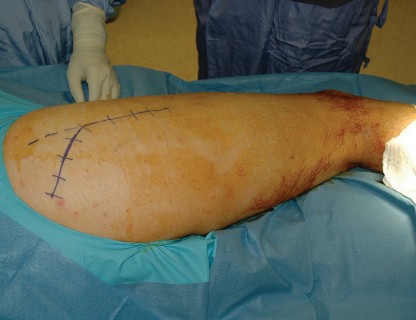
1. Center the incision over the posterior third of the greater trochanter. Use the old scar if it is in a reasonable position.
2. If revising a previous lateral approach, the surgeon may need to use a new incision or modify the old one (Fig. 1).

FIGURE 1 #### 318
Femoral Stem Revision: Posterior Approach
- Expose the fascia lata or gluteus maximus fascia, which may be significantly scarred.
- Do not create skin flaps or undermine the incision.
- Incise the fascia or split the gluteus maximus in the line of the skin incision (Fig. 2).
- Blunt dissection with a finger underneath the fascia will expose the vastus lateralis and gluteus medius, which are often adherent to the fascia lata.
P EARLS
- If revising components without significant subsidence, it is desirable to get circumferential exposure of the joint prior to dislocation as, once dislocated, the femur will tend to shorten, obscuring the exposure.
- Heterotrophic bone may be encountered and should be carefully excised, preserving the surrounding soft tissue.
- If the femoral component has subsided significantly, it may be hidden from view behind the greater trochanter. In this case, dislocation of the hip may be facilitated with a bone hook placed around the prosthesis neck, reducing torque on the femur and minimizing the risk of fracture.
- Maintain maximum length of the posterior soft tissue flap for subsequent repair.
P ITFALLS
- Be aware that the sciatic nerve may not be in its usual position.
- Internally rotate the femur to put posterior tissue under tension.
- Place a retractor deep to the gluteus medius and gently retract the muscle (Fig. 3).
- If revising a previous lateral approach, with anatomy similar to that in a primary hip replacement (see Fig. 3), the posterior capsule is seen after division of the short rotators (Fig. 4).
- If revising a previous posterior approach, the piriformis and short rotators may not be evident. In this case, divide short rotators and capsule from the femoral insertion as one layer, proximally from the free edge of the gluteus medius distally to the quadratus femoris or gluteus maximus tendon if the former muscle is poorly defined scar tissue.
- The sciatic nerve is less apparent than in primary hip replacement and is palpated but not explored.
-
After incising the posterior capsule, with internal rotation to maintain tissue tension, the femoral head and acetabular component will come into view. Preserve the posterior tissue for later repair.

 FIGURE 2


Femoral Stem Revision: Posterior Approach


319 FIGURE 3 FIGURE 4 #### 320
Femoral Stem Revision: Posterior Approach
- Place retractors anterosuperiorly over the superior acetabulum, and inferiorly over the posterior column of the acetabulum. Excise inferior and anterior scar tissue and/or capsule and place a further retractor anterior to the anterior acetabular wall.
- Once the acetabulum is exposed, posterior dislocation of the hip is usually straightforward (Fig. 5).
- Remove the modular femoral head if present and confirm head size.
- Retractors placed around the inferior femoral neck allow the neck to be dissected free.
-
Dissect soft tissue from the bone-prosthesis interfaces of the femur and acetabulum to assess bone loss and implant stability.

 FIGURE 5
Procedure
FIGURE 5
Procedure
Step 1: Removal of Femoral Stem
- Dislocate the hip and remove the modular head.
- Excise tissue at the bone-prosthesis interface.
- Remove bone and/or cement from the shoulders of the prosthesis.
- Remove proximal cement (if revising a cemented prosthesis).
- Use flexible osteotomes to cut the proximal bone-prosthesis interface.
321
P EARLS
- Ensure adequate exposure of the shoulder of the prosthesis prior to attempted removal (Fig. 7). A significant amount of bone may need to be removed, particularly if the component has subsided (Fig. 8A and 8B).
Femoral Stem Revision: Posterior Approach
- A cemented or loose uncemented stem can then be removed with an extraction device with force directed along the line of the intramedullary canal (Fig. 6).
-
If the stem can’t be removed, consider an extended trochanteric osteotomy.

 FIGURE 6

 FIGURE 7


322
Femoral Stem Revision: Posterior Approach
A B
P ITFALLS
1.
Incomplete removal of proximal bone and/or cement may result in a trochanteric fracture occurring during component removal (Fig. 9A and 9B).
FIGURE 8


---
FIGURE 9 A B
#### 323
P EARLS
- Ensure that the window has an oval shape with no notches, to avoid femoral fracture.
- The window can be enlarged distally or proximally if required.
Femoral Stem Revision: Posterior Approach
Step 2: Removal of Cement Restrictor/ Distal Bone Pedestal with Bone Window
- In certain circumstances we use a cortical window on the lateral femoral cortex.
- A cement restrictor and adjacent cement are often well fixed, even if proximal cement is loose (Fig. 10A and 10B).
-
In uncemented stem revision, a sclerotic pedestal may have formed at the tip of the prosthesis. This can make finding the distal canal difficult.


A B
FIGURE 10
#### 324
Femoral Stem Revision: Posterior Approach
- If the prosthesis tip is close to the anterior femoral cortex (Fig. 11), anterior cortical perforation may occur when drilling into the distal canal.
- The window should be at the junction of the cement restrictor and the distal canal. (Its distance from the greater trochanter should be determined during preoperative planning.)
- Elevate the vastus lateralis at the chosen level.
- Use a high-speed burr to cut an oval window 10–20 mm long 10 mm wide (less than one-third the circumference of the femur); remove the bone fragment and set aside.
- Small fragments of cement and restrictor are removed through the window.
- The window can facilitate drilling down the femoral canal and through the obstruction. At this point you can see that the drill is inside the distal femur.
- Bone and cement can be removed with hooks and cement removal instruments as preferred (Fig. 12).
- Prophylactic cerclage wire is placed distal to the window prior to stem insertion.
-
Trials and the definitive stem are inserted under direct vision through the window (with 5 cm stem fixation distal to the window).


325
Femoral Stem Revision: Posterior Approach FIGURE 11 Femur Retractor
Vastus lateralis muscle
2 cm x 1 cm oval window
Intramuscular septum

---
FIGURE 12
#### 326
Femoral Stem Revision: Posterior Approach
- Resected bone window and graft obtained during femoral preparation can be replaced and held with cerclage wire (Fig. 13).
-
A reinforcing strut allograft can be used if distal stem fixation is unsatisfactory (Fig. 14).


 FIGURE 13 FIGURE 14
#### 327
FIGURE 13 FIGURE 14
#### 327
P EARLS
- Ensure that enough distal femoral diaphysis is present to allow press-fitting of the revision stem (ideally 5 cm of isthmus distal to the osteotomy).
- Trephine drills rapidly overheat and require frequent irrigation and removal to clean debris from the teeth.
- Ensure access to allograft to reinforce the repair, particularly if bone stock is poor.
P ITFALLS
- If fracture occurs, strut grafts and/or cable plate should be available for fixation.
Femoral Stem Revision: Posterior Approach
Step 3: Extended Trochanteric Osteotomy to Remove Well-Fixed Stem
- The length of the osteotomy is determined from preoperative templates.
-
A standard posterior approach is used. Distal wound extension is made to the extent of the planned osteotomy (Fig. 15).


 FIGURE 15
#### 328
FIGURE 15
#### 328
Femoral Stem Revision: Posterior Approach
- Divide the gluteus maximus tendon midsubstance and elevate the vastus lateralis from the intramuscular septum, leaving a small cuff of muscle (Fig. 16).
- Use a saw or burr to release the lateral femoral cortex and greater trochanter (one third the diameter of the femoral shaft).
- Distally, curve the osteotomy to minimize risk of fracture propagation.
-
Use osteotomes to carefully displace osteotomized bone (Fig. 17A and 17B).

 FIGURE 16


Femoral Stem Revision: Posterior Approach


329
A
B
FIGURE 17
#### 330


Femoral Stem Revision: Posterior Approach FIGURE 18 1. Despite care, existing osteoporosis often results in a fragile residual shell after implant removal (Fig. 18).
-
Removal of cylindrical sections of the prosthesis is done with thin curved osteotomes (Fig. 19A) and trephine drills (Fig. 19B).


Femoral Stem Revision: Posterior Approach


331
A
B
FIGURE 19
#### 332
Femoral Stem Revision: Posterior Approach
- Once advanced beyond the prosthesis tip, the drill pitch changes and the prosthesis usually comes out with removal of the trephine (Fig. 20).
- Following insertion of the definitive stem, the osteotomy is closed using cerclage wires (Fig. 21).
-
Strut allograft can be used to restore bone stock and ensure that the stem is covered (Fig. 22). If allograft is used for mechanical strength, we use cables to fix it to the femur (Fig. 23A–23C).

--- FIGURE 20

Femoral Stem Revision: Posterior Approach


333 FIGURE 21 FIGURE 22


Femoral Stem Revision: Posterior Approach


334
A
B
FIGURE 23
C
335
Femoral Stem Revision: Posterior Approach
Step 4: Insertion of Definitive Component
- We prefer an extensively coated monoblock stem with distal flutes to improve rotational stability. This stem design is initially reliant on distal fixation and bypasses the abnormal bone.
- Stems greater than 190 mm are usually curved to prevent perforation of the anterior cortex. They require flexible reamers or narrow-stem reamers (Fig. 24) to negotiate the anterior femoral bow.
- We plan for 5 cm of diaphyseal press-fit beyond the femoral defect.
-
Using straight hand reamers, ream sequentially to the desired size (usually 1–1.5 mm less than the final stem diameter). If good cortical “chatter” is not encountered, increase the diameter of the reamer and subsequent stem.

 FIGURE 24
1. Insert undersized trial stems to get a “feel” for the stability. The final trial is usually slightly smaller than the prosthesis to facilitate removal.
FIGURE 24
1. Insert undersized trial stems to get a “feel” for the stability. The final trial is usually slightly smaller than the prosthesis to facilitate removal.
2. Check for stability as for primary replacement.
3. Insert the definitive component and selected head.
4. Closure is similar to that for primary hip replacement (Fig. 25A and 25B), but in revision surgery the increased risk of dislocation is even more important.


336
Femoral Stem Revision: Posterior Approach
A


B
FIGURE 25


337
P EARLS
- Use prophylactic cerclage wire below the defect to prevent fracture.
- Insertion of long stems is easier following an extended trochanteric osteotomy.
- In an intact femur, start with the stem in 90° of anteversion. During impaction, correct this to the normal 15° of anteversion. Anteversion is determined as for primary hip replacement using the axis of the thigh and leg (Fig. 26).
- Chronic stem retroversion during loosening creates a void in the posterior calcar (Fig. 27), making femoral neck geometry unreliable in determining version. Bone graft can fill these defects after definitive stem insertion (Fig. 28).
- Choosing a prosthesis with a high femoral offset increases hip stability.
- If the femoral component will not go down, remove and ream one size larger.
Femoral Stem Revision: Posterior Approach FIGURE 26

 FIGURE 27


338
Femoral Stem Revision: Posterior Approach FIGURE 28 ## P ITFALLS
- Fractures of the femur can occur.
- Undisplaced fractures of the greater trochanter can usually be fixed with a simple figure-of-eight heavy cerclage wire passed around the femur inferior to the lesser trochanter and behind the insertion of the gluteus medius (Fig. 29; see also Fig. 25A and 25B)
- Displaced greater trochanteric fractures require more substantial fixation with cables and a staple or plate (Fig. 30).
-
Fractures of the shaft can be treated with a longer bypass stem or femoral strut grafts/ plates or a combination (Fig. 31).


 FIGURE 29


339
Femoral Stem Revision: Posterior Approach FIGURE 30 FIGURE 31
P EARLS
-
Hip precautions are taken to protect the posterior repair while scar tissue forms, as for the primary approach.
Postoperative Care and Expected Outcomes - Weight bearing is determined by fixation of the femoral stem.
- Possible complications include thromboembolism and heterotrophic ossification.
- Antibiotic prophylaxis is instituted.
