INTRODUCTION TO THE FEMORAL NERVE
The femoral nerve is the largest branch of the lumbar plexus, serving as the primary motor supply to the anterior compartment of the thigh and providing critical sensory innervation to the anteromedial thigh, medial knee, and medial leg. In the realm of operative orthopaedics and peripheral nerve surgery, injuries to the femoral nerve—whether traumatic, iatrogenic, or neoplastic—present complex reconstructive challenges.
Due to its deep retroperitoneal origin and its transition beneath the inguinal ligament into the femoral triangle, surgical exposure requires a masterful understanding of pelvic and proximal thigh anatomy. This comprehensive guide delineates the surgical anatomy, biomechanics, diagnostic evaluation, and step-by-step operative management of femoral nerve injuries, including advanced techniques for closing massive nerve gaps.
SURGICAL ANATOMY AND NEUROBIOMECHANICS
Origin and Proximal Course
The femoral nerve is formed by the union of the posterior divisions of the ventral rami of the L2, L3, and L4 nerve roots. Within the retroperitoneal space, these roots converge to form the nerve trunk within the substance of the psoas major muscle. The nerve emerges from the lower lateral border of the psoas major and descends in the groove between the psoas and the iliacus muscle, deep to the iliac fascia.
As it approaches the thigh, the femoral nerve passes deep to the inguinal ligament. Crucially, it remains lateral to the femoral artery, separated from the vascular bundle by the robust iliopectineal arch (a fascial band extending from the inguinal ligament to the iliopectineal eminence). This anatomical separation is a vital landmark during surgical exploration of the femoral triangle.
Terminal Branches and Innervation
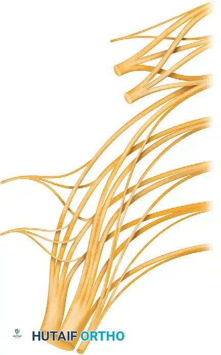
Just distal to the inguinal ligament, the femoral nerve arborizes rapidly into anterior and posterior divisions, separated by the lateral circumflex femoral artery.
The Anterior Branch:
* Sensory Contributions: Divides into the intermediate cutaneous nerve of the thigh and the medial cutaneous nerve of the thigh, supplying the anteromedial aspect of the thigh down to the knee.
* Motor Contributions: Supplies the pectineus muscle (often receiving dual innervation from the obturator nerve) and the sartorius muscle.
The Posterior Branch:
* Sensory Contributions: Gives rise to the saphenous nerve, the largest and longest cutaneous branch of the femoral nerve. The saphenous nerve continues distally alongside the femoral vessels within the subsartorial (Hunter’s) canal. It pierces the deep fascia along the medial side of the knee to become subcutaneous, supplying the skin on the anteromedial aspect of the leg distally to the medial malleolus and the medial arch of the foot.
* Motor Contributions: Provides the critical motor innervation to the quadriceps femoris muscle group (rectus femoris, vastus lateralis, vastus medialis, and vastus intermedius) and the articularis genus.
Clinical Pearl: When exploring the femoral nerve in the proximal thigh, the rapid arborization ("cauda equina of the thigh") makes distal mobilization tedious. The motor branches to the quadriceps must be meticulously identified and preserved using intraoperative nerve stimulation.
ADJACENT NERVES OF THE LUMBAR PLEXUS
Understanding the femoral nerve requires a holistic grasp of the surrounding lumbosacral plexus, as these adjacent nerves are frequently encountered—and at risk of injury—during pelvic and proximal thigh exposures.
The Iliohypogastric, Ilioinguinal, and Genitofemoral Nerves
- Iliohypogastric Nerve (T12-L1): Supplies a small area of skin over the superolateral gluteal region and an area just superior to the pubic bones on the anterior abdominal wall.
- Ilioinguinal Nerve (L1): Supplies a segmental strip of skin along the inguinal ligament overlying the symphysis pubis, the skin of the upper scrotum (or labia majora), the root and dorsal aspect of the penis, and the medial aspect of the proximal thigh.
- Genitofemoral Nerve (L1-L2): Traverses the inguinal canal and supplies the cremaster muscle, the skin of the scrotum, and the adjacent part of the thigh.
Surgical Warning: From a surgical standpoint, these three nerves are highly significant because they may be inadvertently injured or entrapped during herniorrhaphy, appendectomy, or anterior approaches to the pelvic ring (e.g., the ilioinguinal approach to the acetabulum). Such injuries result in persistent, debilitating neuralgic discomfort that may require secondary surgical exploration, neurolysis, or neurectomy.
The Lateral Femoral Cutaneous Nerve (LFCN)
The lateral femoral cutaneous nerve is formed from the posterior divisions of the L2 and L3 nerve roots. It courses obliquely across the iliacus muscle toward the region of the anterior superior iliac spine (ASIS) to exit the pelvis between the lateral attachments of the inguinal ligament. Entrapment of this nerve as it exits the pelvis results in meralgia paresthetica.
MECHANISMS OF INJURY
Femoral nerve injuries, while less common than sciatic or peroneal nerve injuries, carry profound functional morbidity due to the loss of knee extension (quadriceps paralysis).
Traumatic Injuries
- Pelvic Ring Fractures and Sacroiliac Dislocations: The lumbar nerve roots and the lumbosacral plexus are occasionally injured by violent traction forces during high-energy pelvic trauma.
- Penetrating Trauma: The plexus and the femoral nerve proper may be directly transected or contused by missile wounds (gunshot wounds) or stab wounds to the groin or lower abdomen.
Iatrogenic Injuries
- Total Hip Arthroplasty (THA): Particularly during anterior or anterolateral approaches, aberrant retractor placement over the anterior rim of the acetabulum can compress the femoral nerve against the iliopsoas muscle.
- Pelvic Surgery: Prolonged lithotomy positioning or aggressive retraction during gynecological, urological, or general surgical procedures in the pelvis.
CLINICAL EVALUATION AND DIAGNOSTICS
A meticulous physical examination is the cornerstone of diagnosis. Patients typically present with an inability to actively extend the knee, buckling of the knee during the stance phase of gait, and sensory deficits over the anteromedial thigh and medial leg.
Diagnostic Modalities
- Electromyography (EMG) and Nerve Conduction Studies (NCS): Crucial for localizing the lesion, determining the severity of axonal loss, and monitoring for early signs of reinnervation. EMG should typically be delayed until 3 to 4 weeks post-injury to allow for Wallerian degeneration to become electrically apparent.
- Magnetic Resonance Neurography (MRN): High-resolution MRI can visualize nerve continuity, neuroma formation, and surrounding hematomas or compressive lesions.
- Myelography: Historically used, and sometimes still employed in conjunction with CT or MRI, to evaluate for root avulsions in severe pelvic trauma.
Diagnostic Pitfall: In contrast to cervical root avulsions, where myelographic evidence of dural diverticula (pseudomeningoceles) is highly diagnostic, myelographic evidence of dural diverticula in the lumbar spine does not correlate well with the actual avulsion of lumbar roots.
Although the direct microsurgical repair of avulsed lumbar roots within the spinal canal would seem futile due to the lack of perineurium and the central nervous system environment, surgical exploration of the plexus may still be highly helpful in establishing a definitive prognosis and planning subsequent tendon transfers.
SURGICAL INDICATIONS AND TIMING
- Immediate Exploration: Indicated for open, sharp transections (e.g., knife wounds) where the nerve ends can be identified and repaired primarily before retraction and scarring occur. Also indicated in the presence of an expanding retroperitoneal or femoral triangle hematoma causing acute compressive neuropathy.
- Delayed Exploration (3 to 6 months): Indicated for closed traction injuries, contusions, or missile wounds where the nerve is in continuity but failing to show clinical or electromyographic signs of recovery. This delay allows the zone of injury to declare itself, facilitating accurate resection of the neuroma-in-continuity back to healthy fascicles.
OPERATIVE APPROACH AND SURGICAL TECHNIQUE
The surgical exposure of the femoral nerve must be extensile, allowing access to both its retroperitoneal origin and its distal arborization in the thigh.
Patient Positioning and Preparation
- The patient is placed in the supine position on a radiolucent operating table.
- A bump may be placed under the ipsilateral hemipelvis to slightly elevate the operative side.
- The entire lower extremity, hemipelvis, and lower abdomen are prepped and draped free to allow for intraoperative manipulation, specifically acute hip flexion.
- A sterile tourniquet is generally not feasible due to the proximal nature of the dissection.
Incision and Superficial Dissection
- The incision begins 3 to 5 cm proximal to the anterior superior iliac spine (ASIS), curving distally and medially parallel to the inguinal ligament.
- As it crosses the midpoint of the inguinal ligament, the incision turns distally, extending vertically down the anterior thigh over the femoral triangle.
- In the thigh, the deep fascia (fascia lata) is incised. The femoral artery is identified by palpation. The femoral nerve lies immediately lateral to the artery, separated by the iliopectineal fascia.
Deep Dissection and Proximal Exposure
To expose the nerve proximal to the inguinal ligament:
1. The external oblique aponeurosis is incised parallel to the inguinal ligament.
2. The internal oblique and transversus abdominis muscles are divided or retracted to enter the retroperitoneal space.
3. The peritoneum is bluntly swept medially, exposing the iliacus and psoas major muscles.
4. The femoral nerve is identified emerging from the lateral border of the psoas major and is traced distally toward the inguinal ligament.
Methods of Closing Nerve Gaps
One of the most formidable challenges in femoral nerve reconstruction is overcoming segmental defects resulting from trauma or neuroma resection. Remarkably, gaps of 8 to 10 cm can be closed without excessive difficulty if meticulous mobilization techniques are employed.
Step-by-Step Gap Closure:
1. Proximal Mobilization: The nerve is mobilized proximally deep into the retroperitoneum, up to the point where it emerges from the lateral border of the psoas muscle. Care must be taken not to avulse the delicate lumbar roots.
2. Distal Mobilization: The nerve is mobilized distally by carefully freeing the individual branches of the nerve in the proximal thigh. The epineurium can be longitudinally incised (internal neurolysis) to allow the fascicular groups to stretch slightly without tethering.
3. Joint Positioning: The hip is flexed acutely. This maneuver significantly shortens the distance between the retroperitoneum and the anterior thigh, providing massive relief of tension on the nerve ends.
4. Neurorrhaphy: Under operating microscope magnification, the nerve ends are prepared by resecting damaged tissue until healthy, pouting fascicles are visualized. An epineurial or group fascicular repair is performed using 8-0 or 9-0 non-absorbable monofilament sutures. The repair must be entirely tension-free in the flexed position.
5. Reconstruction and Closure: The inguinal ligament, if divided for exposure, is meticulously reconstructed to prevent subsequent herniation. The abdominal wall and thigh wounds are closed in layers over a suction drain.
Surgical Pearl: If the gap exceeds 10 cm, or if primary repair cannot be achieved without tension even with acute hip flexion, nerve grafting is mandatory. Sural nerve autografts are the gold standard, utilized as cable grafts to bridge the defect.
Postoperative Care and Immobilization
Postoperative management is critical to prevent disruption of the neurorrhaphy. The protocol is similar to that described for major sciatic nerve repairs:
1. Immobilization: A custom hip spica cast or a rigid hinged hip orthosis is applied immediately in the operating room, with the hip locked in the acutely flexed position utilized during the repair.
2. Duration: The hip is maintained in acute flexion for 3 to 4 weeks to allow the nerve repair to gain tensile strength.
3. Graduated Extension: After 4 weeks, the cast or orthosis is modified to allow gradual extension of the hip, typically at a rate of 10 to 15 degrees per week. Aggressive, sudden extension will rupture the repair.
4. Rehabilitation: Once full extension is achieved, aggressive physical therapy focuses on maintaining knee joint mobility and strengthening compensatory muscles while awaiting reinnervation.
RESULTS AND PROGNOSIS
The literature regarding the outcomes of femoral nerve repair, particularly concerning massive defects requiring grafting, remains limited. Historically, no statistically significant information was available regarding the exact success rates of grafting for large defects in the femoral nerve.
However, contemporary microsurgical series indicate that because the femoral nerve is a mixed nerve with a heavy motor component destined for a single large muscle group (the quadriceps), the results of primary repair—and even relatively short cable grafts—can be functionally rewarding.
- Motor Recovery: Recovery of antigravity knee extension (Medical Research Council [MRC] grade 3 or better) is achievable in a majority of patients if the repair is performed within 6 months of injury and the gap is manageable.
- Sensory Recovery: Protective sensation in the saphenous distribution frequently returns, mitigating the risk of neuropathic ulceration.
- Salvage Procedures: In cases of failed nerve repair or delayed presentation, tendon transfers (e.g., transferring the hamstrings or the tensor fasciae latae to the patella) remain viable salvage options to restore knee stability during the stance phase of gait.
