Comprehensive Introduction and Patho-Epidemiology
The management of metacarpal nonunion, particularly when complicated by segmental bone loss, represents one of the most formidable challenges in reconstructive hand surgery. Unlike simple, closed fractures of the tubular bones of the hand—which typically proceed to uneventful union with conservative management or standard internal fixation—metacarpal defects are almost exclusively the sequelae of devastating trauma. These injuries usually result from high-energy mechanisms, including severe crush injuries, industrial accidents, gunshot wounds, blast injuries, or aggressive post-infectious surgical debridement. Such etiologies leave behind a profoundly hostile local environment characterized by compromised vascularity, dense cicatrix formation, loss of the gliding paratenon, and significant structural osseous voids.
The pathophysiology of metacarpal nonunion in the setting of segmental loss fundamentally differs from that of a simple hypertrophic or atrophic nonunion. In a standard nonunion, the bone ends are present but fail to bridge due to inadequate stability (hypertrophic) or inadequate biology (atrophic). In contrast, a segmental metacarpal defect presents a complete absence of the osteoconductive scaffold and osteoinductive mediators necessary for bone healing. The intervening space rapidly fills with dense, avascular fibrous tissue that not only prevents osseous bridging but also tethers the adjacent extensor tendons, leading to profound functional impairment of the entire ray.
The ultimate success of structural bone grafting in metacarpal defects is predicated upon two non-negotiable pillars: the absolute vitality of the surrounding soft tissue envelope and the biomechanical precision of the osseous reconstruction. Sterling Bunnell, a pioneer of modern hand surgery, aptly coined the term "bone carpentry" to describe the exacting tolerances required in this domain. A structurally sound, perfectly fashioned graft placed into a poorly vascularized, scarred bed will inevitably resorb, succumb to infection, or fail to incorporate. Conversely, a robust, well-vascularized soft tissue envelope cannot compensate for a poorly fashioned, mechanically unstable graft that fails to resist the immense deforming forces of the hand.
This comprehensive chapter details the surgical correction of metacarpal nonunions utilizing the classic Littler technique. Conceived by J. William Littler, this technique remains a masterclass in structural autografting. It provides orthopedic and hand surgeons with an evidence-based, highly reproducible framework for restoring digital length, correcting rotational and angular malalignment, and ultimately salvaging the biomechanical function of the traumatized hand.
Detailed Surgical Anatomy and Biomechanics
A profound understanding of the surgical anatomy and the unique biomechanical forces acting upon the metacarpals is an absolute prerequisite for executing a durable reconstruction. The metacarpals are not merely static struts; they are dynamic intercalated segments that must withstand complex, multi-planar forces during every motion of the hand.
Osseous and Vascular Anatomy
The metacarpals are tubular bones consisting of a base, shaft (diaphysis), neck, and head. The diaphysis is characterized by a thick cortical shell, particularly along the volar concavity, which is designed to resist the immense compressive loads generated during power grip. The medullary canal is relatively narrow in the diaphysis but expands significantly at the metaphyses. The vascular supply to the metacarpals is dual: an endosteal supply via the nutrient artery (which typically enters the volar aspect of the proximal third of the shaft and is directed distally) and a robust periosteal supply derived from the dorsal and volar metacarpal arteries. In high-energy trauma with segmental loss, the endosteal supply is completely obliterated, rendering the healing of any intercalary graft entirely dependent on the periosteal blood supply creeping from the surrounding soft tissue envelope.
The Soft Tissue Envelope
The dorsum of the hand possesses a naturally thin, highly mobile subcutaneous layer. Unlike the volar aspect, which is padded by thick fascia and palmar skin, the dorsal skin provides minimal cushioning over the extensor tendons and metacarpals. This makes the region highly susceptible to breakdown over prominent hardware or bulky bone grafts. Furthermore, the extensor tendons are enveloped by a delicate paratenon, which provides the necessary gliding surface. In the setting of a metacarpal nonunion, this paratenon is frequently destroyed and replaced by a dense pseudarthrosis that adheres the tendon directly to the bone ends, obliterating the excursion necessary for digital flexion.
Pathoanatomy and Deforming Forces
Understanding the deforming forces acting upon a metacarpal nonunion is critical for designing a graft that will not fail under physiological loads. A metacarpal defect inevitably leads to a predictable, tri-planar pattern of deformity.
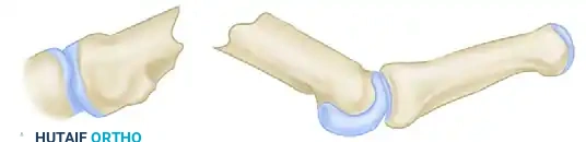
As illustrated in the pathoanatomy, the deformity is driven by the following forces:
1. Longitudinal Shortening: The intrinsic muscles (interossei and lumbricals) and the extrinsic flexors (flexor digitorum superficialis and profundus) exert a constant longitudinal compressive force. Without the strut of the metacarpal diaphysis, the distal fragment migrates proximally. This shortens the ray, disrupts the resting cascade of the hand, leads to an extensor lag at the metacarpophalangeal (MCP) joint, and profoundly weakens grip strength by altering the length-tension relationship of the extrinsic tendons.
2. Dorsal Apex Angulation: The volar anatomical location of the intrinsic muscles creates a constant flexion moment on the distal metacarpal fragment. Without the structural continuity of the intact metacarpal, the bone collapses into dorsal apex angulation. This forces the metacarpal head into the palm, creating a painful prominence and altering the mechanics of the MCP joint.
3. Rotational Malalignment: The lack of osseous continuity allows the distal fragment to rotate freely. Because the deep transverse metacarpal ligament tethers the metacarpal heads, rotation often manifests as digital scissoring or overlapping during active flexion, which can severely impede the function of adjacent, uninjured digits.
Exhaustive Indications and Contraindications
The decision to proceed with a structural bone graft utilizing the Littler technique must be made judiciously. The procedure is technically demanding and requires a compliant patient capable of participating in a rigorous, months-long postoperative rehabilitation protocol.
Indications for the Littler Technique
The primary indication for the Littler structural bone grafting technique is a symptomatic metacarpal nonunion complicated by a segmental bone defect measuring greater than 1.0 to 1.5 centimeters. Defects smaller than this can often be managed with cancellous interposition grafting and rigid plate fixation. However, larger defects require a structural cortico-cancellous strut to bridge the gap and resist the compressive forces of the hand.
Additional indications include the salvage of failed previous metacarpal fixations where hardware failure or infection has resulted in substantial bone loss. It is also highly indicated in cases of atrophic nonunion where the native bone ends are sclerotic, avascular, and incapable of generating a healing response without the introduction of a robust, osteoconductive and osteogenic autograft. The technique is particularly well-suited for the central rays (long and ring fingers), where the adjacent intact metacarpals provide a natural splinting effect that enhances the stability of the press-fit construct.
Soft Tissue Prerequisites
First and foremost, the dorsum of the hand must be well covered by pliable, well-vascularized skin and subcutaneous tissue. If the native tissue has been lost to trauma, or if the skin is paper-thin, adherent, and heavily scarred from multiple previous surgeries, soft tissue reconstruction must precede or accompany the bone grafting procedure.
Never attempt structural bone grafting under a split-thickness skin graft or severely scarred, ischemic skin. If local tissue is inadequate, the surgeon must employ local rotational flaps, pedicled flaps (such as a groin or abdominal pedicle flap), or free tissue transfer (e.g., anterolateral thigh, lateral arm, or radial forearm free flap) to establish a robust biological bed. This "Reconstructive Elevator" approach ensures that the graft will have the necessary vascular ingrowth to incorporate and that the hardware/graft will not extrude through the skin.
Contraindications
| Contraindication Type | Specific Condition | Clinical Rationale and Management |
|---|---|---|
| Absolute | Active or Latent Infection | Structural grafts will rapidly become infected, acting as a nidus for osteomyelitis. Requires aggressive serial debridement, antibiotic spacers, and infectious disease clearance prior to definitive grafting. |
| Absolute | Inadequate Soft Tissue Envelope | Grafts placed under split-thickness skin grafts or dense, avascular scar will fail to incorporate and inevitably extrude. Requires preliminary flap coverage. |
| Absolute | Severe Vascular Compromise | An ischemic digit or hand lacking adequate arterial inflow cannot support graft incorporation. Vascular reconstruction is paramount. |
| Relative | Severe Joint Destruction | If the adjacent MCP or CMC joint is completely destroyed, isolated diaphyseal grafting may be futile. Arthrodesis or staged joint reconstruction may be required. |
| Relative | Patient Non-Compliance | The postoperative protocol requires strict adherence to splinting and staged mobilization. Non-compliant patients are at high risk for graft displacement or severe stiffness. |
Pre-Operative Planning, Templating, and Patient Positioning
Meticulous preoperative planning is the cornerstone of a successful Littler reconstruction. The surgeon must enter the operating room with a precise understanding of the defect size, the required graft dimensions, and the state of the soft tissues.
Advanced Imaging and Defect Mapping
Standard posteroanterior, lateral, and oblique radiographs of the injured hand are mandatory. However, plain films often underestimate the true extent of the osseous defect due to the presence of non-viable, sclerotic bone at the fracture margins that must be resected. A comparative radiograph of the contralateral, uninjured hand should be obtained to serve as a template for restoring the anatomical length of the involved ray.
In complex cases, particularly those involving multiple rays or significant three-dimensional deformity, a computed tomography (CT) scan with 3D reconstructions is invaluable. The CT scan allows the surgeon to accurately map the defect, assess the medullary canal diameter of the distal fragment, and evaluate the integrity of the proximal articular base. If there is any suspicion of latent infection—common in gunshot wounds or open crush injuries—a preoperative workup including inflammatory markers (ESR, CRP), a tagged white blood cell scan, or an MRI with contrast may be indicated to rule out occult osteomyelitis.
Templating and Graft Calculation
Preoperative templating involves measuring the anticipated defect after the planned resection of all sclerotic, avascular bone. The surgeon must calculate the required length of the structural graft. The fundamental rule of the Littler technique is that the harvested graft must be at least 1.3 to 1.5 centimeters longer than the measured intercalary defect. This additional length is absolutely critical; it provides the necessary bone stock to fashion the distal cylindrical dowel (peg) and the proximal 30-degree oblique cut without compromising the length required to restore the digital cascade.
Patient Positioning and Anesthesia
The procedure is typically performed under regional anesthesia (brachial plexus block) combined with intravenous sedation or general anesthesia, depending on patient preference and the anticipated duration of the surgery. The patient is positioned supine with the operative arm extended on a radiolucent hand table. A well-padded pneumatic tourniquet is applied to the proximal arm.
Simultaneously, the graft harvest site must be prepared. While the iliac crest provides excellent cortico-cancellous bone, the tibial crest or the medial face of the proximal tibia is often preferred for the Littler technique. The tibial bone provides a dense, highly structural cortical strut that can be precisely machined into the required geometric shapes without crumbling. The ipsilateral leg is prepped and draped to allow access to the proximal tibia.
Step-by-Step Surgical Approach and Fixation Technique
The Littler technique is the epitome of "bone carpentry." It relies on precise geometric cuts to maximize bone-to-bone contact, resist deforming forces, and utilize the hand's natural longitudinal compression to stabilize the construct.
1. Approach and Soft Tissue Dissection
The defective metacarpal is exposed utilizing a longitudinal or gently curved dorsal incision. The exact placement of the incision should be dictated by the location of existing scars; the surgeon must avoid creating narrow, ischemic skin bridges that are prone to necrosis. Full-thickness fasciocutaneous flaps are elevated to expose the extensor mechanism.
Meticulous dissection is required to free the extensor tendons from the underlying pseudarthrosis. It is absolutely critical to preserve the paratenon intact during this dissection. Stripping the paratenon will result in severe postoperative tendon adhesions, tethering the extensor mechanism directly to the new bone graft and severely limiting digital flexion. Once the tendons are retracted, the fibrous nonunion tissue is dissected en bloc from between the osseous fragments. Complete excision of this dense scar tissue is necessary to mobilize the distal fragment, allowing longitudinal traction to restore normal finger length.
2. Preparation of the Host Bone
The host bone must be prepared to receive the graft with exacting geometric precision. All sclerotic, avascular bone must be resected until punctate bleeding (the "paprika sign") is observed, ensuring a vascularized bed for the graft.
- Proximal Fragment: The proximal fragment is often sclerotic and must be sacrificed as far as its base. Using a sharp osteotome or an oscillating saw under continuous saline irrigation, the proximal fragment is resected at an angle of exactly 30 degrees, creating a dorsal-facing recess or step-cut in the bone. This 30-degree angle is not arbitrary; it is biomechanically designed to directly oppose and resist the dorsal apex angulation forces exerted by the intrinsic muscles.
- Distal Fragment: The end of the distal fragment is cut transversely, perpendicular to its long axis. A high-speed burr or a sharp awl is then used to open and gently ream the medullary canal, preparing it to receive the doweled end of the graft. The inner diameter of the canal should be measured to guide the fashioning of the graft.
3. Graft Harvesting and Fashioning
With the defect measured under longitudinal traction, attention turns to the graft harvest site. A cortico-cancellous block is harvested from the medial face of the proximal tibia, ensuring it is at least 1.3 cm longer than the prepared defect.
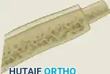

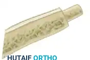
As demonstrated in the detailed intraoperative and schematic images above, the fashioning of the graft requires meticulous attention to detail:
1. Distal End (The Dowel): Using a rongeur and a fine burr, the distal end of the cortical graft is fashioned into a perfectly cylindrical dowel (peg). This dowel must match the inner diameter of the prepared distal metacarpal medullary canal to achieve a tight, interference press-fit.
2. Proximal End (The Oblique Cut): The opposite end of the graft is cut obliquely at a 30-degree angle. This cut must perfectly mate with the recess created in the proximal metacarpal base. The cortical surface of the graft should face dorsally to provide maximum structural rigidity against flexion forces.
4. Graft Insertion and Fixation
The insertion of the graft is a critical maneuver that relies on the inherent tension of the hand's soft tissues.
* Insertion: The doweled end of the graft is first impacted into the medullary canal of the distal fragment. Next, an assistant applies strong longitudinal traction to the digit, distracting the fragments. The surgeon then toggles the 30-degree proximal end of the graft into the prepared proximal recess.
* Biomechanical Stability: Once traction is released, the natural resting tension of the intrinsic and extrinsic muscles actively compresses the graft between the two fragments. This longitudinal compression locks the 30-degree oblique cut into place, providing remarkable inherent stability to the construct.
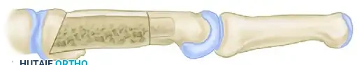
- Supplemental Fixation: While the geometric press-fit provides excellent primary stability, supplemental fixation is almost always prudent to prevent rotational displacement or dislodgement during early postoperative swelling. The construct is typically stabilized by passing one or two smooth Kirschner wires (K-wires) obliquely or transversely through the graft and into the adjacent, uninvolved metacarpals. Care must be taken not to split the cortical graft during K-wire insertion.
- Closure: If a viable periosteal sheath is present, it is meticulously closed over the graft to provide a vascularized cambium layer. The tourniquet is deflated, hemostasis is achieved, and the soft tissues are closed in layers.
Management of Secondary Contractures
Prolonged metacarpal shortening frequently leads to secondary contractures of the adjacent joints, most notably the MCP joint. The MCP joint collateral ligaments are eccentric; they are lax in extension and taut in flexion. When a metacarpal is shortened, these ligaments rapidly contract and fibrose in the shortened position. If intraoperative assessment reveals that MCP joint flexion is severely limited despite restoration of bone length, a formal dorsal capsulotomy and selective release of the dorsal fibers of the collateral ligaments must be performed concurrently to achieve 70 to 90 degrees of passive MCP flexion.
Complications, Incidence Rates, and Salvage Management
Surgeons undertaking the Littler technique must be acutely aware of the potential complications and possess the reconstructive armamentarium to manage them. The hostile nature of the initial trauma makes complication rates higher than in standard elective hand surgery.
| Complication | Estimated Incidence | Etiology and Pathophysiology | Salvage Management and Revision Strategy |
|---|---|---|---|
| Graft Resorption / Nonunion | 10% - 15% | Usually the result of inadequate soft tissue coverage, thermal necrosis during bone preparation (failure to irrigate while sawing), or failure to completely resect the avascular pseudarthrosis. | Requires a comprehensive revision. The non-viable graft must be excised. A fresh cortico-cancellous graft is harvested, and rigid internal fixation (locking plate) is usually required. Preliminary flap coverage may be necessary. |
| Deep Infection | 3% - 8% | Reactivation of dormant bacteria from the initial open trauma, or acute surgical site infection. The avascular cortical graft acts as a nidus for biofilm formation. | A catastrophic complication requiring immediate return to the OR. The graft and hardware must be removed. Aggressive serial debridement, placement of an antibiotic cement spacer, and targeted IV antibiotics are required. Staged reconstruction is delayed until inflammatory markers normalize. |
| Extensor Tendon Adhesions | 20% - 30% | Failure to preserve the paratenon, excessive surgical trauma to the soft tissues, or prolonged postoperative immobilization leading to dense scar formation binding the tendon to the graft. | Prevention via early mobilization of uninvolved digits is key. If severe stiffness persists despite aggressive hand therapy, a secondary tenolysis and capsulotomy may be required 6 to 12 months after radiographic bone healing is confirmed. |
| Rotational Malunion | 5% - 10% | Failure to assess the digital cascade intraoperatively, or loss of fixation postoperatively due to inadequate K-wire stabilization. Leads to digital scissoring. | Intraoperative prevention is paramount: all flexed fingertips must point toward the scaphoid tubercle. Postoperative rotational malunion requires a corrective corrective osteotomy once the graft has fully incorporated. |
| Donor Site Morbidity | 5% - 12% | Hematoma, infection, or tibial stress fracture at the graft harvest site. | Meticulous hemostasis at the donor site, prophylactic avoidance of sharp corners during cortical harvest (to prevent stress risers), and protected weight-bearing postoperatively. |
Phased Post-Operative Rehabilitation Protocols
The postoperative protocol following a Littler reconstruction is a delicate balancing act. The surgeon must provide rigid immobilization to allow the avascular graft to incorporate and revascularize, while simultaneously preventing irreversible joint stiffness and tendon adhesions in the traumatized hand.
Phase I: Immediate Postoperative Immobilization (Days 0-12)
Immediately following surgery, the hand is placed in a bulky, compressive dressing and a rigid plaster splint.
* Positioning: The hand must be immobilized in the classic "position of function" (the intrinsic-plus position). The wrist is extended 20 to 30 degrees, the MCP joints are flexed to 70 to 90 degrees (to place the collateral ligaments on maximal stretch and prevent contracture), and the interphalangeal (IP) joints are left fully extended.
* Edema Control: Postoperative swelling following structural bone grafting and extensive soft tissue dissection can be severe. The cast or splint must be immediately split or bivalved in the operating room to accommodate this swelling and prevent compartment syndrome or devastating soft tissue necrosis over the dorsum of the hand. Strict elevation is mandated.
* Pharmacology: Given the high-risk nature of the procedure and the history of open trauma, prophylactic intravenous antibiotics administered during surgery are typically transitioned to a short course of oral antibiotics for 3 to 5 days postoperatively.
Phase II: Focused Immobilization (Day 12 to 2 Months)
At approximately the 12th postoperative day, the initial bulky dressings are removed. The surgical incisions are inspected, and sutures are removed if the wound is adequately healed.
* Targeted Casting: A new, meticulously molded fiberglass cast or a custom-fabricated rigid thermoplastic splint is applied. The critical transition in this phase is to immobilize only the grafted metacarpal and its corresponding proximal phalanx. The adjacent, uninvolved digits are left completely free.
* Early Motion: The patient is instructed to begin active range of motion (ROM) exercises of the uninvolved digits. This early motion is crucial for preventing diffuse hand stiffness and encouraging excursion of the common extensor muscle belly, which helps prevent adhesions at the surgical site.
* Duration: This targeted immobilization is strictly maintained for a total of 8 weeks, or until definitive radiographic evidence of bridging trabecular bone is confirmed at both the proximal and distal graft-host interfaces.
Phase III: Rehabilitation and Strengthening (Month 2 Onwards)
Once clinical stability (absence of pain at the graft site) and radiographic union are achieved, rigid immobilization is discontinued.
* Hardware Removal: If smooth K-wires were utilized for supplemental transverse fixation, they are typically removed in the clinic between 6 to 8 weeks postoperatively, just prior to the initiation of aggressive mobilization.
* Hand Therapy: Aggressive, formal hand therapy is initiated. The focus shifts to active and passive ROM of the MCP and IP joints, specific tendon gliding exercises to overcome early adhesions, and progressive grip strengthening. Dynamic or static-progressive splinting may be introduced if residual joint contractures persist. Maximum medical improvement is often not reached until 9 to 12 months postoperatively.
Summary of Landmark Literature and Clinical Guidelines
The foundation of structural metacarpal reconstruction rests heavily on the early 20th-century work of Sterling Bunnell, who first articulated the necessity of atraumatic soft tissue handling and precise "bone carpentry" in the hand. However, it was J. William Littler who formalized the specific geometric grafting technique that bears his name. Littler's original descriptions emphasized the biomechanical superiority of the doweled distal end and the 30-degree proximal cut in resisting the intrinsic deforming forces of the hand without the need for bulky, soft-tissue-compromising plates.
Modern clinical guidelines and contemporary literature continue to validate the Littler technique, particularly in environments where advanced locking plate technology is either unavailable or contraindicated due to poor soft tissue envelopes. Comparative studies have demonstrated that while plate fixation provides superior immediate rigidity, the biological footprint of a perfectly executed Littler autograft—relying on longitudinal compression and minimal hardware—often results in superior extensor tendon excursion and fewer hardware-related complications.
Current consensus dictates that the success of the Littler technique is heavily dependent on patient selection, absolute eradication of latent infection, and the prerequisite establishment of a robust soft tissue envelope. When executed with precision, it remains an indispensable, highly effective procedure in the reconstructive orthopedic surgeon's armamentarium for salvaging the severely traumatized hand.
