- Cellular biology of bone ( Fig. 1.2)
- Osteoblasts
- Appear as cuboid cells aligned in layers along immature osteoid
- Are derived from undifferentiated mesenchymal stem cells
- These stem cells line haversian canals, endosteum, and periosteum.
- Become osteoblasts under conditions of low strain and increased oxygen tension
-
Transcription factor RUNX2 and bone morphogenetic protein (BMP) direct mesenchymal cells to the osteoblast lineage.
- Core-binding factor α-1 and β-catenin also stimulate differentiation into osteoblast
- Become cartilage under conditions of intermediate strain and low oxygen tension
- Become fibrous tissue under conditions of high strain
- Have more endoplasmic reticulum, Golgi apparatus, and mitochondria than do other cells (for synthesis and secretion of matrix)
- Bone surfaces lined by more differentiated, metabolically active cells
- Entrapped cells: less active cells in resting regions; maintain the ionic milieu of bone
- Disruption of the active lining cell layer activates entrapped cells.
- Receptor-effector interactions in osteoblasts are summarized in Table 1.2 . 7. Osteoblasts produce the following:
- Alkaline phosphatase
- Osteocalcin (stimulated by 1,25dihydroxyvitamin D [1,25(OH)2D3])
- Type I collagen
- Bone sialoprotein
- Receptor activator of nuclear factor (NF)-κβ ligand (RANKL)
- Osteoprotegerin—binds RANKL to limit its activity
-
Osteoblast activity stimulated by intermittent
(pulsatile) exposure to parathyroid hormone (PTH)

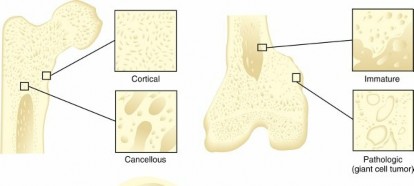
 FIG. 1.1 Types of bone. Cortical bone consists of tightly packed osteons. Cancellous bone consists of a meshwork of trabeculae. In immature bone, unmineralized osteoid lines the immature trabeculae. Pathologic bone is characterized by atypical osteoblasts and architectural disorganization. Colorized from Brinker MR, Miller MD:
Fundamentals of
FIG. 1.1 Types of bone. Cortical bone consists of tightly packed osteons. Cancellous bone consists of a meshwork of trabeculae. In immature bone, unmineralized osteoid lines the immature trabeculae. Pathologic bone is characterized by atypical osteoblasts and architectural disorganization. Colorized from Brinker MR, Miller MD:
Fundamentals of
orthopaedics, Philadelphia, 1999, Saunders, p 2. Microscopic Subtypes Characteristics Examples Appearance --- Lamellar | Cortical
| Structure is oriented along lines of stress
Strong
| Femoral shaft Cancellous | More elastic than cortical bone
| Distal femoral metaphysis Woven | Immature
| Not stress oriented
| Embryonic
skeleton Fracture callus Pathologic | Random
organization Increased turnover Weak
Flexible
| Osteogenic
sarcoma Fibrous
dysplasia
Table 1.1 Types of Bone
Modified from Brinker MR, Miller MD:
Fundamentals of orthopaedics,
Philadelphia, 1999, Saunders, p 1.
9. Osteoblast activity inhibited by TNF-α
10. Wnts are proteins that promote osteoblast survival and proliferation.
1. Deficient Wnt causes osteopenia; excessive Wnt expression causes high bone mass.
2. #### Wnts can be sequestered by other secreted molecules such as sclerostin (Scl) and Dickkopf-related protein 1 (Dkk-1).
2.
Osteocytes (see
Fig. 1.1)
1. Maintain bone
1. Inhibiting sclerostin or Dkk-1 will lead to increased bone mass
1. Constitute 90% of the cells in the mature skeleton
2. Former osteoblasts surrounded by newly formed matrix
3. High nucleus/cytoplasm ratio
4. Long interconnecting cytoplasmic processes projecting through the canaliculi
5. Less active in matrix production than osteoblasts
6. Important for control of extracellular calcium and phosphorus concentration

 FIG. 1.2 Cellular origins of bone and cartilage cells.
### Table 1.2
Bone Cell Types, Receptor Types, and Effects Cell Type
|
Receptor
|
Effect
| ---|---|---|
Osteoblast
| PTH
FIG. 1.2 Cellular origins of bone and cartilage cells.
### Table 1.2
Bone Cell Types, Receptor Types, and Effects Cell Type
|
Receptor
|
Effect
| ---|---|---|
Osteoblast
| PTH
| Releases a secondary messenger (exact mechanism unknown) to stimulate osteoclastic activity
Activates adenylyl cyclase
1,25(OH) 2 vitamin D 3
| Stimulates matrix and alkaline phosphatase synthesis and production of bone-specific proteins (e.g., osteocalcin)
Glucocorticoids
| Inhibits DNA synthesis, collagen production, and osteoblast protein synthesis
Prostaglandins
| Activates adenylyl cyclase and stimulates bone resorption
Estrogen
| Has anabolic (bone production) and anticatabolic (prevents bone resorption) properties
Increases mRNA levels for alkaline phosphatase
Inhibits activation of adenylyl cyclase
Osteoclast
| Calcitonin
| Inhibits osteoclast function (inhibits bone resorption)

---
FIG. 1.3 Paracrine crosstalk between osteoblasts and osteoclasts.
From Kumar V et al, editors: Bones, joints, and soft tissue tumors. In
Robbins and Cotran pathologic basis of disease,
ed 9, Philadelphia, 2014, Saunders, Fig. 26-5.
7. Directly stimulated by calcitonin, inhibited by PTH
8. #### Sclerostin secreted by osteocytes helps negative
feedback on osteoblasts’ bone deposition (**
Fig. 1.3**
).
1. Osteoclasts
2. Differentially regulated according to mechanical loading, with decreased sclerostin in areas of concentrated strain
3. Downregulation is associated with increased bone formation (via sclerostin antibody).
4. Potential for use in fracture healing, bone loss, osseous integration of implants, and genetic bone diseases via upregulation of sclerostin
1. Multinucleated irregular giant cells
2. #### Derived from hematopoietic cells in macrophage lineage
3. Monocyte progenitors form giant cells by fusion
4. Function
1. Bone resorption
1. #### Bone formation and resorption are linked
2. #### Stimulated primarily by RANKL binding to RANK receptor on cell surface
3.
Osteoblasts (and tumor cells) express RANKL (
Fig. 1.4 ):
1. #### Binds to receptors on osteoclasts
2. #### Stimulates differentiation into mature osteoclasts
3. #### Inhibited by osteoprotegerin (OPG) binding to RANKL
4. Occurs both normally and in certain conditions, including multiple myeloma and metastatic bone disease
5. Resorption mechanism
1. #### Denosumab is a monoclonal antibody that targets and inhibits RANKL binding to the RANK receptor
1. Signaling
2. Osteoclasts possess a ruffled (brush) border and surrounding clear zone
1. Border consists of plasma membrane enfoldings that increase surface area
2. #### Bind to bone surfaces through cell attachment (anchoring) proteins
1. Integrin (αvβ3 or vitronectin receptor)
3. Bone resorption occurs in depressions: Howship lacunae.
1. Effectively seal the space below the osteoclast
2. Synthesize tartrate-resistant acid phosphate
3. Produce hydrogen ions through carbonic anhydrase
4. Lower pH
5. Increase solubility of hydroxyapatite crystals
4. Organic matrix then removed by proteolytic digestion through activity of the lysosomal enzyme
cathepsin K
3. Have calcitonin receptors, which inhibit osteoclastic resorption
4. Interleukin-1 (IL-1): potent stimulator
of osteoclast differentiation and bone resorption
hole zones and pores.

 FIG. 1.4 Control and function of the osteoclast.
Vit,
FIG. 1.4 Control and function of the osteoclast.
Vit,
vitamin.
