Patient Presentation & History
A 35-year-old male presented to the emergency department following a high-energy motor vehicle collision (MVC). He was the unrestrained driver involved in a head-on collision at approximately 50 mph, experiencing direct impact to the lateral aspect of his left knee against the dashboard. Paramedics reported significant deformity and ecchymosis of the left knee at the scene.
On arrival, the patient reported immediate, severe, excruciating pain in his left knee, rendering him unable to bear any weight. He denied loss of consciousness but reported transient numbness and paresthesia in his left foot and ankle immediately after the impact, which had partially resolved. There were no other overt injuries reported in the primary survey by the trauma team, though secondary survey was ongoing.
AMPLE History:
*
A
llergies: No known drug allergies (NKDA).
*
M
edications: None.
*
P
ast Medical/Surgical History: Healthy, no prior surgeries, no chronic medical conditions.
*
L
ast Meal: Approximately 3 hours prior to injury.
*
E
vents: Detailed as above.
Social History: Non-smoker, occasional alcohol use. Employed as a construction worker, indicating a high functional demand.
Clinical Examination
Upon focused orthopedic examination after primary and secondary surveys by the trauma team, the following was noted:
General: Patient was alert, oriented, and hemodynamically stable. Pain localized to the left knee.
Left Lower Extremity:
*
Inspection:
Gross deformity of the left knee, with significant swelling and ecchymosis extending from the distal thigh to the proximal calf. There was a large, tense effusion. Skin integrity was compromised with a large, non-blanchable blister developing laterally over the tibial plateau, approximately 3x2 cm, and impending fracture blisters around the anterolateral aspect. No open wounds were noted initially. The limb appeared subtly shortened and in slight valgus angulation.
*
Palpation:
Markedly tender globally around the knee joint, particularly over the lateral and medial tibial condyles. Palpable crepitus was elicited with gentle manipulation. The patella was ballotable, confirming a significant hemarthrosis. Distal pulses (dorsalis pedis and posterior tibial) were present but faintly palpable compared to the contralateral limb; capillary refill was sluggish at 3-4 seconds. Compartments of the lower leg felt firm but not tense on initial examination.
*
Range of Motion (ROM):
Actively and passively severely limited due to pain and guarding. Estimated active ROM was 0-10 degrees of flexion. Attempts at passive ROM were met with extreme pain.
*
Neurological Assessment:
*
Peroneal Nerve:
Weakness in ankle dorsiflexion (grade 3/5) and great toe extension (grade 3/5). Sensory deficit noted in the first web space and dorsum of the foot. This represented a concerning partial peroneal nerve palsy.
*
Tibial Nerve:
Intact motor function (plantarflexion grade 5/5) and sensation in the sole of the foot.
*
Femoral Nerve:
Intact motor and sensory function.
*
Vascular Assessment:
Distal pulses were weak. Ankle-Brachial Index (ABI) was performed bilaterally. Left ABI was 0.82, right ABI was 1.0. The diminished ABI raised significant concern for a popliteal artery injury, despite palpable pulses.
*
Ligamentous Stability:
Due to extreme pain, guarding, and gross swelling, a comprehensive assessment of ligamentous stability was deferred. However, gross instability was noted with gentle varus stress in extension, suggesting significant lateral collateral ligament (LCL) or posterolateral corner (PLC) involvement, or severe bone loss.
*
Associated Injuries:
No obvious hip or ankle injuries detected on clinical examination. Spine was stable to palpation.
Given the high-energy mechanism, gross deformity, significant soft tissue compromise, neurological deficit, and concerning vascular status, an emergent workup was initiated.
Imaging & Diagnostics
Initial Radiographs (AP, Lateral, Oblique views of the Left Knee):
* Demonstrated a complex, comminuted intra-articular fracture involving both the medial and lateral tibial condyles, with significant articular depression and metaphyseal-diaphyseal dissociation.
* The lateral condyle showed a large split component with central depression.
* The medial condyle appeared more fragmented, with a coronal split extending distally.
* Significant widening of the proximal tibia was noted.
* The overall appearance was highly suggestive of a Schatzker Type VI tibial plateau fracture.
* No obvious distal femur or patella fracture.
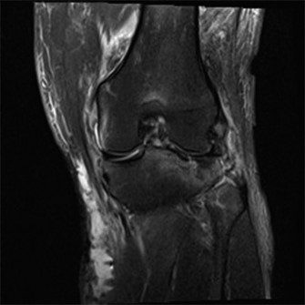
Figure 1: Initial AP radiograph of the left knee demonstrating a comminuted, bicondylar tibial plateau fracture with significant articular depression and metaphyseal-diaphyseal dissociation, consistent with a Schatzker Type VI injury.
Computed Tomography (CT) Scan of the Left Knee (with 3D Reconstructions):
*
Crucial for operative planning
, the CT scan confirmed the Schatzker Type VI bicondylar fracture pattern.
*
Lateral Plateau:
Demonstrated a comminuted articular segment with approximately 8 mm of depression and a 12 mm step-off. A large split component extended into the metaphysis.
*
Medial Plateau:
Showed a highly comminuted fracture with a significant coronal component involving the posterior aspect of the medial condyle (posterior meniscocapsular injury often associated), along with a metaphyseal split extending proximally from the medial cortex. The medial articular surface was depressed by approximately 6 mm.
*
Metaphyseal-Diaphyseal Dissociation:
Extensive comminution in the metaphyseal region, connecting the articular segments to the diaphysis.
*
Soft Tissue Windowing:
Confirmed severe soft tissue swelling and a large hemarthrosis. No overt signs of acute compartment syndrome on CT, but clinical monitoring remained paramount.
*
3D reconstructions
were invaluable in appreciating the complex geometry of the fracture and planning reduction strategies, particularly for the posterior fragments.
CT Angiography (CTA) of the Left Lower Extremity:
* Performed urgently due to the abnormal ABI (0.82) and high-energy mechanism.
* Revealed intimal dissection of the popliteal artery in the region immediately posterior to the knee joint, with compromised luminal flow but without complete occlusion. This explained the diminished, but still palpable, distal pulses.
Magnetic Resonance Imaging (MRI):
* Given the vascular injury and the need for immediate surgical intervention (vascular repair and provisional fixation), MRI was deferred initially.
* It would typically be indicated pre-operatively (after provisional fixation) or later if meniscal or cruciate ligament injuries were highly suspected and not adequately assessed during surgery, or if the patient developed persistent pain/instability post-fixation. High suspicion for meniscal tears and possibly ACL/PCL injuries with this fracture pattern.
Pre-Operative Templating:
* Utilizing the CT scans (2D slices and 3D reconstructions), meticulous pre-operative templating was performed. This involved selecting appropriate plate lengths (e.g., locking plates for lateral and medial columns), screw trajectories to capture articular fragments and avoid hardware collision, and identifying potential areas for bone grafting. The significant metaphyseal void suggested the need for bone graft (autograft from iliac crest or allograft).
Differential Diagnosis
The patient's presentation with a high-energy knee injury, gross deformity, and neurovascular compromise necessitates a broad differential diagnosis. While imaging quickly pointed to a specific diagnosis, it's crucial to consider other potential pathologies for similar presentations.
| Feature / Condition | Tibial Plateau Fracture (Schatzker VI) | Knee Dislocation (Multiligamentous Injury) | Distal Femur Fracture (Type C3) |
|---|---|---|---|
| Mechanism of Injury | High-energy axial load with valgus/varus component (e.g., MVC, fall). | High-energy direct trauma or hyperextension/hyperflexion with rotation. | High-energy (MVC, fall from height) or low-energy (osteoporotic). |
| Clinical Presentation | Gross deformity, severe pain, inability to bear weight, swelling, ecchymosis, crepitus. | Gross deformity (often reducible/reduced), severe pain, significant instability, swelling. | Gross deformity, severe pain, inability to bear weight, often with soft tissue compromise. |
| Palpation | Localized tenderness over tibial condyles, joint effusion, crepitus. | Joint line tenderness, often laxity with gentle stress (if not reduced). | Localized tenderness over distal femur, effusion often less prominent initially. |
| Neurological Deficit | Peroneal nerve palsy common (stretch/compression). | Peroneal nerve palsy common due to stretch. | Sciatic or femoral nerve injury less common but possible with severe displacement. |
| Vascular Compromise | Popliteal artery injury less common than knee dislocation but possible with severe displacement or bicondylar patterns. ABI <0.9 requires CTA. | HIGH RISK of popliteal artery injury (up to 40%). ABI <0.9 mandates CTA. | Popliteal artery injury possible but less frequent than knee dislocation. ABI still important. |
| Radiographs | Intra-articular fracture of proximal tibia (bicondylar involvement, articular depression, metaphyseal comminution). | Often normal bone anatomy (unless associated avulsion fx), joint incongruity. | Fracture lines in distal femur, often intra-articular, epiphyseal-metaphyseal extension. |
| CT Scan | Defines fracture pattern, articular depression, comminution, fragment displacement. Essential for Schatzker classification. | Rule out occult fractures, assess bony avulsions, patellofemoral tracking. | Defines fracture lines, articular involvement, comminution. Essential for AO/OTA classification. |
| MRI Scan | Later to assess meniscal/ligamentous injuries, or if soft tissue injury suspected but not seen on CT. | CRITICAL for assessing integrity of cruciate and collateral ligaments, menisci, and capsule. | Assess chondral injury, meniscal injury (if associated), or ligamentous injury. |
| Urgent Management | Provisional external fixation, vascular repair (if needed), monitoring for compartment syndrome. | Urgent reduction (often closed), external fixation, vascular repair (if needed), compartment syndrome monitoring. | Provisional external fixation, vascular repair (if needed), compartment syndrome monitoring. |
| Long-Term Sequelae | Post-traumatic arthritis, malunion, non-union, stiffness, chronic pain. | Chronic instability, arthrofibrosis, post-traumatic arthritis, persistent nerve palsy. | Stiffness, post-traumatic arthritis, non-union, malunion. |
Surgical Decision Making & Classification
Based on the clinical presentation, neurological deficit, vascular injury, and advanced imaging confirming a Schatzker Type VI bicondylar tibial plateau fracture with significant articular depression and metaphyseal-diaphyseal dissociation, the decision for urgent operative intervention was unequivocal.
Indications for Operative Fixation:
1.
Intra-articular fracture with articular incongruity:
More than 2-3 mm of articular depression or step-off requires anatomical reduction to prevent post-traumatic arthritis. Our patient had 8mm and 6mm depression.
2.
Significant fracture displacement and instability:
The bicondylar involvement with metaphyseal comminution leads to gross instability of the knee joint.
3.
Vascular compromise:
The popliteal artery intimal dissection required immediate surgical repair to prevent limb ischemia.
4.
Neurological deficit:
The peroneal nerve palsy, though incomplete, required evaluation during surgery and decompression if entrapped.
5.
Soft tissue compromise:
Impending fracture blisters and severe swelling mandated a staged approach to allow for soft tissue recovery.
6.
Open fracture:
While not initially open, severe soft tissue compromise could progress to an open injury.
Classification:
*
Schatzker Classification:
Type VI (Bicondylar fracture with metaphyseal-diaphyseal dissociation). This is a high-energy injury involving both medial and lateral condyles with extension into the metaphysis, often implying severe soft tissue disruption.
*
AO/OTA Classification:
41-C3 (Complete articular fracture, multifragmentary articular and metaphyseal). This provides a more detailed description of the fracture complexity.
Surgical Timing and Staging:
Given the emergent vascular injury, the surgical plan was a
staged approach
:
1.
Immediate surgical exploration for vascular repair:
This takes precedence over definitive fracture fixation. The popliteal artery intimal dissection required immediate repair to restore optimal distal perfusion.
2.
Provisional External Fixation:
Following vascular repair, a spanning external fixator across the knee joint would be applied. This would provide immediate stability, restore length and alignment, and allow for the injured soft tissues to recover from swelling and reduce the risk of further skin breakdown or compartment syndrome. The fixator would also achieve indirect reduction via ligamentotaxis.
3.
Delayed Definitive Open Reduction and Internal Fixation (ORIF):
After approximately 7-14 days, once the soft tissue envelope had sufficiently recovered (wrinkle sign present, blisters healed, swelling decreased), definitive ORIF would be performed. This delay is critical to minimize wound complications such as infection, dehiscence, and necrosis.
Surgical Technique / Intervention
Stage 1: Emergent Vascular Repair & External Fixation (Day 0)
A. Vascular Repair:
*
Patient Positioning:
Supine with the left leg prepped and draped from hip to toes.
*
Approach:
Medial approach to the popliteal artery via a longitudinal incision over the posteromedial aspect of the knee.
*
Procedure:
The popliteal artery was exposed. Intraoperative findings confirmed an intimal dissection. Vascular surgery performed an arteriotomy and primary repair of the intimal tear, followed by an interposition saphenous vein graft from the contralateral leg to bypass the damaged segment and ensure robust flow. Completion angiogram confirmed excellent flow.
B. Provisional External Fixation:
*
Patient Positioning:
Supine with the leg supported on a radiolucent table.
*
Technique:
A spanning knee external fixator (e.g., Stryker Hoffmann II MRI-compatible) was applied.
*
Proximal Pins:
Two pins were inserted into the distal femur, parallel to the knee joint, ensuring bicortical purchase.
*
Distal Pins:
Two pins were inserted into the distal tibia, approximately 10-15 cm below the knee joint, in the sagittal plane, avoiding the fracture zone.
*
Frame Assembly:
The frame was constructed to allow for distraction and correction of the gross angulation and length. Ligamentotaxis provided initial indirect reduction of the articular fragments.
*
Post-Procedure:
Compartment pressures were monitored hourly following the vascular repair due to the risk of reperfusion injury and subsequent compartment syndrome. The leg was kept in a slightly flexed position to minimize tension on the vascular repair.
Stage 2: Definitive Open Reduction and Internal Fixation (Day 9)
After 9 days, the patient's soft tissue envelope had improved significantly, with resolution of most blisters and a positive "wrinkle sign."
- Patient Positioning: Supine on a radiolucent table with a bump under the ipsilateral hip. A high thigh tourniquet was applied. The knee was flexed to 90 degrees using a leg holder (e.g., femoral distractor setup for visualization and aid in reduction).
-
Approaches: Given the bicondylar nature (Schatzker VI), a dual incision approach was chosen for optimal visualization and fixation of both medial and lateral columns.
- Anterolateral Approach: A longitudinal incision centered over Gerdy's tubercle, extending proximally and distally. The iliotibial band was split longitudinally. Subperiosteal dissection exposed the lateral tibial plateau. The anterior tibialis muscle was elevated from the proximal tibia.
- Modified Posteromedial Approach: A separate longitudinal incision made along the posteromedial aspect of the proximal tibia, posterior to the saphenous vein and nerve. This approach allows direct access to the posterior medial condyle fragments and buttress plating. Care taken to protect the saphenous nerve and vein.
-
Reduction Techniques:
-
Lateral Plateau Reduction:
- An arthrotomy was made, allowing visualization of the articular surface.
- The lateral meniscus was inspected; a peripheral tear was identified and tagged for later repair.
- Using an osteotome or elevator, the depressed articular fragments were gently elevated back to their anatomical position under direct visualization and fluoroscopic guidance.
- The resultant metaphyseal void beneath the elevated articular segment was packed with corticocancellous autograft harvested from the ipsilateral iliac crest to provide structural support and promote healing. Allograft cancellous chips were also available if needed.
- Temporary fixation with K-wires was used to hold the reduced articular surface.
-
Medial Plateau Reduction:
- Access via the posteromedial incision.
- Using osteotomes and clamps, the medial condyle fragments, including the coronal split component, were anatomically reduced.
- Indirect reduction through ligamentotaxis from the external fixator (which was removed immediately prior to definitive fixation) helped restore overall alignment.
- Fluoroscopy confirmed reduction of the articular surface and overall alignment.
- Temporary K-wires were used for provisional stabilization.
- Metaphyseal-Diaphyseal Realignment: Attention was turned to restoring the overall length, alignment, and rotation of the proximal tibia relative to the diaphysis. This was achieved with traction and careful manipulation, guided by fluoroscopy.
-
Lateral Plateau Reduction:
-
Fixation Construct:
- Lateral Fixation: A pre-contoured locking plate (e.g., LCP Proximal Lateral Tibia Plate) was applied to the lateral aspect of the tibia. Screws were carefully inserted to achieve bicortical purchase and capture the articular fragments (raft screws) while avoiding intra-articular penetration. Fluoroscopy confirmed optimal screw placement and length.
- Medial Fixation: A medial buttress plate (e.g., LCP Medial Proximal Tibia Plate) was applied via the posteromedial incision. This plate provided strong support against varus collapse and helped neutralize the coronal split components. Screws were placed strategically, ensuring stable fixation without compromising the lateral construct.
- Meniscal Repair: The lateral meniscal tear identified earlier was repaired using an all-inside suture technique.
- Final Checks: The knee joint was thoroughly irrigated. ROM was checked to ensure no hardware impingement and stability. All reductions and fixation were confirmed with final fluoroscopic images in multiple planes.
-
Closure: Layered closure of both surgical incisions. A drain was placed in the deep lateral wound, typically removed post-operative day 1 or 2.
Post-Operative Protocol & Rehabilitation
Phase 1: Acute Protection and Early Motion (Weeks 0-6)
*
Weight Bearing:
Non-weight bearing (NWB) on the operated limb using crutches or a walker. Strict adherence is critical.
*
Immobilization:
A hinged knee brace locked in full extension for ambulation and transfers to protect the fracture fixation.
*
Range of Motion (ROM):
* Continuous Passive Motion (CPM) machine initiated immediately post-op, aiming for 0-30 degrees initially, progressing to 0-90 degrees by week 6, as tolerated.
* Gentle active and passive ROM exercises within the brace limits while supine.
*
Exercises:
* Quad sets, gluteal sets, ankle pumps (for DVT prophylaxis).
* Straight leg raises (SLRs) with the brace locked in extension.
* Upper body and core strengthening.
*
Pain Management:
Multimodal approach including NSAIDs, acetaminophen, and opioid analgesics as needed.
*
DVT Prophylaxis:
Low molecular weight heparin (LMWH) or aspirin per protocol.
*
Wound Care:
Daily dressing changes, monitor for signs of infection. Drain removed typically POD 1-2.
Phase 2: Progressive Weight Bearing and Strengthening (Weeks 6-12)
*
Weight Bearing:
Gradual progression from NWB to partial weight bearing (PWB) (e.g., 25% of body weight) around week 6-8, increasing as tolerated, guided by radiographic evidence of healing. Full weight bearing (FWB) typically by week 10-12, assuming adequate radiographic union.
*
Brace:
Hinged knee brace continued, gradually increasing ROM limits as directed by the surgeon (e.g., unlock for specific exercises, then remove for ambulation).
*
ROM:
Aim for full knee flexion and extension by week 12. Continued use of CPM or self-stretching techniques.
*
Exercises:
* Initiate light closed-chain exercises (mini-squats, leg presses with limited depth).
* Isometric strengthening of quadriceps and hamstrings.
* Balance and proprioception exercises (e.g., single-leg stance with support).
* Stationary cycling with low resistance.
*
Imaging:
X-rays at 6 weeks and 12 weeks to assess fracture healing.
Phase 3: Advanced Strengthening and Return to Activity (Weeks 12-24+)
*
Weight Bearing:
Full weight bearing as tolerated, without support if able.
*
Brace:
Discontinued once cleared by the surgeon and physical therapist.
*
Exercises:
* Progressive resistive exercises (PREs) for all major muscle groups around the knee.
* Open-chain exercises (leg extensions, hamstring curls) can be added carefully.
* Advanced balance and proprioception.
* Agility drills, sport-specific training (if applicable, with caution).
* Focus on cardiovascular fitness.
*
Goals:
Restore full strength, endurance, and function. Return to work (construction worker) will be carefully phased based on physical demands. High-impact activities or return to sport may be delayed for 6-12 months or longer, depending on healing and knee function.
*
Potential for Hardware Removal:
If hardware becomes symptomatic (e.g., irritation, bursitis), removal may be considered 12-18 months post-op, typically after complete fracture healing.
Monitoring for Complications:
*
Infection:
Vigilant monitoring of surgical sites.
*
Arthrofibrosis/Stiffness:
Early, controlled ROM is key. Aggressive physical therapy. Manipulation under anesthesia or even arthroscopic lysis of adhesions may be required if severe.
*
Post-Traumatic Arthritis:
A significant risk with intra-articular fractures, even with anatomical reduction. Long-term follow-up is necessary.
*
Non-union/Malunion:
Monitored with serial X-rays. May require revision surgery.
*
Hardware Complications:
Breakage, prominence, loosening.
*
Chronic Pain/Neuropathy:
Particularly relevant given the initial peroneal nerve palsy. Nerve gliding exercises, pain management.
Pearls & Pitfalls (Crucial for FRCS/Board Exams)
Pearls:
- Vascular Assessment is Paramount: Always perform an Ankle-Brachial Index (ABI) on any high-energy knee injury. An ABI < 0.9 mandates urgent CT Angiography to rule out popliteal artery injury. Even palpable pulses do not rule out intimal damage. Vascular injury takes precedence.
- Soft Tissue Envelope Dictates Timing: For severe tibial plateau fractures, particularly Schatzker V/VI, a staged approach with provisional external fixation is crucial to allow soft tissue swelling to subside and blisters to heal. Never rush definitive fixation into a compromised soft tissue envelope ("wait for the wrinkle sign").
- CT Scan is Mandatory: Plain radiographs are initial screening, but a high-resolution CT scan with 3D reconstructions is non-negotiable for accurate fracture mapping, understanding articular depression, comminution, and pre-operative templating.
- Anatomical Reduction of the Articular Surface: This is the primary goal to minimize the risk of post-traumatic arthritis. Even 1-2 mm of articular step-off can significantly impact long-term outcomes. Use direct visualization (mini-arthrotomy), reduction clamps, and temporary K-wires.
- Bone Grafting for Metaphyseal Voids: After elevating depressed articular fragments, fill the metaphyseal defect with bone graft (autograft, allograft, or synthetic substitute) to provide structural support and prevent collapse.
- Dual Plating for Bicondylar Fractures: Schatzker Type V and VI fractures often require dual plating (lateral locking plate, medial buttress plate) to provide adequate stability against axial loads, valgus/varus stress, and to capture complex fragment patterns.
- Address Meniscal Injuries: High-energy tibial plateau fractures frequently involve meniscal tears (especially lateral). Inspect the menisci during surgery and repair if indicated and feasible, as they are crucial for knee kinematics and load distribution.
- Early, Controlled Range of Motion: Once stable fixation is achieved, initiate early, controlled ROM within safe limits (often with a hinged brace and CPM) to prevent arthrofibrosis and improve cartilage nutrition, balancing this with fracture stability.
- Careful Nerve Dissection: Identify and protect the common peroneal nerve laterally and the saphenous nerve and great saphenous vein medially during exposure.
Pitfalls:
- Missing Vascular or Neurological Injuries: Failure to perform a thorough neurovascular assessment, especially ABI, can lead to devastating consequences (limb loss, permanent nerve deficits).
- Operative Fixation Through Compromised Soft Tissues: Operating on a swollen, blistered knee significantly increases the risk of wound dehiscence, infection, and flap necrosis, potentially jeopardizing the entire construct.
- Inadequate Articular Reduction: Accepting an imperfect articular reduction will almost guarantee early onset and progression of post-traumatic osteoarthritis, leading to chronic pain and potentially requiring early knee arthroplasty.
- Insufficient Fixation: Using an inadequate construct for complex bicondylar fractures (e.g., single plating for a Schatzker VI) can lead to early fixation failure, loss of reduction, and non-union.
- Failure to Graft Voids: Not filling metaphyseal defects leaves unsupported articular cartilage, which will inevitably collapse over time.
- Premature Weight Bearing: Starting weight bearing too early, before radiographic signs of union are present, can lead to collapse of the articular surface, loss of reduction, and hardware failure.
- Neglecting Concomitant Ligamentous Injuries: While the fracture is the primary focus, severe multi-ligamentous injuries can coexist. While some are addressed acutely, others may require delayed reconstruction. Failure to recognize or address these can lead to chronic instability.
- Compartment Syndrome: Always maintain a high index of suspicion, especially with high-energy trauma, vascular repair, or delayed surgery. Clinical monitoring is key, and prompt fasciotomy is limb-saving.
- Hardware Impingement: Poorly placed screws or prominent plates can cause significant soft tissue irritation or impede joint motion, necessitating early hardware removal.
