Advanced Sacral Fracture Management: Orthopedic Cases & Viva Exam Guide
Key Takeaway
Sacral fracture management involves precise diagnosis, understanding injury patterns like Denis or AO classifications, and tailored treatment. Challenging cases often require multidisciplinary approaches, considering neurovascular integrity and spinopelvic stability. Our guide details advanced surgical and non-surgical strategies crucial for orthopedic specialists.
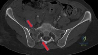
A 24-year-old male presents following a high-energy motor vehicle collision. He is haemodynamically stable but complains of severe lower back pain. No focal neurological deficits are detected on clinical examination. An axial CT scan is obtained as shown below.
Describe the fracture pattern and comment on the associated risk of neurological injury.
Candidate: The image shows a sacral fracture passing through the sacral foramen, which classifies it as a Denis Type II fracture. The risk of neurological injury in this specific pattern is approximately 28%.
Failing to classify the fracture accurately using the Denis system or guessing the neurological deficit percentages. Candidates often confuse Type I, II, and III, or fail to mention that the L5 nerve root is specifically at risk in these patterns.
The candidate should state: "This is a Denis Type II sacral fracture, involving the sacral alae and extending through the foramina. Neurological injury risk is approximately 28%. For completeness, Denis Type I (alar) carries a 6% risk, and Type III (trans-foraminal/central canal) carries a 57% risk due to the potential for cauda equina injury."
The decision is made to stabilize this fracture surgically using percutaneous iliosacral screws. What specific radiographic view is critical to minimize the risk of iatrogenic foot drop, and what structure are you primarily protecting?
Candidate: The pelvic inlet view is essential. It confirms the anterior-to-posterior trajectory of the screw to prevent an anterior cortical breach, which would endanger the L5 nerve root as it drapes over the anterior sacral ala.
Confusing the utility of the Inlet vs. Outlet view. The Outlet view is for cephalad-caudad positioning and looking for sacral dysmorphism; the Inlet view is specifically for avoiding anterior/posterior breach.
Identify the Inlet view clearly for anterior/posterior safety. Explicitly name the L5 nerve root (the "lumbosacral trunk") as the high-risk structure sitting on the ventral ala. Mentioning the "safe zone" of the sacral ala is a bonus high-scoring detail.
Briefly outline how you would screen for "sacral dysmorphism" on plain radiographs prior to screw placement, and why this is clinically significant.
Candidate: Sacral dysmorphism refers to variants that preclude standard safe-zone screw placement. Screening involves checking the outlet view for a non-recessed sacrum, mammillary processes, or a residual disc space between S1-S2. These are significant because the traditional S1 corridor may be narrowed or anatomically altered, necessitating an S2 or trans-sacral screw path.
Focusing only on the fracture and forgetting that the anatomy itself might be abnormal. Failing to recall specific signs of dysmorphism like the "tongue-in-groove" SI joint or mammillary processes.
Provide a structured approach: Identify radiographic signs (residual S1-S2 disc space, mammillary processes, non-recessed sacral ala on outlet view). Explain that dysmorphism signifies an acute alar slope, significantly increasing the risk of screw exit if a standard S1 trajectory is blindly followed. Emphasize that CT is the gold standard for final trajectory planning in these cases.
