Bilateral Cervical Facet Dislocation: Comprehensive Review of Anatomy, Biomechanics, & Surgical Management

Key Takeaway
Bilateral cervical facet dislocation (BCFD) is a severe cervical spine injury involving complete posterior ligamentous disruption and often significant anterior vertebral subluxation, typically from hyperflexion-distraction forces. It carries a high spinal cord injury risk. Management is primarily surgical, focusing on spinal cord decompression, alignment restoration, and stable fixation to optimize patient outcomes.
A 28-year-old male presents following a high-speed motorcycle accident. He is conscious and alert. On examination, he has bilateral C6 radiculopathy. His lateral cervical spine radiograph shows a C5-C6 bilateral facet dislocation with 50% anterolisthesis. Describe the initial management priorities and your decision-making process regarding the timing of MRI.
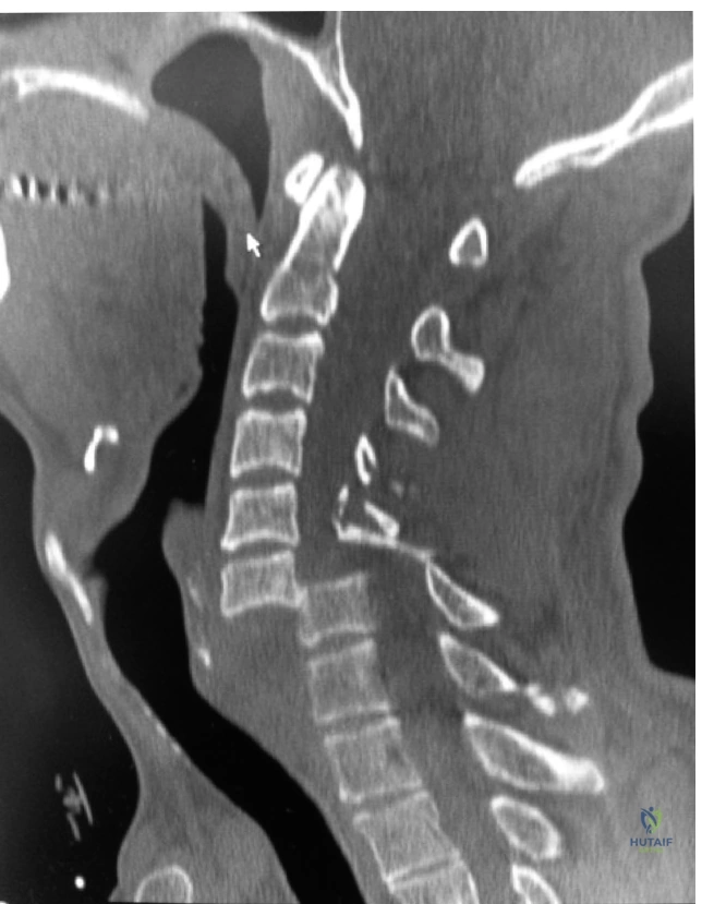
Candidate: I would immediately stabilize the cervical spine using a collar and maintain spinal precautions. Given the bilateral dislocation, this is an unstable injury. Because the patient is awake and cooperative, I would attempt a closed reduction using Gardner-Wells tongs under continuous neurological monitoring. I would obtain an MRI before surgery, but in an awake patient, I could potentially proceed with reduction before the MRI if the patient is neurologically stable and the reduction is monitored carefully. I would plan for definitive stabilization, likely via an anterior or posterior approach depending on findings.
Candidates often fail to explicitly state the "awake and alert" threshold for closed reduction. They might suggest MRI in every case, which delays potentially life-saving decompression, or conversely, suggest reduction in an unconscious patient without an MRI, which is a major red flag as it risks driving a hidden disc fragment into the spinal cord.
The candidate must structure the answer: 1. ATLS/Primary Survey (ABCDE). 2. Rigid stabilization and neurological assessment (ASIA scale). 3. Decision-making on MRI: "In this awake, alert, and cooperative patient, a trial of closed reduction via Gardner-Wells tongs is appropriate if neurological function is monitored throughout. However, if the patient were comatose or intoxicated, an MRI is mandatory to rule out a traumatic disc herniation prior to any reduction maneuver to prevent secondary cord injury." 4. Mention of the STASCIS trial protocol (early decompression within 24 hours). 5. Definitive management involving surgical stabilization (ACDF or posterior fusion).
You have decided to proceed with a posterior surgical approach for a persistent bilateral facet dislocation. Describe the anatomical landmarks and the specific technique for lateral mass fixation at the involved level.
Candidate: I would perform a midline incision and expose the lateral masses of the cervical vertebrae. For the Magerl technique, the starting point is 1mm medial and 1mm cephalad to the center of the lateral mass. The screw should be directed 25 degrees laterally and 20-30 degrees cephalad, staying parallel to the superior facet. This avoids the vertebral artery and the nerve root.
Failing to mention the trajectory risks (vertebral artery injury) or confusing the Magerl (start point slightly medial/cephalad) with the Roy-Camille (center of the lateral mass) technique. Candidates often forget to mention "preserving the facet capsule" of adjacent non-fused levels to avoid iatrogenic instability.
A systematic technical description: 1. Approach: Subperiosteal dissection to the lateral border of the lateral mass. 2. Identification: Confirm levels under fluoroscopy. 3. Technique: Specifically detail Magerl (25° lateral, 20-30° cephalad) vs Roy-Camille. 4. Safety: Mention the avoidance of the vertebral artery and exiting nerve roots. 5. Arthrodesis: Crucially emphasize the need for decortication and placement of bone graft to ensure fusion occurs across the lateral masses and facet joints.
