-
-
-
Arm
-
-
-
Osteology
- Humerus
- Posterior spiral groove (for radial nerve) adjacent to the deltoid tuberosity that runs obliquely from proximal medial to distal lateral
- Trochlea: medial spool-shaped structure; articulates with olecranon of the ulna
-
Capitellum: lateral globe-shaped structure; opposes radial head
Articular surface of distal humerus has 7-degree valgus tilt relative to shaft (carrying angle of elbow).


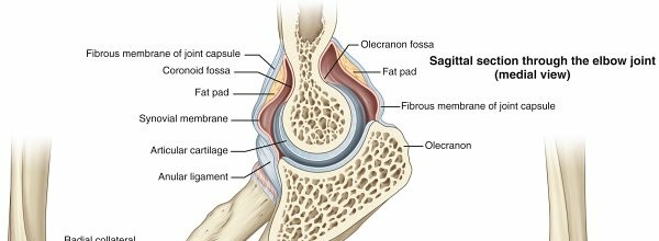
FIG. 2.5 Elbow joint including fibrous membranes of joint capsule and ligaments. From Drake RL et al, editors: Gray’s atlas of anatomy, ed 2, Philadelphia, 2015, Churchill Livingstone.
Table 2.4
Elbow Ligaments Ligament Components Comment(S) --- Medial or ulnar collateral | Anterior band
| 1. Anteroinferior portion of the medial humeral epicondyle to sublime tubercle (18 mm distal to coronoid tip)
2. Strongest elbow ligament and primary restraint to valgus stress
3. Taut from 60 degrees of flexion to full extension
4. Reconstructed in UCL reconstruction (Tommy John surgery)
Posterior band
| 1. Taut from 60 to 120 degrees of flexion
2. Greatest change in length from flexion to extension
Transverse band
| 1. Cooper ligament
Lateral collateral
| Lateral ulnar collateral ligament
| 1. Lateral humeral epicondyle to supinator crest
2. Deficiency results in posterolateral rotator instability
| Anular ligament, quadrate (anular ligament to radial neck), and oblique cord
| ### Arthrology
Table 2.5
Muscles of the Arm/Elbow Muscle | Origin | Insertion | Action | Innervation | ---|---|---|---|---| Coracobrachialis | Coracoid
| Midhumerus (medial)
| Flexion,
adduction
| Musculocutaneous
Biceps brachii
| Coracoid (short head)
Supraglenoid (long head)
| Radial
tuberosity
| Supination, flexion
| Musculocutaneous
Brachialis
| Anterior humerus
| Ulnar tuberosity (anterior)
| Flexing
forearm
| Musculocutaneous radial
Triceps brachii
| Infraglenoid (long head)
Posterior
humerus (lateral head)
Posterior
humerus (medial head)
| Olecranon
| Extending
forearm
| Radial
1. Elbow: hinge (ulnohumeral articulation) and pivot joint (radiocapitellar articulation)
1. ### Radial head should line up with capitellum at all arm positions on all radiographic views.
2. Tensile forces at medial elbow, compressive forces at lateral elbow
Capsuloligamentous structures of elbow are a key source of testable material (
Fig. 2.5).
1. Capsule allows maximum distension at approximately 70 to 80 degrees of flexion (patients with effusion most comfortable in this position).
2. Anterior capsule attaches at a point approximately 6 mm distal to the tip of the
coronoid.
1. Coronoid tip is an intraarticular structure that is visualized during elbow arthroscopy.
3.
Elbow ligaments (
Table 2.4)
1. Medial or ulnar collateral ligament primary valgus stabilizer
2. Lateral ulnar collateral ligament posterolateral stabilizer
3. Osborne’s ligament stabilizes ulnar nerve in cubital tunnel.
Ligament of Struthers: variant anatomy arising from supracondylar process to attach to medial epicondyle; potential site of median nerve compression
1. **
Muscles of the arm/elbow (**
Table 2.5)
2. Brachialis strongest elbow flexor and attaches to the coronoid 11 mm
