العربية
Part of the Master Guide

AAOS Orthopedic MCQs (Set 1): Spine Disorders, Sports Medicine & Arthroplasty | 2026 Board Review

AAOS Anatomy Board Review (Set 2): Upper, Lower Extremity & Spine MCQs | 2008 Exam Prep

23 Apr 2026 52 min read 102 Views
Anatomy 2008 MCQs - Part 2

Key Takeaway

This high-yield question set for the AAOS/ABOS 2008 anatomy board review specifically targets musculoskeletal anatomy. Questions delve into complex aspects of upper extremity, lower extremity, and spine anatomy, essential for comprehensive orthopedic knowledge and exam preparation.

AAOS Anatomy Board Review (Set 2): Upper, Lower Extremity & Spine MCQs | 2008 Exam Prep

Comprehensive 100-Question Exam


00:00

Start Quiz

Question 1

A 25-year-old tennis player has shoulder pain and weakness to external rotation. MRI scans are shown in Figures 16a and 16b. What is the most likely cause of his weakness?


Explanation

The MRI scans show a paralabral cyst, which is most commonly associated with labral tears. Compression of the suprascapular nerve results in weakness of the supraspinatus and/or infraspinatus depending on the level of compression. Piatt BE, Hawkins RJ, Fritz RC, et al: Clinical evaluation and treatment of spinoglenoid notch ganglion cysts. J Shoulder Elbow Surg 2002;11:600-604.

Question 2

The posterior approach to the proximal radius uses what intermuscular interval?


Explanation

Knowledge of intermuscular and internervous planes allows safe exposures throughout the body. The posterior (Thompson) approach to the proximal forearm uses the interval between the extensor carpi radialis brevis and extensor digitorum communis. The anterior (Henry) approach to the proximal forearm uses the interval between the brachioradialis and the flexor carpi radialis. Spinner M: Injuries to the Major Branches of Peripheral Nerves of the Forearm, ed 2. Philadelphia, PA, WB Saunders, 1978, pp 66-77.


Question 3

Which of the following statements best describes the anatomic considerations of the popliteal artery posterior to the knee joint?


Explanation

Popliteal artery injury during total knee arthroplasty is relatively rare. Knee flexion, the position that occurs during most of the arthroplasty procedure, allows the popliteal vessels to fall posteriorly, further away from harm. Anatomically, the popliteal artery lies anterior to the popliteal vein and 9 mm posterior to the posterior aspect of the tibial plateau in 90 degrees of flexion. Barrack RL, Booth RE Jr, Lonner JH, et al (eds): Orthopaedic Knowledge Update: Hip and Knee Reconstruction 3. Rosemont, IL, American Academy of Orthopaedic Surgeons, 2006, p 151.


Question 4

A 62-year-old woman reports diffuse aches and pains of the hip and pelvis. She denies any significant trauma but does have a history of chronic anemia. Figure 17a shows a radiograph of the pelvis, and Figures 17b and 17c show T2-weighted MRI scans. What is the most likely diagnosis?


Explanation

The radiograph reveals diffuse osteopenia and areas in the proximal femora that are moth-eaten in appearance. The extent of the marrow-replacing process is evident on the MRI scans, which reveal signal abnormality throughout the entire pelvis and both proximal femora. This represents a marrow-packing process, of which multiple myeloma is the best choice. This diagnosis is also supported by the anemia noted on the patient's history. Metastatic carcinoma and lymphoma also may have a similar presentation.


Question 5

Involvement of what single muscle best distinguishes an L5 radiculopathy from a peroneal neuropathy?


Explanation

All of the muscles are innervated by the peroneal nerve with the exception of the tibialis posterior which is innervated by the tibial nerve. Tibialis posterior function is best tested with resistance to plantar flexion and inversion.


Question 6

What structure is located at the tip of the arrow in Figure 18?


Explanation

The structure shown is the exiting nerve root at the L3-4 disk, which is the right L3 root.


Question 7

A patient undergoes the procedure shown in Figure 19. An important part of this procedure is preservation of what wrist ligament?


Explanation

Proximal row carpectomy is a salvage wrist procedure that yields a surprisingly stable construct. This has been attributed to two factors: 1) the congruency of the head of the capitate in the lunate fossa (this articulation is less congruent than the native lunate/lunate fossa relationship, but surprisingly stable), and 2) preservation of the radioscaphocapitate ligament, the most radial of the palmar extrinsic ligaments, which prevents ulnar subluxation after proximal row carpectomy.


Question 8

A 23-year-old woman reports right knee pain and fullness. The pain is worse with activity but also present at rest. Radiographs are shown in Figures 20a and 20b. What is the most likely diagnosis?


Explanation

The radiographs reveal a predominantly lytic, destructive lesion of the distal femur, although there is a hint of some blastic change as well. The lesion has violated the cortex, and there is mineralization outside the cortex laterally. The lateral radiograph suggests a soft-tissue density. These aggressive changes on radiographs in this age group are strongly suggestive of osteosarcoma. Sanders TG, Parsons TW: Radiographic imaging of musculoskeletal neoplasia. Cancer Control 2001;8:221-231.


Question 9

What is the structure indicated by the letter "A" in Figure 21?


Explanation

The ligaments shown are the components of the lateral collateral ligament complex, and the structure indicated by the letter "A" is the radial collateral ligament. The lateral ulnar collateral ligament is the structure indicated by the letter "C" and the annular ligament is indicated by the letter "B." The transverse ligament is a component of the medial collateral ligament complex. Morrey BF: Anatomy of the elbow joint, in Morrey BF (ed): The Elbow and Its Disorders. Philadelphia, PA, WB Saunders, 1993, p 30.


Question 10

A 16-year-old boy sustains a twisting injury to the left knee while wrestling. MRI scans are shown in Figures 22a through 22c. What is the most likely diagnosis?


Explanation

The MRI scans show a displaced bucket-handle medial meniscus tear that can be visualized on coronal, sagittal, and axial views. The sagittal view shows the typical "double posterior cruciate ligament sign," in which the low-signal bucket-handle fragment parallels the normal low-signal posterior cruciate ligament. The coronal and axial images both show the displaced medial meniscus in the notch. Sanders TG, Miller MD: A systematic approach to magnetic resonance imaging interpretation of sports medicine injuries of the knee. Am J Sports Med 2005;33:131-148.


Question 11

A 48-year-old woman reports bilateral thigh pain that is limiting her function as a librarian. A radiograph and a bone scan are shown in Figures 23a and 23b. What is the most likely diagnosis?


Explanation

The radiograph reveals bilateral severe acetabular protrusio. The bone scan and history confirm involvement of multiple joints, including the knees and the hindfoot. Although the first four choices can all cause the acetabular protrusio, the associated multiple joint involvement suggests the diagnosis of rheumatoid arthritis. Arthrokatadysis, or primary protrusio acetabuli, is often associated with osteomalacia but not other joint disease. Developmental dysplasia is a common cause of bilateral hip pathology but does not have acetabular protrusio. Resnick D: Diagnosis of Bone and Joint Disorders, ed 3. Philadelphia, PA, WB Saunders, 1995, pp 956-957. Wheeless' Textbook of Orthopaedics: Acetabular Protrusio. www.wheelessonline.com/ortho/acetabular_protrusio


Question 12

At the level of tibial bone resection in total knee arthroplasty, where does the common peroneal nerve lie?


Explanation

At the level of tibial bone resection in total knee arthroplasty, the common peroneal nerve lies superficial to the lateral head of the gastrocnemius and is therefore protected by this structure. In an MRI study of 60 knees, the mean distance from the bony posterolateral corner of the tibia to the nerve was 1.49 cm, with no distance less than 0.9 cm. The distance from the bone to nerve was greater in larger legs. Clarke HD, Schwartz JB, Math KR, et al: Anatomic risk of peroneal nerve injury with the "pie crust" technique for valgus release in total knee arthroplasty. J Arthroplasty 2004;19:40-44.


Question 13

Figures 24a through 24c show the coronal T1-weighted, T2-weighted fat-saturated, and T1-weighted fat-saturated gadolinium MRI scans of the proximal thigh of a 52-year-old woman who reports a mass in the medial thigh and groin area. She notes that the fullness has grown in size over the course of many months. Based on these findings, what is the most likely diagnosis?


Explanation

The images show a complex, lobular lesion of the thigh that has signal characteristics that follow fat. The size of the lesion, the areas of stranding within the mass, along with mild uptake on the gadolinium sequences and the mild edema within the lesion on the T2-weighted image make liposarcoma the most likely diagnosis and simple intramuscular lipoma far less likely. All other diagnoses listed would not follow fat characteristics shown on the MRI sequences.


Question 14

The arrows in the axial T1-weighted MRI scan shown in Figure 25 show which of the following structures?


Explanation

The arrows in the figure show the deep branch of the ulnar nerve (more radial) and the superficial branch of the ulnar nerve within Guyon's canal. Guyon's canal is approximately 4 cm long beginning at the proximal extent of the transverse carpal ligament and ends at the aponeurotic arch of the hypothenar muscles. Many structures comprise the boundaries of Guyon's canal. The floor, for example, consists of the transverse carpal ligament, the pisohamate and pisometacarpal ligaments, and the opponens digiti minimi. Within Guyon's canal, the ulnar nerve bifurcates into the superficial and deep branches. The ulnar artery is immediately adjacent and radial to the ulnar nerve. The median nerve is visualized within the carpal tunnel, and the palmar cutaneous branch is more radial to Guyon's canal and volar to the carpal tunnel. The radial and ulnar digital nerves to the little finger are branches off of the superficial branch of the ulnar nerve distal to its emergence from Guyon's canal. The ulnar artery is the round structure located radial to the branches of the ulnar nerve within Guyon's canal. Adjacent to the ulnar artery are two small veins. The dorsal cutaneous branch of the ulnar nerve branches from the ulnar nerve in the distal forearm, well proximal to Guyon's canal. The common digital artery to the fourth web branches from the superficial palmar arch distal to Guyon's canal. The hook of the hamate is clearly seen in the figure, orienting the observer to the ulnar side of the wrist. Gross MS, Gelberman RH: The anatomy of the distal ulnar tunnel. Clin Orthop Relat Res 1985;196:238-247.


Question 15

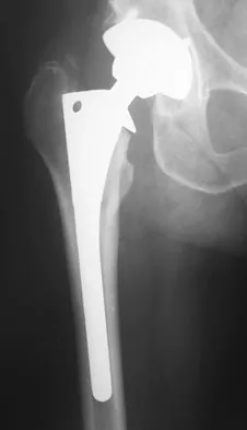
An 82-year-old man has had episodic right thigh pain after undergoing a total hip arthroplasty 10 years ago. Initial postoperative radiographs are shown in Figures 26a and 26b, and current radiographs are shown in Figures 26c and 26d. What is the most likely cause of his pain?


Explanation

These radiographs are dominated by the subsidence of the femoral component. There is also evidence of polyethylene wear and femoral osteolysis in the region of the greater trochanter. There is no evidence of proximal (calcar) stress shielding, and there is a thick distal pedestal. Engh and associates defined two major signs of osseointegration - the absence of radiolucent lines around the porous-surfaced portion of the implant and new bone bridging the gap between the endosteal surface and the porous portion of the implant. Implant migration indicates failure of ingrowth. Osteolysis is a periprosthetic loss of bone secondary to particulate debris and it is often clinically silent unless it is accompanied by pathologic fracture. It is often globular. Acetabular loosening is based on radiolucent lines and implant migration. The current radiographs demonstrate subsidence of the stem with pedestal formation. Engh CA, Massin P, Suthers KE: Roentgenographic assessment of biologic fixation of porous-surface femoral components. Clin Orthop Relat Res 1990;257:107-128.


Question 16

A 37-year-old patient with type I diabetes mellitus has a flexor tenosynovitis of the thumb flexor tendon sheath following a kitchen knife puncture wound to the volar aspect of the thumb. Left unattended, this infection will likely first spread proximally creating an abscess in which of the following spaces of the palm?


Explanation

Flexor tenosynovitis of the thumb flexor tendon sheath can spread proximally and form an abscess within the thenar space of the palm. The flexor pollicis longus tendon does not pass through the central space of the palm or the hypothenar space of the palm. The flexor pollicis longus tendon does pass through the carpal tunnel, but this is not a palmar space. The three palmar spaces include the hypothenar space, the thenar space, and the central space. The posterior adductor space would likely only be involved secondarily after spread from a thenar space infection. Hollinshead W: Anatomy for Surgeons: The Back and Limbs, ed 3. Philadelphia, PA, Harper and Row, 1982, vol 3, pp 478-479.


Question 17

What tendon is closest to an appropriately placed anterolateral portal for ankle arthroscopy?


Explanation

The appropriate placement of the anterolateral portal provides access to the lateral gutter of the joint while avoiding the superficial peroneal nerve. The safest location for the portal is approximately 4 mm lateral to the peroneus tertius tendon, the closest of the tendons listed to the anterolateral portal. Because the superficial peroneal nerve location is variable, attempts to visualize, palpate, or transilluminate the nerve are mandatory.


Question 18

A 52-year-old woman reports nagging shoulder pain that has been present for months and is slowly progressive in nature. The patient also reports nocturnal pain and notes that the pain is not activity related. Figures 27a and 27b show the radiograph and bone scan, and Figures 27c through 27e show T1-weighted, T2-weighted, and gadolinium MRI scans, respectively. Based on these findings, what is the most likely diagnosis?


Explanation

The radiograph reveals a metaphyseal lesion with some stippled mineralization suggesting a chondroid tumor. The bone scan shows increased uptake, beyond what is expected for a simple enchondroma, and beyond the limits of the lesion. The MRI sequences shows a lobular lesion on the T1- and T2-weighted (bright on the T2 sequence) images with inhomogeneous uptake of gadolinium; both findings are typical for a chondroid lesion. The history of pain, the positive bone scan, the age of the patient, the size of the lesion, and the central location (enostotic) of the lesion all suggest a malignant cartilage tumor. The images are not consistent with the other diagnoses. In particular, plasmacytoma is more uniformly bright on T2-weighted images and often has a negative bone scan. Menendez LR (ed): Orthopaedic Knowledge Update: Musculoskeletal Tumors. Rosemont, IL, American Academy of Orthopaedic Surgeons, 2002, pp 187-194.


Question 19

Figure 28 shows an arthroscopic view of a right shoulder in the lateral position through a posterior portal. What is the area between structure B (biceps) and SS (subscapularis tendon)?


Explanation

The rotator cuff interval is located between the supraspinatus and subscapularis and the biceps tendon is deep to the interval. It is a triangular area where the base is the coracoid process and the apex is the transverse humeral ligament at the biceps sulcus. Closure or tightening of this area is often helpful in patients with shoulder instability. Conversely, this area is often contracted in patients with adhesive capsulitis and may need to be released. Selecky MT, Tibone JE, Yang BY, et al: Glenohumeral joint translation after arthroscopic thermal capsuloplasty of the rotator interval. J Shoulder Elbow Surg 2003;12:139-143.


Question 20

New painful paresthesias near the site of the incision after an ulnar nerve transposition is the result of injury to what nerve?


Explanation

Branches of the medial antebrachial cutaneous nerve can often be identified during routine ulnar nerve surgery crossing the medial aspect of the elbow. It should be preserved to avoid development of painful paresthesias.


Question 21

A 23-year-old man reports pain on the superior aspect of his right shoulder with repetitive overhead activities and when lying on his right side. Figure 29 shows an axial MRI scan. What is the most likely diagnosis based on the MRI findings?


Explanation

Os acromiale represents a failure of fusion of the anterior acromial apophysis and has been reported in approximately 8% of the population. Patients with a symptomatic os acromiale often report impingement-type symptoms with pain over the superior acromion, especially with overhead activities or sleeping. When nonsurgical management is unsuccessful, surgical options include excision, open reduction and internal fixation, and arthroscopic decompression. Kurtz CA, Humble BJ, Rodosky MW, et al: Symptomatic os acromiale. J Am Acad Orthop Surg 2006;14:12-19.


Question 22

Following a chevron bunionectomy performed through a dorsal approach, a patient has persistent numbness on the dorsal and medial aspect of the hallux. What nerve has most likely been injured?


Explanation

The dorsomedial cutaneous nerve of the hallux, which is a distal branch of the superficial peroneal nerve, supplies sensation to the skin on the dorsal and medial half of the hallux and may be injured during a chevron bunionectomy. Injury to the nerve leads to particularly painful neuromas that directly impinge on the shoe. For this reason, direct medial approaches are typically preferred for access to the medial aspect of the metatarsophalangeal joint.


Question 23

A 74-year-old man reports progressive left hip pain with weight-bearing activities. A radiograph is shown in Figure 30. What is the most likely underlying diagnosis?


Explanation

The radiograph shows enlargement of the bone, coarse trabeculation, a blastic appearance, and thickening of the cortex, revealing the classic appearance of Paget's disease in the sclerotic phase, the most common presentation. While lymphoma may present as a blastic lesion, it will not have the same enlargement, coarse trabeculation of bone, and the significant sclerosis seen here. Menendez LR (ed): Orthopaedic Knowledge Update: Musculoskeletal Tumors. Rosemont, IL, American Academy of Orthopaedic Surgeons, 2002, pp 211-215.


Question 24

The anatomy of the sciatic nerve as it exits the pelvis is best described as exiting through the


Explanation

The sciatic nerve is formed by the roots of the lumbosacral plexus. It exits the pelvis through the greater sciatic notch and appears in the buttock anterior to the piriformus. From that point, the sciatic nerve passes posteriorly over the superior gemellus, obturator internus, inferior gemellus, and quadratus femoris before it passes deep to the biceps femoris. The tendon of the obturator internus passes through the lesser sciatic notch. Hoppenfeld S, deBoer P: Surgical Exposures in Orthopaedics: The Anatomic Approach. Philadelphia, PA, JB Lippincott, 1984, p 347. Anderson JE: Grant's Atlas of Anatomy, ed 7. Baltimore, MD, Lippincott Williams & Wilkins, 1978, pp 4-34, 4-36.


Question 25

What complication is more likely following excessive medial retraction of the anterior covering structures during the anterolateral (Watson-Jones) approach to the hip?


Explanation

The femoral nerve is the most lateral structure in the anterior neurovascular bundle. The femoral artery and vein lie medial to the nerve. Retractors placed in the anterior acetabular lip should be safe, although neurapraxia of the femoral nerve may occur if retraction is prolonged or forceful leading to quadriceps weakness. The femoral artery and nerve are well protected by the interposed psoas muscle. Damage to the lateral femoral cutaneous nerve, causing numbness over the anterolateral thigh, can occur while developing the interval between the tensor fascia latae and sartorious in the anterior (Smith-Petersen) approach but less likely in the Watson-Jones approach. Superior gluteal injury and accompanying abductor insufficiency may occur during excessive splitting of the glutei during the direct lateral (Hardinge) approach. Foot drop secondary to sciatic injury is more common with a posterior exposure or posterior retractor placement. Hoppenfeld S, deBoer P: Surgical Exposures in Orthopaedics: The Anatomic Approach. Philadelphia, PA, JB Lippincott, 1984, p 325.


Question 26

During a posterior cervical approach, the surgeon dissects laterally. At which level does the vertebral artery typically enter the transverse foramen and become at risk during lateral mass screw placement?


Explanation

The vertebral artery typically enters the cervical spine at the C6 transverse foramen, bypassing C7. It then ascends through the foramina to the foramen magnum.

Question 27

In reconstruction of the posterolateral corner of the knee, understanding the popliteus anatomy is crucial. Where does the popliteus tendon insert on the femur relative to the lateral collateral ligament (LCL) femoral attachment?


Explanation

The popliteus tendon inserts into the popliteal sulcus of the lateral femoral condyle. This footprint is located anterior and distal to the femoral attachment of the LCL.

Question 28

The anterior (Smith-Petersen) approach to the hip utilizes a true internervous plane. Which two nerves supply the muscles that form the superficial boundary of this interval?


Explanation

The superficial interval of the Smith-Petersen approach is between the tensor fasciae latae (supplied by the superior gluteal nerve) and the sartorius (supplied by the femoral nerve).

Question 29

When performing a volar (Henry) approach to the proximal radius, the surgeon must mobilize the supinator. How should the supinator be managed to protect the posterior interosseous nerve (PIN)?


Explanation

To safely protect the PIN, the supinator should be detached from its insertion on the radius and reflected ulnarly. The nerve runs within the substance of the muscle belly.

Question 30

A patient presents with acute weakness of the quadriceps and an absent patellar reflex. MRI shows a far lateral (extraforaminal) disc herniation at L4-L5. Which nerve root is most likely compressed?


Explanation

A far lateral disc herniation at L4-L5 compresses the exiting L4 nerve root. This leads to quadriceps weakness and a diminished patellar reflex.

Question 31

During surgical release of the tarsal tunnel, the structures posterior to the medial malleolus are encountered. What is the correct order of these structures from anterior to posterior?


Explanation

The mnemonic "Tom, Dick, And Very Nervous Harry" dictates the order from anterior to posterior: Tibialis posterior, Flexor Digitorum Longus, Artery, Vein, Nerve, Flexor Hallucis Longus.

Question 32

A patient develops posterior shoulder pain and weakness in external rotation following a direct blow to the posterior axilla. Compression in the quadrangular space is suspected. Which of the following structures pass through this space?


Explanation

The quadrangular space contains the axillary nerve and the posterior circumflex humeral artery. It is bounded by the teres minor, teres major, long head of triceps, and the surgical neck of the humerus.

Question 33

In the surgical treatment of stenosing tenosynovitis (trigger finger), a release of the affected pulley is planned. To prevent bowstringing of the flexor tendons, which adjacent pulley MUST remain intact?


Explanation

Release of the A1 pulley is the standard treatment for trigger finger. The A2 and A4 pulleys are biomechanically critical for preventing bowstringing and must be preserved.

Question 34

Which portion of the medial ulnar collateral ligament complex is the primary restraint to valgus stress at the elbow during the late cocking phase of throwing?


Explanation

The anterior bundle is the primary valgus stabilizer of the elbow. Within it, the anterior band is tight in extension, which is critical during the late cocking and early acceleration phases.

Question 35

During an ilioinguinal approach to the acetabulum, massive bleeding occurs upon dissection near the superior pubic ramus. This is most likely due to injury to the "corona mortis," which is an anastomosis between the:


Explanation

The corona mortis is an arterial or venous anastomosis between the external iliac (or inferior epigastric) and the obturator vessels. It rests on the posterior aspect of the superior pubic ramus.

Question 36

The precarious blood supply of the scaphoid makes it prone to avascular necrosis following fracture. The primary blood supply enters the scaphoid at which location?


Explanation

The primary blood supply to the scaphoid arises from branches of the radial artery. It enters the bone dorsally at the distal pole, perfusing the proximal pole in a retrograde fashion.

Question 37

A posterior approach to the hip is performed. To protect the main blood supply to the adult femoral head, careful handling of which vessel is required, and where does it course?


Explanation

The medial femoral circumflex artery provides the primary blood supply to the adult femoral head. It courses posterior to the obturator externus and anterior to the triceps coxae/quadratus femoris.

Question 38

During an anterior cervical discectomy and fusion (ACDF), self-retaining retractors are placed. Which anatomical structure is at greatest risk of injury leading to Horner's syndrome if the longus colli muscles are retracted too far laterally?


Explanation

The sympathetic trunk lies on the anterior surface of the longus colli muscles laterally. Placing retractors too far laterally can damage it, causing Horner's syndrome (ptosis, miosis, anhidrosis).

Question 39

A 24-year-old athlete sustains a severe high ankle sprain. Anatomically, which ligament provides the strongest restraint to diastasis of the distal tibiofibular syndesmosis?


Explanation

The PITFL provides the greatest resistance (approx. 40%) to lateral displacement of the fibula. While the AITFL is the most commonly injured in syndesmotic sprains, it is weaker than the PITFL.

Question 40

When performing a lateral approach to the distal humerus, the radial nerve is identified as it pierces the lateral intermuscular septum. At what average distance proximal to the lateral epicondyle does this occur?


Explanation

The radial nerve pierces the lateral intermuscular septum to move from the posterior compartment to the anterior compartment approximately 10 cm proximal to the lateral epicondyle.

Question 41

A patient suffers a laceration to the volar forearm, completely transecting the median nerve proximal to the elbow. Which muscle belly of the flexor digitorum profundus (FDP) will primarily lose its innervation?


Explanation

The FDP has dual innervation. The anterior interosseous branch of the median nerve supplies the index and middle fingers, while the ulnar nerve supplies the ring and small fingers.

Question 42

A patient develops compartment syndrome of the leg following a highly comminuted tibial shaft fracture. Decompression of the deep posterior compartment is essential. Which muscle is located within this specific compartment?


Explanation

The deep posterior compartment of the leg contains the tibialis posterior, flexor hallucis longus (FHL), and flexor digitorum longus (FDL).

Question 43

During a posterior approach to the cervical spine at C1-C2, the surgeon must be careful to avoid injury to the vertebral artery. At this level, the vertebral artery lies directly superior to which anatomical structure?


Explanation

The vertebral artery exits the C1 transverse foramen and courses medially along the superior surface of the C1 posterior arch in the vertebral groove before piercing the atlanto-occipital membrane.

Question 44

An orthopedic surgeon is performing a lateral approach to the hindfoot for an intra-articular calcaneal fracture fixation. Which nerve is most at risk during the standard lateral extensile approach?


Explanation

The sural nerve courses posterior to the lateral malleolus and provides sensation to the lateral aspect of the foot, making it highly susceptible to injury during a lateral extensile approach to the calcaneus.

Question 45

The anterior (volar) approach to the radius (Henry approach) proximally exploits the internervous plane between which two muscles?


Explanation

The proximal internervous plane of the Henry approach to the radius is between the brachioradialis (innervated by the radial nerve) and the pronator teres (innervated by the median nerve).

Question 46

A surgeon performs a direct anterior approach (Smith-Petersen) for a total hip arthroplasty. The superficial internervous plane is between the sartorius and tensor fasciae latae. What is the deep internervous plane?


Explanation

The deep internervous plane in the direct anterior approach to the hip is between the rectus femoris (femoral nerve) and the gluteus medius (superior gluteal nerve).

Question 47

During a retroperitoneal approach to the anterior lumbar spine at L4-L5, which of the following vascular structures must be mobilized from left to right to safely expose the disc space?


Explanation

The iliolumbar vein typically crosses the L4-L5 disc space or the body of L5. It tethers the left common iliac vein and must be ligated and divided to allow safe medial retraction of the major vasculature.

Question 48

A 45-year-old male undergoes arthroscopic rotator cuff repair. During portal placement, the surgeon places a portal 5 cm distal to the lateral edge of the acromion. Which nerve is most at risk?


Explanation

The axillary nerve courses circumferentially around the surgical neck of the humerus, approximately 5 cm distal to the lateral acromial edge. Portals placed too distally risk transecting its branches.

Question 49

The medial collateral ligament (MCL) of the knee has a superficial and deep component. The superficial MCL attaches distally to the medial aspect of the proximal tibia deep to which structure?


Explanation

The superficial MCL inserts on the medial tibia approximately 4-5 cm distal to the joint line, situated deep to the pes anserinus tendons (sartorius, gracilis, and semitendinosus).

Question 50

When inserting a pedicle screw at the T8 level, what is the anatomical relationship of the exiting nerve root to the corresponding pedicle?


Explanation

In the thoracic and lumbar spine, the numbered nerve root exits through the intervertebral foramen below the corresponding numbered pedicle. For example, the T8 nerve root exits below the T8 pedicle.

Question 51

De Quervain's tenosynovitis involves the first dorsal compartment of the wrist. Which of the following tendons are located in this compartment?


Explanation

The first dorsal compartment of the wrist contains the abductor pollicis longus (APL) and extensor pollicis brevis (EPB) tendons. Inflammation of the sheath surrounding these tendons causes De Quervain's tenosynovitis.

Question 52

The corona mortis is an anastomotic vascular connection at risk during the ilioinguinal approach to the acetabulum. It connects which two vessel systems?


Explanation

The corona mortis is a vascular anastomosis between the external iliac (or inferior epigastric) system and the obturator system. It crosses the superior pubic ramus and is highly vulnerable during pelvic trauma and anterior surgical approaches.

Question 53

The deep branch of the ulnar nerve supplies all of the following muscles EXCEPT:


Explanation

The opponens pollicis is a thenar muscle innervated by the recurrent motor branch of the median nerve. The deep branch of the ulnar nerve supplies the hypothenar muscles, all interossei, the adductor pollicis, and the deep head of the flexor pollicis brevis.

Question 54

During a fasciotomy for acute compartment syndrome of the leg, a double-incision technique is used. Which nerve is most at risk of injury during the distal extent of the lateral incision used to decompress the anterior and lateral compartments?


Explanation

The superficial peroneal nerve exits the lateral compartment to become subcutaneous in the distal third of the leg, placing it at significant risk of iatrogenic injury during the distal portion of a lateral fasciotomy incision.

Question 55

When placing an iliosacral screw for a zone II sacral fracture, the surgeon must aim to keep the screw within the "safe zone" of the S1 vertebral body. The superior boundary of this safe zone is defined by which structure?


Explanation

The upper boundary of the S1 safe zone for iliosacral screw placement is the L5 nerve root, which courses over the sacral ala. The anterior boundary is the iliac vessels, and the inferior boundary is the S1 neuroforamen.

Question 56

A 30-year-old male sustains a Monteggia fracture-dislocation. During surgical fixation via a posterior (Boyd) approach, the surgeon elevates the supinator off the proximal radius. Which nerve lies within the substance of the supinator and is at risk?


Explanation

The posterior interosseous nerve (PIN), a branch of the radial nerve, passes between the superficial and deep heads of the supinator muscle. It is at significant risk during posterior and lateral approaches to the proximal radius.

Question 57

The "watershed" area of the Achilles tendon, which is prone to rupture and represents an area of relative hypovascularity, is typically located:


Explanation

The watershed zone of the Achilles tendon is an area of relative hypovascularity located roughly 2 to 6 cm proximal to its insertion on the calcaneus, making it the most common site for degenerative tears and ruptures.

Question 58

In the deltopectoral approach to the shoulder, the cephalic vein is typically identified and retracted laterally. This interval marks the internervous plane between which two nerves?


Explanation

The deltopectoral interval lies between the deltoid (innervated by the axillary nerve) and the pectoralis major (innervated by the medial and lateral pectoral nerves). Therefore, the plane is between the axillary and lateral pectoral nerves.

Question 59

The posterolateral approach to the femur utilizes the internervous plane between the vastus lateralis and the biceps femoris. What is the innervation of these two muscles, respectively?


Explanation

The vastus lateralis is innervated by the femoral nerve, while the biceps femoris is innervated by the sciatic nerve (the long head by the tibial division, and the short head by the common peroneal division).

Question 60

In the lumbar spine, the facet joints are oriented primarily in which plane, thereby allowing for significant flexion and extension but heavily limiting axial rotation?


Explanation

The lumbar facet joints are aligned predominantly in the sagittal plane. This unique orientation facilitates flexion and extension while strongly restricting axial rotation, in contrast to the more coronally oriented thoracic facets.

Question 61

A patient presents unable to extend the interphalangeal joint of the thumb, but has normal wrist extension with radial deviation. Sensation over the dorsal web space is completely intact. Compression of which structure is most likely?


Explanation

The posterior interosseous nerve (PIN) is a pure motor branch supplying the thumb and finger extensors, frequently entrapped at the Arcade of Frohse. The superficial radial nerve is spared (intact sensation), and the ECRL is intact (causing radial deviation on wrist extension).

Question 62

The posterolateral corner (PLC) of the knee is a complex arrangement of static and dynamic stabilizers. Which of the following structures is considered a primary static stabilizer of the PLC?


Explanation

The primary static stabilizers of the posterolateral corner of the knee include the lateral collateral ligament (LCL), the popliteofibular ligament, and the popliteus tendon. The biceps femoris and popliteus muscle belly act as dynamic stabilizers.

Question 63

A 32-year-old patient sustains an isolated penetrating injury to the medial cord of the brachial plexus. Which of the following muscles will demonstrate normal strength on physical examination?


Explanation

The pronator teres is innervated by the median nerve via fibers originating from C6 and C7, which travel through the lateral cord. The other listed muscles receive innervation entirely or predominantly from the medial cord (C8-T1).

Question 64

When placing pedicle screws in the lumbar spine, which level typically requires the greatest medial angulation?


Explanation

The pedicles in the lumbar spine become wider and require more medial angulation as you progress caudally from L1 to L5. The L5 pedicle typically requires 25 to 30 degrees of medial angulation.

Question 65

A patient presents with weakness in shoulder abduction and external rotation following a posterior shoulder dislocation. The nerve injured passes through a space bordered by which of the following sets of structures?


Explanation

The axillary nerve passes through the quadrangular space. The borders of this space are the teres minor (superior), teres major (inferior), long head of the triceps (medial), and surgical neck of the humerus (lateral).

Question 66

During a plantar approach to the foot for a plantar fibromatosis excision, the surgeon identifies the "Master Knot of Henry". Which two tendons intersect at this anatomic landmark?


Explanation

The Master Knot of Henry is located in the medial plantar aspect of the midfoot. At this location, the flexor hallucis longus (FHL) tendon crosses dorsal to the flexor digitorum longus (FDL) tendon.

Question 67

In the typical cervical spine, the vertebral artery most commonly enters the transverse foramen at which vertebral level?


Explanation

The vertebral artery typically branches from the subclavian artery and enters the transverse foramen at the C6 level. It then travels superiorly through the transverse foramina to the foramen magnum.

Question 68

The volar (Henry) approach to the proximal radius utilizes an internervous plane between which two muscles?


Explanation

The proximal internervous plane for the anterior (Henry) approach to the radius lies between the brachioradialis (innervated by the radial nerve) and the pronator teres (innervated by the median nerve).

Question 69

Which vessel provides the primary blood supply to the weight-bearing portion of the adult femoral head?


Explanation

The deep branch of the medial femoral circumflex artery provides the predominant blood supply to the adult femoral head. It primarily supplies the superolateral weight-bearing dome.

Question 70

The alar ligaments are essential primary stabilizers of the craniocervical junction. What is their primary biomechanical function?


Explanation

The alar ligaments connect the dens to the medial aspect of the occipital condyles. Their primary biomechanical role is to limit axial rotation and lateral bending of the head relative to the cervical spine.

Question 71

Which of the following annular pulleys are considered critical and must be preserved during a trigger finger release to prevent bowstringing of the flexor tendons?


Explanation

The A2 (located over the proximal phalanx) and A4 (located over the middle phalanx) pulleys are the most critical biomechanical pulleys. They must be preserved to prevent bowstringing of the flexor tendons and loss of mechanical advantage.

Question 72

The popliteofibular ligament is a crucial static stabilizer of the posterolateral corner of the knee. It originates from the popliteus musculotendinous junction and inserts on the:


Explanation

The popliteofibular ligament arises from the popliteus tendon and inserts onto the posteromedial aspect of the fibular styloid process. It plays a key role in resisting posterior translation, varus angulation, and external rotation.

Question 73

Posterolateral rotatory instability (PLRI) of the elbow is primarily caused by insufficiency of the lateral ulnar collateral ligament (LUCL). What is the precise insertion of the LUCL?


Explanation

The LUCL originates on the lateral epicondyle of the humerus, blends with the annular ligament, and inserts on the supinator crest of the proximal ulna. Its insufficiency is the primary lesion in posterolateral rotatory instability.

Question 74

During a four-compartment fasciotomy of the leg for compartment syndrome, failure to adequately release the deep posterior compartment is a common cause of poor outcomes. Which nerve courses within this compartment and is at risk if ischemia persists?


Explanation

The tibial nerve runs within the deep posterior compartment of the leg alongside the posterior tibial vessels. Ischemia in this compartment leads to weakness in toe flexion and loss of plantar sensation.

Question 75

A patient presents with weakness in ankle dorsiflexion and numbness in the first dorsal web space. MRI demonstrates a far lateral (extra-foraminal) disc herniation at the L4-L5 level. Which nerve root is most likely compressed?


Explanation

In the lumbar spine, a far lateral (extra-foraminal) disc herniation compresses the exiting nerve root at the same level. Therefore, an L4-L5 far lateral herniation compresses the L4 nerve root.

Question 76

A 28-year-old volleyball player presents with painless weakness of shoulder external rotation. Atrophy is noted in the infraspinatus fossa, while the supraspinatus fossa is normal. Where is the most likely site of nerve compression?


Explanation

Compression of the suprascapular nerve at the spinoglenoid notch affects only the infraspinatus muscle, leading to isolated external rotation weakness. Compression at the suprascapular notch would affect both the supraspinatus and infraspinatus.

Question 77

During an ilioinguinal approach to the acetabulum, the surgeon must identify and ligate the "corona mortis" to prevent massive hemorrhage. This structure is an anastomosis between the:


Explanation

The corona mortis is a critical vascular anastomosis between the obturator vessels and the inferior epigastric or external iliac vessels. It is typically located on the posterior aspect of the superior pubic ramus.

Question 78

A patient presents with medial winging of the scapula after a traumatic injury. The injured nerve originates from which of the following roots of the brachial plexus?


Explanation

Medial winging of the scapula is caused by paralysis of the serratus anterior muscle due to injury of the long thoracic nerve. The long thoracic nerve originates from the anterior rami of the C5, C6, and C7 nerve roots.

Question 79

A surgeon is performing an anterior approach to the cervical spine at the C5-C6 level. The recurrent laryngeal nerve is at risk. Which of the following describes its typical anatomical course on the right side?


Explanation

The right recurrent laryngeal nerve loops around the right subclavian artery, while the left loops around the aortic arch. Both ascend in the tracheoesophageal groove to innervate the larynx.

Question 80

During a posterolateral approach to the hip (Kocher-Langenbeck), the piriformis tendon is identified. What nerve exits the sciatic notch immediately superior to the piriformis?


Explanation

The superior gluteal nerve exits the greater sciatic foramen superior to the piriformis muscle. The sciatic, inferior gluteal, and pudendal nerves exit inferior to the piriformis.

Question 81

A 30-year-old male undergoes a fasciotomy for acute compartment syndrome of the leg. The deep posterior compartment is released. Which of the following structures is found within this compartment?


Explanation

The deep posterior compartment of the leg contains the tibialis posterior, flexor digitorum longus, flexor hallucis longus, and the tibial nerve.

Question 82

In the standard volar (Henry) approach to the distal radius, the flexor carpi radialis (FCR) is retracted. Between which two tendons is the deep dissection carried out?


Explanation

The distal Henry approach exploits the interval between the brachioradialis (radially) and the FCR (ulnarly). The radial artery is mobilized carefully to access the pronator quadratus.

Question 83

A patient sustains a mid-shaft humerus fracture. The radial nerve is at risk as it passes through the intermuscular septum. At what approximate distance from the lateral epicondyle does the radial nerve pierce the lateral intermuscular septum?


Explanation

The radial nerve pierces the lateral intermuscular septum from posterior to anterior approximately 10 cm proximal to the lateral epicondyle.

Question 84

What is the primary arterial supply to the femoral head in a healthy 30-year-old adult?


Explanation

The medial circumflex femoral artery (MFCA) provides the primary blood supply to the adult femoral head via the lateral epiphyseal artery.

Question 85

During an anterior (Smith-Petersen) approach to the hip, the superficial interval is created. Which two muscles define this interval?


Explanation

The superficial interval of the Smith-Petersen approach is between the sartorius (femoral nerve) and the tensor fasciae latae (superior gluteal nerve).

Question 86

A fracture of the medial epicondyle of the humerus often endangers which nerve?


Explanation

The ulnar nerve runs directly posterior to the medial epicondyle in the cubital tunnel, making it highly susceptible to injury in medial epicondyle fractures.

Question 87

In lumbar pedicle screw placement, the intersection of the pars interarticularis, the transverse process, and the superior articular facet serves as a landmark. The exiting nerve root at the L4-L5 level is:


Explanation

In the lumbar spine, the exiting nerve root is named for the pedicle above the disc space. Therefore, the L4 nerve root exits at the L4-L5 foramen.

Question 88

A 22-year-old football player sustains a complete rupture of the anterior cruciate ligament (ACL). The femoral footprint of the anteromedial (AM) bundle of the ACL is best described as being located:


Explanation

The AM bundle of the ACL originates high and deep (proximal and posterior) on the medial aspect of the lateral femoral condyle. It is tight in flexion.

Question 89

A 40-year-old male sustains an injury to the primary stabilizing structure of the distal radioulnar joint (DRUJ). Which structure is most crucial for DRUJ stability?


Explanation

The TFCC is the primary stabilizer of the DRUJ, specifically the deep radioulnar ligaments (ligamentum subcruentum) which attach to the fovea of the ulnar head.

Question 90

A patient presents with winged scapula following a breast lumpectomy and axillary node dissection. The affected nerve innervates which of the following muscles?


Explanation

The long thoracic nerve innervates the serratus anterior muscle. Injury to this nerve leads to medial scapular winging.

Question 91

In evaluating the deltoid ligament of the ankle, the superficial portion crosses two joints. Which of the following is a component of the deep deltoid ligament, the primary medial stabilizer of the ankle?


Explanation

The deep deltoid ligament consists primarily of the deep anterior and deep posterior tibiotalar ligaments, providing the most significant restraint against lateral talar shift.

Question 92

The flexor pulleys of the finger prevent bowstringing of the flexor tendons. Which pulley is located directly over the proximal interphalangeal (PIP) joint?


Explanation

The A3 pulley is located directly volar to the PIP joint. The A2 (over the proximal phalanx) and A4 (over the middle phalanx) are the most important for preventing bowstringing.

Question 93

During a lateral approach to the calcaneus for open reduction internal fixation of a fracture, the sural nerve must be protected. What is its sensory distribution?


Explanation

The sural nerve provides sensory innervation to the posterolateral lower leg and the lateral aspect of the foot and heel.

Question 94

The rotator interval of the shoulder is a triangular anatomical space. What structures form its superior and inferior borders?


Explanation

The rotator interval is bounded superiorly by the anterior edge of the supraspinatus and inferiorly by the superior edge of the subscapularis.

Question 95

In the spine, the vertebral artery typically enters the transverse foramen at which cervical level?


Explanation

The vertebral artery arises from the subclavian artery and typically enters the transverse foramen at C6, though anatomical variations can occur.

Question 96

A surgeon is fixing a displaced scaphoid waist fracture using a volar approach. Blood supply to the scaphoid is primarily provided by branches of which artery?


Explanation

The scaphoid is predominantly supplied by dorsal branches of the radial artery, which enter distally and flow in a retrograde fashion.

Question 97

When performing a posterior approach to the knee, the tibial nerve is identified in the popliteal fossa. Which of the following correctly describes its position relative to the popliteal artery and vein?


Explanation

In the popliteal fossa, the sequence of structures from superficial (posterior) to deep (anterior) is nerve, vein, artery. The tibial nerve is lateral to the vessels proximally and crosses to medial distally.

Question 98

A 45-year-old patient presents with pain and weakness in thumb extension and radial abduction. A diagnosis of De Quervain's tenosynovitis is made. Which tendons are involved?


Explanation

De Quervain's tenosynovitis involves the first dorsal extensor compartment of the wrist, which contains the abductor pollicis longus (APL) and extensor pollicis brevis (EPB) tendons.

Question 99

A 35-year-old avid cyclist presents with profound weakness of the intrinsic muscles of his right hand and isolated numbness of the small finger. Nerve conduction studies confirm ulnar nerve compression at Guyon's canal. Which of the following structures forms the floor of this fibro-osseous anatomic tunnel?


Explanation

The floor of Guyon's canal is formed by the transverse carpal ligament and the pisohamate ligament. The roof is formed by the palmar carpal ligament and palmaris brevis, while the medial and lateral borders are the pisiform and hook of hamate, respectively.

Question 100

A surgeon performs an extensile lateral approach to the calcaneus for open reduction and internal fixation of a joint-depressed fracture. The sural nerve is at high risk of iatrogenic injury during the flap elevation. The sural nerve receives its contributing fibers from which of the following nerve pairs?


Explanation

The sural nerve provides sensation to the posterolateral distal leg and lateral foot. It is formed by the union of the medial sural cutaneous nerve (a branch of the tibial nerve) and the sural communicating branch of the lateral sural cutaneous nerve (from the common peroneal nerve).

None

Clinic OS
Medically Verified Content by
Prof. Clinic OS
Consultant Orthopedic & Spine Surgeon
Chapter Index