العربية
Part of the Master Guide

AAOS Orthopedic MCQs (Set 1): Spine Disorders, Sports Medicine & Arthroplasty | 2026 Board Review

Orthopedic Anatomy MCQs (Set 1): Spine, Upper & Lower Limb | AAOS ABOS 2002 Review

27 Apr 2026 51 min read 104 Views
Anatomy 2002 MCQs - Part 1

Key Takeaway

This high-yield question set (Set 1 from 2002) for AAOS/ABOS exams covers essential orthopedic anatomy. It features challenging MCQs on spinal column structures, upper limb bones and muscles, and lower limb musculature, crucial for board preparation and clinical understanding.

Orthopedic Anatomy MCQs (Set 1): Spine, Upper & Lower Limb | AAOS ABOS 2002 Review

Comprehensive 100-Question Exam


00:00

Start Quiz

Question 1

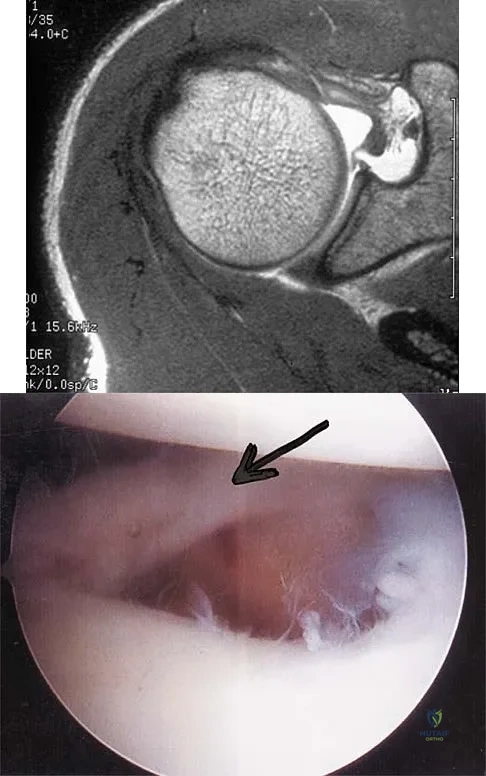
A patient has right shoulder pain. Figure 1a shows a gadolinium-enhanced transverse MRI scan at the level of the coracoid. Figure 1b shows an arthroscopic view of the anterior structures from a posterior portal. These images reveal which of the following findings?


Explanation

The area shown in the arthroscopic view and MRI scan is referred to as a Buford complex and represents a normal labral variant. It consists of a thickened, cord-like middle glenohumeral ligament, a superior labral attachment of the middle glenohumeral ligament just anterior to the biceps tendon, and absence of the anterosuperior labrum. This combination of findings can be confusing and may simulate labral pathology. Mistaken repair of the lesion back to the glenoid rim can result in significant loss of external rotation. A Bankart lesion would be located at the inferior anterior glenoid rim. The subscapularis is seen anterior to the labrum. Normal variations that occur in the anterosuperior labrum can simulate pathology. Gusmer PB, Potter HG, Schatz JA, et al: Labral injuries: Accuracy of detection with unenhanced MR imaging of the shoulder. Radiology 1996;200:519-524. Griffin LY (ed): Orthopaedic Knowledge Update: Sports Medicine. Rosemont, IL, American Academy of Orthopaedic Surgeons, 1994, pp 47-63.

Question 2

What muscle attaches to the site shown by the arrow in Figure 2?

Anatomy 2002 Practice Questions: Set 1 (Solved) - Figure 3


Explanation

The latissimus dorsi inserts on the humerus metaphysis between the pectoralis major (posterior) and teres major (anterior). Teres minor inserts on the base of the greater tuberosity. Pectoralis minor does not insert on the humerus. Williams PL, Warwick R, Dyson M, Bannister LH: Neurology, in Gray's Anatomy, ed 37. Edinburgh, Scotland, Churchill Livingstone, 1989, pp 1131-1132.

Question 3

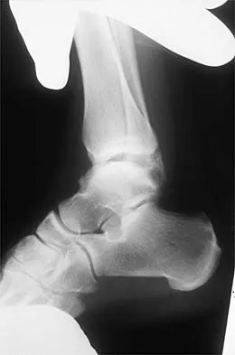
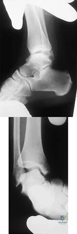
Figures 3a and 3b show the inversion stress radiographs of a patient's ankle. What is the most likely ligament injury pattern?


Explanation

The radiographic findings show 30 degrees of talar tilt (severe) and 10 mm of anterior translation that typically involves laxity of both of the major lateral ligaments of the ankle (anterior talofibular and calcaneofibular). There is no evidence of deltoid laxity. Harper MC: Stress radiographs in the diagnosis of lateral instability of the ankle and hindfoot. Foot Ankle 1992;13:435-438.

Question 4

Posterior sternoclavicular dislocations are most commonly associated with which of the following complications?

Anatomy 2002 Practice Questions: Set 1 (Solved) - Figure 6


Explanation

Posterior sternoclavicular dislocations are commonly associated with tracheal compression, which can be a life-threatening condition requiring immediate reduction. The other listed complications are less common. Brooks AL, Henning GD: Injury to the proximal clavicular epiphysis, abstracted. J Bone Joint Surg Am 1972;54:1347-1348.

Question 5

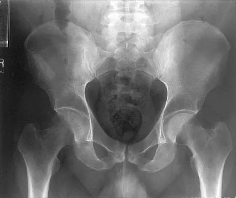
An AP radiograph of the pelvis is shown in Figure 4. What muscle attaches to the avulsed fragment of bone identified by the arrow?

Anatomy 2002 Practice Questions: Set 1 (Solved) - Figure 7


Explanation

The radiograph reveals an avulsion of the ischial apophysis, most likely the result of violent contraction of the attached hamstring tendons (semimembranosus, semitendinosus, and long head of the biceps femoris). The short head of the biceps femoris arises from the linea aspera on the posterior femur. The pectineus and adductor longus attach to the pubic portion of the pelvis. The piriformis runs from the sacrum to the femur. Woodburne RT (ed): Essentials of Human Anatomy. New York, NY, Oxford University Press, 1978, pp 542-545.

Question 6

A patient with an acromioclavicular dislocation has a very prominent distal clavicle. Examination reveals that the deformity increases rather than reduces with an isometric shoulder shrug. Which of the following structures is most likely intact?

Anatomy 2002 Practice Questions: Set 1 (Solved) - Figure 8


Explanation

Severely displaced acromioclavicular injuries disrupt the deltotrapezial fascia and muscular origin in addition to the ligaments (acromioclavicular and coracoclavicular or trapezoid and conoid). When the deltoid is still attached to the clavicle, an isometric shoulder shrug will tend to reduce the displacement. When the deltoid is detached but the trapezius is attached, this manuever will increase the deformity and surgery may be indicated.

Question 7

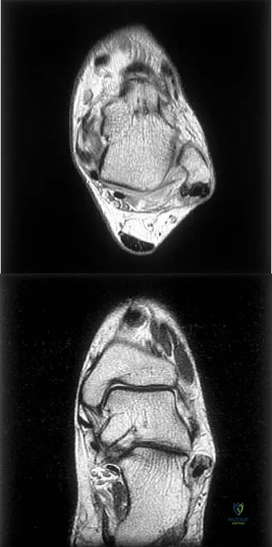
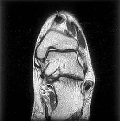
Figures 5a and 5b show axial and coronal MRI images of the left ankle of a patient with lateral ankle pain. What is the most likely diagnosis?


Explanation

The figures show a longitudinal split within the peroneus brevis tendon as it courses posterior to the fibula. The peroneus longus tendon has been driven between the medial and lateral components of the peroneus brevis tendon. Peroneal split syndrome is a cause of lateral ankle pain but may be less asymptomatic in the elderly. It may be associated with tendon subluxation following a tear of the superior peroneal retinaculum.

Question 8

Which of the following anatomic structures is often difficult to visualize during elbow arthroscopy?

Anatomy 2002 Practice Questions: Set 1 (Solved) - Figure 11


Explanation

The ulnar collateral ligament is often difficult to visualize during elbow arthroscopy. It can be seen clearly in only 10% to 30% of elbow arthroscopies. All of the other structures should be easily and thoroughly seen and palpated during elbow arthroscopy. Johnson LL: Arthroscopic Surgery: Principles and Practice. St Louis, MO, CV Mosby, 1988.

Question 9

The quadrilateral space in the shoulder contains which of the following structures?

Anatomy 2002 Practice Questions: Set 1 (Solved) - Figure 12


Explanation

The quadrilateral or quadrangular space of the shoulder is formed laterally by the humerus, proximally by the subscapularis (and teres minor viewed from posterior), distally by the teres major, and medially by the long head of triceps. The posterior humeral circumflex artery and axillary nerve pass through it. The axillary artery is more proximal. The radial nerve and profunda brachii pass through a triangular space more inferior. The circumflex scapular artery passes through a triangular space more medial. Hollinshead WH: Textbook of Anatomy, ed 3. Hagerstown, MD, Harper and Row, 1974, pp 205-206.

Question 10

Based on the MRI scan shown in Figure 6, the abnormal signal is seen in what carpal bone?

Anatomy 2002 Practice Questions: Set 1 (Solved) - Figure 13


Explanation

The MRI scan reveals an abnormal signal in the trapezoid, which lies adjacent to the capitate in the distal carpal row. The tumor is a giant cell tumor of bone. Cooney WP, Linscheid RL, Dobyns JH: The Wrist: Diagnosis and Operative Treatment. St Louis, MO, Mosby-Year Book, 1998, vol 1, pp 278-282. Green DP, Hotchkiss RN, Pederson WC (eds): Operative Hand Surgery, ed 4. New York, NY, Churchill Livingstone, 1999, pp 2238-2240. bar based on these measurements is shown in Figure 54d. Initial treatment should consist of 1- bony bar resection and distal fibula epiphysiodesis. 2- bony bar resection and corrective osteotomy. 3- bony bar resection and physiodesis of the opposite distal tibial physis. 4- corrective osteotomy and a limb-lengthening procedure. 5- corrective osteotomy and physiodesis of the opposite distal tibial physis. 2 54a 54b 54c 54d Mapping of a physeal bar from biplane polytomography or CT helps to identify lesions that should be treated surgically and aids in planning the surgical approach and resection. Criteria for surgical excision are at least 2 years of longitudinal growth remaining and involvement of no more than 50% of the physis. Osteotomy is required if angular deformity is greater than 20 degrees. Although this physeal bar is large, it is slightly less than 50% of the total area of the physis. Limb lengthening in this case should be reserved for failure of bar resection. Physiodesis of the opposite distal tibia at this age would result in disproportionate shortening of both tibiae. Carlson WO, Wenger DR: A mapping method to prepare for surgical excision of a partial physeal arrest. J Pediatr Orthop 1984;4:232-238.

Question 11

The recurrent motor branch of the median nerve innervates which of the following muscles?

Anatomy 2002 Practice Questions: Set 1 (Solved) - Figure 14


Explanation

The recurrent motor branch of the median nerve supplies the thenar muscles (abductor pollicis brevis, flexor pollicis brevis, and opponens pollicis) that are primarily responsible for thumb opposition. The nerve can be injured in carpal tunnel release. A branch of the nerve also supplies the first lumbrical. The adductor pollicis and the interossei are supplied by the ulnar nerve. Last RJ: Anatomy: Regional and Applied, ed 6. London, England, Churchill Livingstone, 1978, p 109.

Question 12

Which of the following nerves innervates the muscle that originates from the middle third of the dorsal surface of the lateral border of the scapula, as shown in Figure 7?

Anatomy 2002 Practice Questions: Set 1 (Solved) - Figure 15


Explanation

Teres minor originates from the middle third of the dorsal surface of the lateral border of the scapula. It is supplied by the axillary nerve (C5). Williams PL, Warwick R, Dyson M, Bannister LH: Myology, in Gray's Anatomy, ed 37. Edinburgh, Scotland, Churchill Livingstone, 1989, pp 611-615.

Question 13

Based on the MR arthrogram of the elbow shown in Figure 8, which of the following structures is torn?

Anatomy 2002 Practice Questions: Set 1 (Solved) - Figure 16


Explanation

Based on the MR arthrogram in which gadolinium (bright on T1-weighted images) was injected into the joint space prior to imaging, the study shows a tear of the anterior band of the ulnar collateral ligament (UCL). The disruption in the distal end of the UCL is outlined by contrast. A small collection of contrast extravasation into the flexor musculature further confirms the presence of a tear. The UCL has a broad-based attachment on the medial epicondyle and has a pointed or tapered attachment distally on the ulna. Most UCL tears occur distally at the ulnar (coronoid) attachment. MR arthrography provides improved sensitivity compared to conventional MRI, without contrast, for the detection of UCL pathology, particularly in the subacute or chronic setting. After the soft-tissue edema and joint fluid associated with the injury have resolved, the torn end of the ligament may lie in contact with its adjacent attachment and create a false-negative appearance. In this patient, a noncontrasted MR arthrogram showed no tear, yet the tear is apparent with intra-articular contrast and distention. MR arthrography of the elbow also may be useful in detecting intra-articular bodies or in evaluation for loose osteochondral fragments or flaps. Morrey BF: Acute and chronic instability of the elbow. J Am Acad Orthop Surg 1996;4:117-128.

Question 14

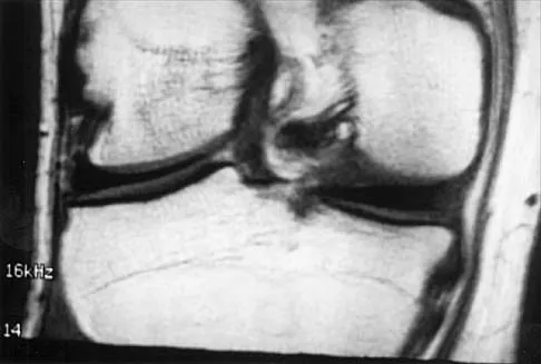
A 26-year-old man has recurrent right knee pain. Figures 9a and 9b show consecutive sagittal T2-weighted MRI scans, and Figure 9c shows a coronal T1-weighted MRI scan. What is the most likely diagnosis?
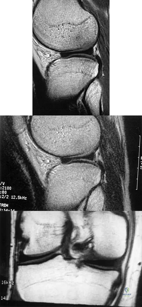


Explanation

A discoid meniscus is a large disk-like meniscus. It is seen in the lateral meniscus in 3% of the population; a discoid medial meniscus is much less common. It can be identified on the coronal view by noting meniscal tissue extending into the tibial spine at the intercondylar notch. The average width of a normal meniscus is less than 11 mm. A bow-tie appearance should not be seen on more than two consecutive sagittal images because the conventional thickness of the sagittal slices is 3 mm and the interval between two consecutive slices is 1.5 mm. Two sagittal slices will cover a 9-mm thickness. A discoid meniscus can be diagnosed on the sagittal views by noting a bow-tie appearance on more than two consecutive images. Helms CA: MR image of the knee, in Fundamentals of Skeletal Radiology, ed 2. Philadelphia, PA, WB Saunders, 1995, pp 172-191.

Question 15

The gluteus maximus is innervated by which of the following nerves?

Anatomy 2002 Practice Questions: Set 1 (Solved) - Figure 20


Explanation

The inferior gluteal nerve supplies the gluteus maximus muscle. The superior gluteal nerve supplies the gluteus medius, gluteus minimus, and tensor fascia lata muscles. The femoral nerve supplies the quadriceps, sartorius, and pectineus muscles. The pudendal nerve is primarily a sensory nerve.

Question 16

The dorsal (Thompson) approach to the proximal forearm uses which of the following intermuscular intervals?

Anatomy 2002 Practice Questions: Set 1 (Solved) - Figure 21


Explanation

The Thompson posterior approach is used in treatment of fractures of the proximal radius. Dissection is carried out through the interval between the extensor carpi radialis brevis (radial nerve) and the extensor digitorum communis (posterior interosseous nerve). To identify this interval, the forearm is pronated and the mobile lateral wad of muscles (the ulnar-most belly is the extensor carpi radialis brevis) is grasped with the thumb and finger and pulled from the much less mobile mass of the extensor digitorum communis. The furrow created is marked with a skin marker for subsequent skin incision. The skin incision follows a line from the lateral epicondyle of the humerus to a point corresponding to the middle of the posterior aspect of the wrist. Distally, the intermuscular plane is between the extensor carpi radialis brevis and the extensor pollicis longus. Crenshaw AH Jr: Surgical techniques and approaches, in Canale ST (ed): Campbell's Operative Orthopaedics, ed 9. St Louis, MO, Mosby-Year Book, 1998, vol 1, pp 128-129. Hoppenfeld S, deBoer P: Posterior approach to the radius, in Surgical Exposures in Orthopaedics: The Anatomic Approach, ed 2. Philadelphia, PA, Lippincott-Raven, 1992, pp 136-146.

Question 17

A 45-year-old man who smokes reports the rapid onset of color changes and coolness in the fingers. Examination shows an abnormal Allen test. Plain radiographs of the hand and wrist are normal. Which of the following studies will best aid in diagnosis?

Anatomy 2002 Practice Questions: Set 1 (Solved) - Figure 22


Explanation

The patient has symptoms typical of Raynaud's phenomenon secondary to underlying vascular disease. The next most appropriate step in the management of this patient should be to perform contrast angiography on the involved upper extremity to look for proximal or distal arterial lesions or insufficiencies. MRI and contrast CT are not as specific as angiography for the identification of vascular lesions of the upper extremity. Although patients with primary Raynaud's vasospastic disease can have normal angiographic findings, they typically are younger than age 40 years, are female, and have normal results on an Allen test. Green DP, Hotchkiss RN, Pederson WC (eds): Operative Hand Surgery, ed 4. New York, NY, Churchill Livingstone, 1999, pp 2288-2290.

Question 18

A purulent flexor tenosynovitis of the thumb may communicate with the small finger flexor through which of the following structures?

Anatomy 2002 Practice Questions: Set 1 (Solved) - Figure 23


Explanation

Only the flexor sheaths of the thumb and small finger are continuous from the digit through the carpal canal and into the distal forearm. If one of the sheaths ruptures from synovitis, it may contaminate the other sheath through Parona's space in the distal forearm. This potential space lies superficial to the pronator quadratus and deep to the flexor tendons. Green DP, Hotchkiss RN, Pederson WC (eds): Operative Hand Surgery, ed 4. New York, NY, Churchill Livingstone, 1999, pp 1044-1045.

Question 19

Which of the following nerves travels with the deep palmar arch?

Anatomy 2002 Practice Questions: Set 1 (Solved) - Figure 24


Explanation

The ulnar nerve divides alongside the pisiform, and the deep branch supplies the three hypothenar muscles and crosses the palm with the deep palmar arch to supply the two ulnar lumbricals, all interossei, and finally the adductor pollicis. The superficial branch supplies the ulnar digital branches to the small and ring fingers. The median nerve branches are more superficial in the palm near the superficial palmar arch. Last RJ: Anatomy: Regional and Applied, ed 6. London, England, Churchill Livingstone, 1978, p 109.

Question 20

Figures 10a through 10c show the plain radiograph and MRI scans of a 41-year-old man who has right hip pain. What is the most likely diagnosis?
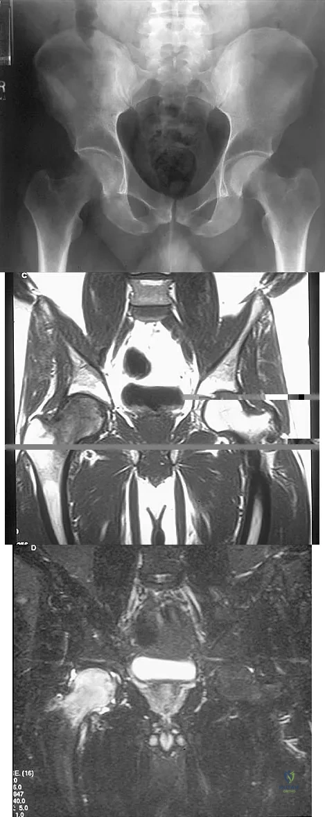


Explanation

Transient osteoporosis is a self-limited painful but reversible disorder. Although first described in pregnant women, it is more common in young to middle-aged men. The radiograph shows loss of mineralization in the right hip relative to the left side. There is no osseous destruction or cortical expansion typical of metastasis or giant cell tumor. The process is confined to the femoral side of the joint unlike rheumatoid arthritis, which would be centered in the joint. Osteonecrosis is better defined with sharp but irregularly shaped margins, and there is no double-line sign. The MRI scans reveal diffuse edema in the femoral head and neck that is atypical for osteonecrosis. Transient osteoporosis may recur in the same or opposite hip.

Question 21

Figure 11 shows the anatomic dissection of the medial side of the knee joint after removal of the superficial fascia. The arrow is pointing to what structure?

Anatomy 2002 Practice Questions: Set 1 (Solved) - Figure 28


Explanation

The semitendinosus and gracilis tendons lie beneath the superficial fascia and superficial to the medial collateral ligament. The semitendinosus is located more inferior to the gracilis tendon. The sartorius is more posterior and distal as is the medial collateral ligament. The semimembranosus is posterior. Pagnani MJ, Warner JJ, O'Brien SJ, Warren RF: Anatomic considerations in harvesting the semitendinosus and gracilis tendons and a technique of harvest. Am J Sports Med 1993;21:565-571.

Question 22

Figure 12 shows a lateral radiograph of the elbow. What is the most likely diagnosis?

Anatomy 2002 Practice Questions: Set 1 (Solved) - Figure 29


Explanation

The figure shows a supracondylar process, which is a normal anatomic variant. An osteochondroma tends to occur more toward the end of bones, and the medullary space of the underlying bone extends into the base of the osteochondroma. The presence of a supracondylar process is usually asymptomatic. However, the ligament of Struthers that always extends from the supracondylar process to the medial epicondyle can result in median nerve entrapment secondary to trauma. Last RJ: Anatomy: Regional and Applied, ed 6. London, England, Churchill Livingstone, 1978, pp 132-133.

Question 23

Which of the following nerves is most likely responsible for symptoms associated with plantar fasciitis?

Anatomy 2002 Practice Questions: Set 1 (Solved) - Figure 30


Explanation

The first branch of the lateral calcaneal nerve innervates the abductor digiti minimi. It is reported to be trapped at the interval between the abductor hallucis and the quadratus plantae muscles.

Question 24

A 16-year-old cheerleader reports an ache in the right shoulder and arm that is worse after activity. She denies any history of acute trauma. Examination reveals a positive sulcus sign and an AP glide test with a posterior and anterior apprehension sign. To confirm a diagnosis of multidirectional instability, which of the following imaging studies is most appropriate?

Anatomy 2002 Practice Questions: Set 1 (Solved) - Figure 31


Explanation

Multidirectional instability is a common finding in young female athletes. The anatomic structures are all intact but are hypermobile; therefore, CT and bone scans and scapular Y-views are often normal. Obtaining a weighted or AP stress view while applying downward traction on the arm will document instability and hypermobility of the joint. MRI generally is not indicated in this condition. Ultrasound is used primarily for rotator cuff pathology. Neer CS II, Foster CR: Inferior capsular shift for involuntary inferior and multidirectional instability of the shoulder: A preliminary report. J Bone Joint Surg Am 1980;62:897-908.

Question 25

Which of the following findings is seen in the chest radiograph shown in Figure 13?

Anatomy 2002 Practice Questions: Set 1 (Solved) - Figure 32


Explanation

Orthopaedic surgeons are often responsible for interpreting radiographs of general examinations such as the chest radiograph shown. For accurate interpretation, it is important to systematically review all of the information available on the radiograph. Using this approach, the fracture of the left proximal humerus is readily recognized. Linear air soft-tissue density at the lung periphery would suggest a pneumothorax, but this finding is not shown on the radiograph. The upper thoracic spine is well aligned. The sternoclavicular and distal clavicles are normal.

Question 26

In the standard anterior Smith-Robinson approach to the cervical spine, the dissection interval relies on an internervous plane. Which of the following structures constitutes the medial boundary of this plane?


Explanation

The standard anterior approach to the cervical spine utilizes the internervous plane between the carotid sheath laterally and the visceral structures (trachea and esophagus) medially. The recurrent laryngeal nerve lies in the tracheoesophageal groove and must be protected.

Question 27


Figure 10 represents a cross-section of the shoulder. Which nerve exits the axilla posteriorly through the quadrilateral space?


Explanation

The axillary nerve and the posterior circumflex humeral artery exit the axilla posteriorly through the quadrilateral space. The boundaries of this space include the teres minor (superior), teres major (inferior), long head of triceps (medial), and surgical neck of the humerus (lateral).

Question 28

The blood supply to the adult femoral head is predominantly provided by the lateral epiphyseal artery. This artery is a direct terminal branch of which of the following vessels?


Explanation

The lateral epiphyseal artery, which provides the majority of the blood supply to the adult femoral head, is a terminal branch of the medial femoral circumflex artery (MFCA). The MFCA courses posterior to the femoral neck and is protected during posterior approaches by releasing the short external rotators near their insertion.

Question 29

When placing lumbar pedicle screws, accurate identification of the starting point is critical to avoid nerve root injury. The medial border of the lumbar pedicle correlates with which anatomic landmark on the posterior elements?


Explanation

The medial border of the lumbar pedicle corresponds roughly to the lateral edge of the superior articular process. The classic starting point for a lumbar pedicle screw is at the intersection of the pars, the midpoint of the transverse process, and the superior articular facet.

Question 30

During a lateral approach to the proximal tibia and knee, the surgeon isolates a nerve that passes posterior to the biceps femoris tendon and wraps around the fibular neck. Injury to this nerve leads to severe weakness in which of the following movements?


Explanation

The common peroneal nerve wraps around the fibular neck and is vulnerable during lateral knee approaches. Injury to this nerve causes paralysis of the anterior and lateral compartments of the leg, resulting in foot drop and weakness in ankle dorsiflexion.

Question 31


Based on a normal cross-section of the forearm (as seen in Figure 6), the anterior interosseous nerve (AIN) courses distally along the interosseous membrane. Which of the following muscles is NOT innervated by the AIN?


Explanation

The anterior interosseous nerve (AIN) innervates the flexor pollicis longus, pronator quadratus, and the radial half of the flexor digitorum profundus. The flexor digitorum superficialis is innervated by the main trunk of the median nerve.

Question 32

In the standard posterolateral approach to the ankle for fixation of a posterior malleolus fracture, the internervous plane lies between the flexor hallucis longus and which of the following structures?


Explanation

The posterolateral approach to the ankle exploits the internervous plane between the flexor hallucis longus (innervated by the tibial nerve) and the peroneal muscles, including the peroneus brevis (innervated by the superficial peroneal nerve). This approach provides excellent exposure to the posterior malleolus.

Question 33

During a sacral laminectomy or decompression, the surgeon must be acutely aware of the termination of the dural sac. In the normal adult spine, the dural sac typically terminates at which vertebral level?


Explanation

In an adult, the spinal cord typically ends at the conus medullaris around L1-L2, but the dural sac and subarachnoid space usually extend further, terminating at the S2 vertebral level. Knowledge of this anatomy is vital to prevent cerebrospinal fluid leaks during sacral surgery.

Question 34

A patient presents with a deep penetrating injury to the palm and demonstrates an inability to forcefully cross the index and middle fingers. Which of the following nerves is most likely injured?


Explanation

Crossing the index and middle fingers requires the function of the dorsal and volar interossei muscles, which are responsible for digit abduction and adduction. These intrinsic muscles are innervated by the deep motor branch of the ulnar nerve.

Question 35


Figure 4 demonstrates a cross-section of the posterior thigh. The sciatic nerve generally divides into the tibial and common peroneal nerves proximally. Which muscle is exclusively innervated by the common peroneal division of the sciatic nerve?


Explanation

The short head of the biceps femoris is the only muscle in the posterior compartment of the thigh that receives its innervation from the common peroneal division of the sciatic nerve. The other hamstring muscles are innervated by the tibial division.

Question 36

During the Kocher approach to the radial head, the surgeon dissects through an internervous plane. This plane lies between the extensor carpi ulnaris and which other muscle?


Explanation

The Kocher approach to the lateral elbow utilizes the internervous plane between the anconeus (innervated by the radial nerve) and the extensor carpi ulnaris (innervated by the posterior interosseous nerve). Care must be taken distally to avoid injuring the LCL complex and the PIN.

Question 37

The ilioinguinal approach is commonly used for anterior column acetabular fractures. Which nerve is at greatest risk of iatrogenic injury during the mobilization of the structures in the "middle window"?


Explanation

The femoral nerve lies within the middle window of the ilioinguinal approach. It is situated just lateral to the external iliac vessels and anterior to the iliopsoas muscle, making it highly susceptible to traction injury during deep retraction.

Question 38

When approaching the thoracic spine via a costotransversectomy, the artery of Adamkiewicz must be considered to avoid spinal cord ischemia. This critical vessel typically enters the spinal canal through an intervertebral foramen at which levels?


Explanation

The artery of Adamkiewicz (great anterior radiculomedullary artery) provides the major blood supply to the anterior spinal artery for the lower two-thirds of the spinal cord. It typically enters the canal between T9 and L2, most commonly on the left side.

Question 39

The deep motor branch of the ulnar nerve passes between the pisiform and the hook of the hamate in Guyon's canal. Which of the following structures forms the roof of this canal?


Explanation

The roof of Guyon's canal is formed by the volar carpal ligament and the palmaris brevis muscle. The floor is formed by the transverse carpal ligament and the pisohamate ligament.

Question 40

During open reduction and internal fixation of a calcaneus fracture via an extensile lateral approach, the surgeon elevates a full-thickness subperiosteal flap. Which nerve is at the highest risk of transection in the proximal vertical limb of the incision?


Explanation

The sural nerve courses distally just posterior to the lateral malleolus. It is at significant risk of direct injury during the vertical portion of the extensile lateral approach to the calcaneus.

Question 41

A patient exhibits marked lateral winging of the scapula following a lymph node biopsy in the posterior triangle of the neck. The patient struggles to abduct the shoulder past 90 degrees. Which nerve was most likely injured?


Explanation

The spinal accessory nerve (CN XI) innervates the trapezius and is frequently injured during procedures in the posterior cervical triangle. Palsy results in lateral winging of the scapula and profound weakness in overhead shoulder abduction.

Question 42

The vertebral artery is a critical vascular structure that ascends through the cervical spine. In the majority of the population, at which cervical vertebral level does the vertebral artery typically first enter the transverse foramen?


Explanation

The vertebral artery arises from the subclavian artery and typically enters the transverse foramen at C6. It then ascends vertically through the foramina of C6 to C1 before turning medially to enter the foramen magnum.

Question 43


When evaluating injuries to the posterolateral corner of the knee, understanding the intricate capsuloligamentous anatomy is paramount. Which specific structure directly attaches the fibular head to the lateral meniscus?


Explanation

The meniscofibular ligament attaches the inferior peripheral margin of the lateral meniscus directly to the fibular head. It is an integral component of the posterolateral corner, contributing to lateral meniscal stability.

Question 44

During a Bankart repair, the surgeon must address the essential capsulolabral lesion. Which of the following structures serves as the primary static restraint to anterior translation of the humeral head when the shoulder is positioned in 90 degrees of abduction and maximal external rotation?


Explanation

The anterior band of the inferior glenohumeral ligament (IGHL) complex is the primary static restraint against anterior translation of the humeral head when the arm is in the abducted and externally rotated "apprehension" position.

Question 45

During a deltoid-splitting lateral approach to the proximal humerus, the axillary nerve is at significant risk of iatrogenic injury. What is the average anatomical distance from the lateral tip of the acromion to the axillary nerve in an adult?


Explanation

The axillary nerve courses roughly 5 to 7 cm distal to the lateral edge of the acromion. A deltoid split should safely not extend past 5 cm to avoid denervating the anterior deltoid.

Question 46

A patient presents with a severe traction injury to the brachial plexus after a motorcycle collision. Physical examination reveals miosis, ptosis, and anhidrosis on the ipsilateral side. Avulsion of which nerve root is most likely responsible for these specific findings?


Explanation

Horner syndrome (miosis, ptosis, anhidrosis) indicates a preganglionic proximal nerve root avulsion involving the T1 nerve root. The T1 root connects to the sympathetic cervical ganglion.

Question 47

When decompressing Guyon's canal for ulnar nerve entrapment, a surgeon must be intimately aware of its boundaries. Which of the following structures forms the floor of Guyon's canal?


Explanation

The floor of Guyon's canal is formed by the transverse carpal ligament (flexor retinaculum) and the pisohamate ligament. The roof is formed by the volar carpal ligament (palmar carpal ligament).

Question 48

Compression of the posterior interosseous nerve (PIN) most commonly occurs at the Arcade of Frohse. This arcade is formed by the proximal aponeurotic edge of which muscle?


Explanation

The Arcade of Frohse is a fibrous arch formed by the proximal border of the superficial head of the supinator muscle. It is the most common site of PIN entrapment in radial tunnel syndrome.

Question 49

During anterior cervical spine surgery extending to the lower cervical segments, the vertebral artery is at risk. In the vast majority of the population, the vertebral artery enters the transverse foramen at which cervical level?


Explanation

The vertebral artery typically enters the transverse foramen at the C6 level. Rarely, it can enter at C7 or higher levels like C5.

Question 50

To properly place a lumbar pedicle screw, the surgeon must identify the correct starting point to avoid neurological injury. Anatomically, the standard starting point is defined by the intersection of the:


Explanation

The standard starting point for a lumbar pedicle screw is the intersection of the pars interarticularis, the mid-transverse process, and the lateral border of the superior articular facet.

Question 51

When performing a posterior approach to the hip (Kocher-Langenbeck), the medial femoral circumflex artery (MFCA) is at risk. The main ascending branch of the MFCA consistently runs deep to which muscle?


Explanation

The MFCA is the main blood supply to the femoral head. Its ascending branch is protected by running deep (anterior) to the quadratus femoris muscle.

Question 52

The posterolateral corner (PLC) of the knee provides critical rotatory stability. Which structure is the primary restraint to external rotation of the tibia at 30 degrees of knee flexion?


Explanation

The popliteus complex (including the popliteofibular ligament) is the primary restraint to external rotation of the tibia at 30 degrees of flexion. The LCL is the primary restraint to varus stress.

Question 53

During a posterolateral approach to the ankle for fixation of a posterior malleolus fracture, the sural nerve must be protected. The sural nerve typically courses in close proximity to which structure in the distal leg?


Explanation

The sural nerve courses distally in the posterior leg adjacent to the small saphenous vein. It passes posterior to the lateral malleolus.

Question 54

During an ilioinguinal approach for an anterior column acetabular fracture, severe hemorrhage is encountered upon dissection over the superior pubic ramus. This bleeding is most likely from the corona mortis, an anastomosis between the:


Explanation

The corona mortis is a critical vascular anastomosis between the external iliac (or deep inferior epigastric) vessels and the obturator vessels. It crosses the superior pubic ramus.

Question 55

The superficial radial nerve is at risk during the distal extent of the Henry approach to the forearm. Anatomically, it emerges from beneath which muscle in the distal third of the forearm to become subcutaneous?


Explanation

The superficial sensory branch of the radial nerve runs deep to the brachioradialis in the mid-forearm. It emerges between the brachioradialis and ECRL tendons in the distal third.

Question 56

A patient sustains a deep laceration to the palm, severing the deep branch of the ulnar nerve. Which of the following functional deficits is most likely to be observed?


Explanation

The deep branch of the ulnar nerve is purely motor. It supplies the interossei, including the first dorsal interosseous, whose primary function is abduction of the index finger.

Question 57

When placing iliosacral screws for pelvic ring injuries, the surgeon must remain within the osseous safe zone of the sacral ala. The anterior limit of this safe zone in S1 is defined by the risk of injury to which structure?


Explanation

The L5 nerve root runs directly anterior to the sacral ala. Breaching the anterior cortex of the S1 body or ala places the L5 nerve root at significant risk of iatrogenic injury.

Question 58

A 25-year-old male develops acute compartment syndrome of the deep posterior compartment of the leg after a tibial fracture. Which of the following neurovascular structures is contained within this compartment?


Explanation

The deep posterior compartment of the leg contains the tibialis posterior, flexor hallucis longus, flexor digitorum longus, tibial nerve, and posterior tibial vessels.

Question 59

During a medial subvastus approach to the distal femur, the adductor canal (Hunter's canal) is visualized. Which of the following nerves runs within the adductor canal?


Explanation

The adductor canal contains the superficial femoral artery, superficial femoral vein, and the saphenous nerve. The saphenous nerve exits anteriorly before the hiatus.

Question 60

A 28-year-old overhead athlete presents with isolated weakness in external rotation of the shoulder. An MRI reveals a paralabral cyst in the spinoglenoid notch. Which muscle is predominantly denervated?


Explanation

The suprascapular nerve innervates the supraspinatus before passing through the spinoglenoid notch to innervate the infraspinatus. Entrapment at the spinoglenoid notch results in isolated infraspinatus weakness.

Question 61

The adult spinal cord typically terminates distally as the conus medullaris. In the majority of adults, this termination occurs at which vertebral body level?


Explanation

The conus medullaris marks the distal end of the spinal cord proper. In most adults, it terminates at the L1-L2 intervertebral disc level.

Question 62

The Master Knot of Henry is an important anatomical landmark in the plantar aspect of the midfoot. It refers to the intersection where the:


Explanation

At the Master Knot of Henry, the flexor digitorum longus (FDL) tendon crosses plantar (superficial) to the flexor hallucis longus (FHL) tendon in the medial midfoot.

Question 63

During a posterior cervical foraminotomy at C5-C6, the surgeon is mindful of the boundaries of the intervertebral foramen. The vertebral artery is typically located in which relation to the exiting C6 nerve root?


Explanation

The vertebral artery lies anterior to the exiting cervical nerve roots in the intervertebral foramen. Surgical decompression must be performed carefully to avoid injury to this anteriorly situated vascular structure.

Question 64

A 28-year-old volleyball player presents with insidious onset of vague posterior shoulder pain and profound weakness in external rotation, with preserved abduction. At what anatomical site is the injured nerve most likely compressed?


Explanation

Isolated weakness of the infraspinatus (external rotation) with preserved supraspinatus function (abduction) suggests suprascapular nerve entrapment at the spinoglenoid notch. Entrapment at the suprascapular notch would affect both muscles.

Question 65

Figure 4 shows an anatomic dissection of the lateral aspect of the knee.

The primary restraint to external rotation of the tibia at 30 degrees of knee flexion inserts on which of the following structures?


Explanation

The primary restraints to external rotation of the tibia at 30 degrees of flexion are the popliteofibular ligament and the fibular collateral ligament. Both of these structures insert on the fibular head.

Question 66

When placing a pedicle screw at the L4 level, the optimal starting point is at the intersection of the pars interarticularis, the superior articular facet, and the transverse process. What nerve root is most at risk if the screw breaches the pedicle inferiorly?


Explanation

In the lumbar spine, the exiting nerve root travels inferior to the corresponding pedicle (e.g., L4 root exits below the L4 pedicle). A medial breach would place the traversing L5 nerve root at risk.

Question 67

A patient sustains a deep laceration over the volar aspect of the index finger metacarpophalangeal joint. The first lumbrical muscle is injured. What is its precise origin and innervation?


Explanation

The first and second lumbricals are unipennate muscles that originate from the radial sides of their respective flexor digitorum profundus (FDP) tendons. They are innervated by the median nerve.

Question 68

During a Kocher-Langenbeck approach to the hip, the surgeon must protect the primary blood supply to the femoral head. Which structure serves as a critical landmark to protect the deep branch of the medial femoral circumflex artery?


Explanation

The deep branch of the medial femoral circumflex artery runs anterior to the quadratus femoris and posterior to the obturator externus. Preserving the intact obturator externus protects this critical artery.

Question 69

Figure 10 demonstrates standard portals for ankle arthroscopy.

When establishing the posterolateral portal, the sural nerve is at risk. What is its normal anatomical relationship to the portal site?


Explanation

The posterolateral ankle portal is made lateral to the Achilles tendon. The sural nerve runs posterior to the peroneal tendons along with the small saphenous vein and is vulnerable during portal placement.

Question 70

When placing Sacral Alar Iliac (SAI) screws for spinopelvic fixation, the trajectory crosses the SI joint. Which anterior structure is most at risk with an overly anterior and superior trajectory breach of the sacral ala?


Explanation

The L5 nerve root drapes over the sacral ala anteriorly as it joins the sacral plexus. It is at the highest risk with an anterior and superior cortical breach during pelvic fixation.

Question 71

A 45-year-old mechanic complains of chronic, aching pain in the proximal lateral forearm without overt motor weakness, exacerbated by resisted forearm supination. The most common site of compression for the involved nerve is the:


Explanation

Radial tunnel syndrome involves compression of the posterior interosseous nerve (PIN), presenting with lateral forearm pain without motor weakness. The most common compression site is the proximal edge of the superficial supinator, known as the Arcade of Frohse.

Question 72

Figure 13 displays a cross-section of the midfoot.

The true Lisfranc ligament connects which two osseous structures?


Explanation

The Lisfranc ligament is a strong interosseous ligament that connects the lateral aspect of the medial cuneiform to the medial base of the second metatarsal. It is critical for midfoot stability.

Question 73

During a deltopectoral approach for a proximal humerus fracture, identifying and protecting the axillary nerve is paramount. At what distance distal to the lateral acromial edge does the main trunk of the axillary nerve typically cross the humerus?


Explanation

The axillary nerve wraps around the surgical neck of the humerus, running transversely on the deep surface of the deltoid approximately 5 to 7 cm distal to the lateral edge of the acromion.

Question 74

A spine surgeon evaluates a pre-operative CT scan for a T8 burst fracture. Compared to the lumbar spine, which of the following is true regarding the anatomy of mid-thoracic pedicles?


Explanation

Thoracic pedicles originate higher on the vertebral body (near the superior endplate) compared to lumbar pedicles. They are also narrower and have less medial angulation.

Question 75

Figure 19 highlights an arthroscopic view of a meniscus.

Which of the following describes a key anatomical difference between the medial and lateral menisci?


Explanation

The lateral meniscus is more circular, covers a larger percentage of the articular surface area, and is more mobile because it lacks a dense continuous capsular attachment due to the popliteus hiatus.

Question 76

A 21-year-old collegiate baseball pitcher is diagnosed with an ulnar collateral ligament (UCL) tear. The anterior bundle of the UCL inserts on the sublime tubercle. Which specific portion of the anterior bundle is most taut in extension?


Explanation

The anterior bundle of the UCL consists of anterior and posterior bands. The anterior band is taut in extension, while the posterior band becomes more taut in deeper flexion.

Question 77

Figure 25 details the anterior thigh musculature.

A patient requires a femoral nerve block. Which of the following accurately describes the position of the femoral nerve within the femoral triangle?


Explanation

In the femoral triangle, structures from lateral to medial are Nerve, Artery, Vein, Empty space, Lymphatics (NAVEL). The femoral nerve lies lateral to the artery and outside the femoral sheath.

Question 78

In atlas (C1) lateral mass screw fixation, the surgeon must avoid both vascular structures. Which anatomic trajectory and starting point minimizes the risk to both the vertebral artery and the internal carotid artery?


Explanation

Starting at the center of the C1 lateral mass and angling 10-15 degrees medial and 15 degrees cephalad directs the screw safely away from the vertebral artery (lateral) and the internal carotid artery (anterior).

Question 79

A patient complains of ulnar-sided wrist pain after a fall. MRI shows a tear of the foveal attachment of the Triangular Fibrocartilage Complex (TFCC). This specific attachment is critical for:


Explanation

The foveal attachment of the TFCC consists of the deep radioulnar ligaments, which converge on the fovea of the ulnar head. They are the primary stabilizers of the distal radioulnar joint (DRUJ).

Question 80

A patient develops an isolated acute compartment syndrome of the lateral compartment of the lower leg. Which of the following physical exam findings is most expected?


Explanation

The lateral compartment contains the peroneus longus and brevis muscles (ankle eversion) and the superficial peroneal nerve, which provides sensation to the dorsum of the foot.

Question 81

Figure 30 demonstrates the osseous landmarks of the scapula.

The coracoacromial ligament attaches to the acromion and the coracoid process. Which variation of the acromion morphology is most strongly associated with full-thickness rotator cuff tears?


Explanation

The Bigliani Type III (hooked) acromion reduces the subacromial space significantly. It is highly associated with subacromial impingement and full-thickness rotator cuff tears.

Question 82

During an anterior intrapelvic (modified Stoppa) approach for an acetabular fracture, significant hemorrhage occurs near the superior pubic ramus. The most likely source is the corona mortis, which represents an anastomosis between:


Explanation

The corona mortis is a critical vascular anastomosis between the external iliac system (or inferior epigastric) and the obturator system (internal iliac). It crosses the superior pubic ramus and is highly vulnerable during anterior pelvic approaches.

Question 83

A surgeon is performing a lateral transpsoas approach to the L4-L5 disc space. To avoid injury to the lumbar plexus, the retractor should be placed carefully, as the nerve responsible for quadriceps function is located in which region of the psoas muscle at this level?


Explanation

At the L4-L5 disc space, the lumbar plexus (specifically the femoral nerve) typically lies within the posterior half of the psoas major muscle. Approaching anterior to the middle of the psoas helps avoid nerve injury during lateral interbody fusion.

Question 84

During a standard deltopectoral approach to the shoulder, the cephalic vein is identified in the interval. To optimally preserve its venous drainage, what is the standard recommended handling of this structure?


Explanation

The cephalic vein is typically retracted laterally with the deltoid muscle during the deltopectoral approach. This preserves the primary venous tributaries which enter the vein from the lateral side.

Question 85

A 24-year-old athlete requires surgical reconstruction for a posterolateral corner knee injury. Identifying and protecting the common peroneal nerve is critical. Which of the following describes its most reliable anatomical position in the posterolateral knee?


Explanation

The common peroneal nerve courses distally along the posterior and medial border of the biceps femoris tendon. It then wraps around the fibular neck to enter the anterior and lateral compartments of the leg.

Question 86

An anterior cervical discectomy and fusion (ACDF) is planned at the C6-C7 level. A right-sided approach is historically considered to have a higher risk of recurrent laryngeal nerve (RLN) injury compared to a left-sided approach due to which anatomical variant?


Explanation

The right recurrent laryngeal nerve loops around the right subclavian artery and enters the operative field at varying angles, making it more susceptible to injury. A non-recurrent right laryngeal nerve occurs in about 1% of the population.

Question 87

A patient presents with weakness in flexing the interphalangeal joint of the thumb and the distal interphalangeal joint of the index finger. Which anomalous structure is most commonly responsible for compressing the nerve involved?


Explanation

The patient has anterior interosseous nerve (AIN) syndrome, characterized by weakness of the FPL and FDP to the index finger. Gantzer muscle, an accessory head of the flexor pollicis longus, is a frequent cause of AIN compression.

Question 88

During a Kocher-Langenbeck (posterior) approach to the acetabulum, identifying the sciatic nerve is essential. The nerve classically exits the greater sciatic foramen in what relation to the short external rotators?


Explanation

In standard anatomy, the sciatic nerve exits the pelvis inferior to the piriformis muscle. It courses superficially over the superior gemellus, obturator internus, inferior gemellus, and quadratus femoris.

Question 89

The deep palmar arch of the hand provides significant collateral blood flow. It is primarily formed by the anastomosis of which of the following vessels?


Explanation

The deep palmar arch is primarily formed by the terminal portion of the radial artery anastomosing with the deep branch of the ulnar artery. The superficial palmar arch is primarily formed by the terminal ulnar artery.

Question 90

When performing an anterior approach for a thoracolumbar corpectomy, the surgeon must be aware of the artery of Adamkiewicz to prevent anterior spinal cord syndrome. This vessel most commonly arises from the aorta at which levels?


Explanation

The artery of Adamkiewicz provides the dominant blood supply to the anterior lower two-thirds of the spinal cord. It most commonly arises from the left side of the aorta between the T9 and L1 levels in approximately 75% of individuals.

Question 91

An anterolateral approach to the distal tibia is used for pilon fracture fixation. The superficial peroneal nerve is at risk in this exposure. Where does this nerve predictably pierce the deep fascia to become subcutaneous?


Explanation

The superficial peroneal nerve typically pierces the crural fascia to become superficial about 10 to 12 cm proximal to the lateral malleolus. It then divides into medial and intermediate dorsal cutaneous branches.

Question 92

A patient sustains an injury resulting in medial winging of the scapula. Which nerve is injured, and what are its correct nerve root origins?


Explanation

Medial scapular winging is caused by paralysis of the serratus anterior muscle, which is innervated by the long thoracic nerve. This nerve originates from the ventral rami of C5, C6, and C7.

Question 93

A surgeon is performing a midfoot reconstruction and exploring the plantar aspect of the foot. The Master Knot of Henry is identified. This anatomical landmark represents the intersection of which two structures?


Explanation

The Master Knot of Henry is located in the plantar midfoot where the flexor digitorum longus (FDL) tendon crosses over (plantar/superficial to) the flexor hallucis longus (FHL) tendon.

Question 94

During placement of an S1 iliosacral screw for a displaced sacral fracture, an anterior extraosseous screw trajectory risks injuring which neural structure passing over the sacral ala?


Explanation

The L5 nerve root courses inferiorly and anteriorly over the sacral ala. An iliosacral screw that breaches the anterior cortex of the upper sacrum (S1 body) puts the L5 nerve root at high risk of iatrogenic injury.

Question 95

When utilizing a dorsal approach to the wrist, Lister's tubercle is a key osseous landmark. It serves as a mechanical pulley for which tendon?


Explanation

Lister's tubercle (dorsal tubercle of the radius) separates the second and third extensor compartments. The extensor pollicis longus (EPL) tendon uses this tubercle as a pulley to change its line of pull toward the thumb.

None

Clinic OS
Medically Verified Content by
Prof. Clinic OS
Consultant Orthopedic & Spine Surgeon
Chapter Index