Volar Approach to the Carpal Tunnel and Wrist
Volar Approach to the Carpal Tunnel and Wrist
Position of the Patient
Landmarks and Incision
Landmarks
Incision
Internervous Plane
Superficial Surgical Dissection
Deep Surgical Dissection
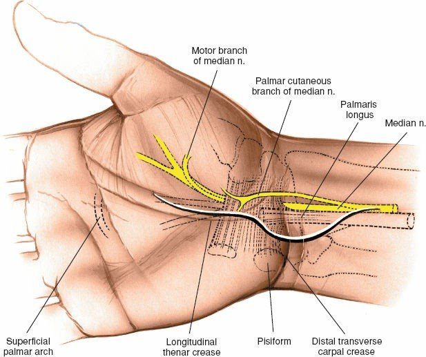
Figure 5-21 The incision for the volar approach to the wrist. The incision should be made on the ulnar side of the palmaris longus tendon to protect the palmar cutaneous branch of the median nerve.
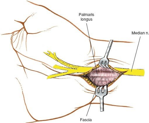
Figure 5-22 The skin is retracted, and the deep fascia and tendon of the palmaris longus are inspected.
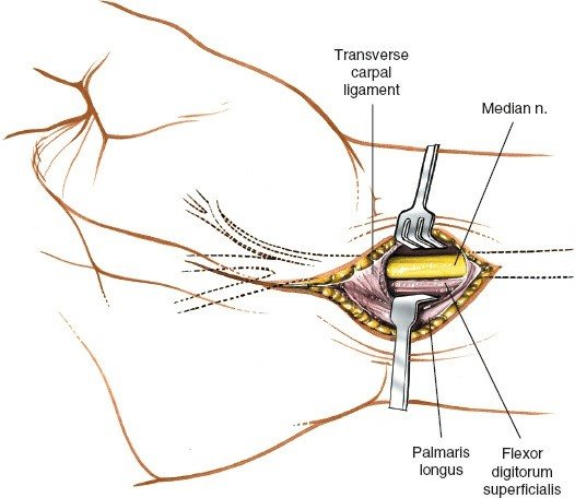
Figure 5-23 The deep fascia is incised. The palmaris longus is retracted toward the ulna, revealing the median nerve as it enters the carpal tunnel.
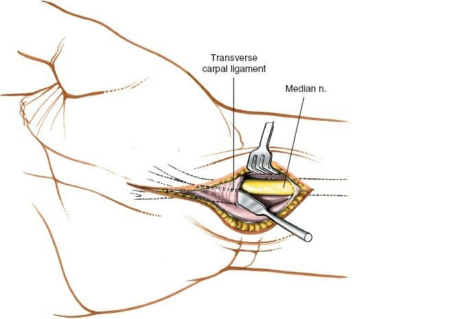
Figure 5-24 A spatula is placed under the transverse carpal ligament to protect the median nerve as the ligament is incised.
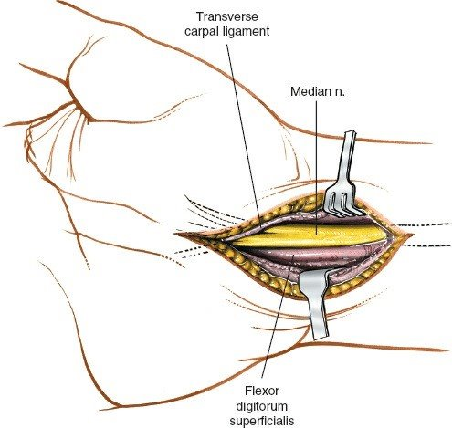
Figure 5-25 The transverse carpal ligament is released on the ulnar side of the nerve to avoid damage to the motor branch of the thenar muscle.
Dangers
Nerves
Vessels
How to Enlarge the Approach
Extensile Measures
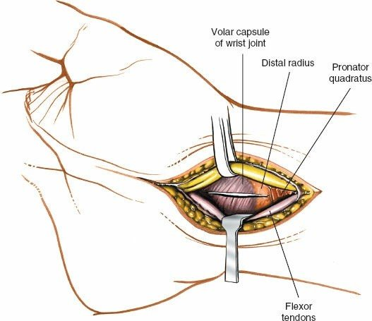
Figure 5-26 The median nerve is retracted radially and the flexor tendons are retracted toward the ulna, revealing the distal radius and joint capsule. An incision then is made into the capsule to expose the carpus.
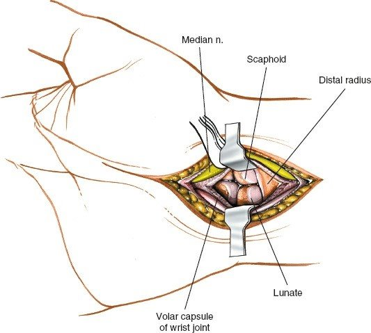
Figure 5-27 Incise the joint capsule to expose the carpus.
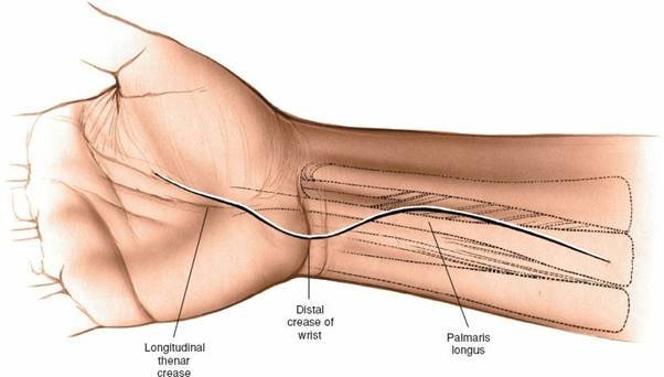
Figure 5-28 Extend the wrist incision proximally to expose the distal forearm and median nerve.
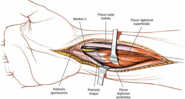
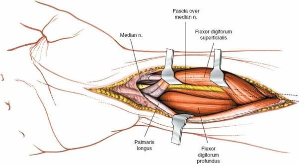
Figure 5-29 Incise the fascia on the forearm between the palmaris longus and the flexor carpi radialis to expose the tendons and muscles of the flexor digitorum superficialis.
