K-Wire Fixation of Distal Radius Fractures With and Without External Fixation
K-Wire Fixation of Distal Radius Fractures With and Without External Fixation
DEFINITION
ANATOMY
PATHOGENESIS
NATURAL HISTORY
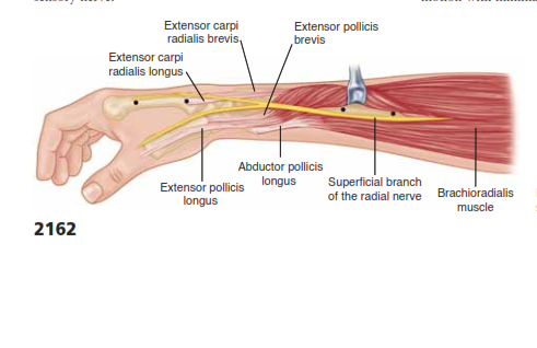
FIG 1 • Anatomy surrounding the radial sensory nerve branch in the forearm.
PATIENT HISTORY AND PHYSICAL FINDINGS
IMAGING AND OTHER DIAGNOSTIC STUDIES
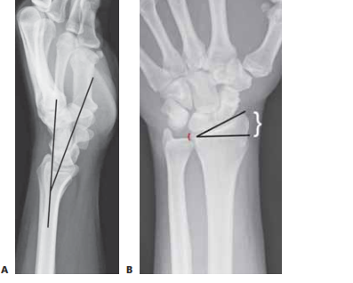
FIG 2 • A. Lateral radiograph of the wrist demonstrating volar tilt (black lines). B. PA radiograph demonstrating radial incli- nation (black lines), ulnar variance (red bracket), and radial height (white bracket).
DIFFERENTIAL DIAGNOSIS
NONOPERATIVE MANAGEMENT
SURGICAL MANAGEMENT
Preoperative Planning
Positioning
Approach
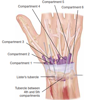
FIG 4 • Areas for K-wire insertion at the distal radius.
CLOSED REDUCTION OF A DISTAL RADIUS FRACTURE
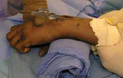
TECH FIG 1 • Closed reduction over a towel bump using trac- tion and palmar translation.
KAPANDJI TECHNIQUE FOR PERCUTANEOUS PINNING
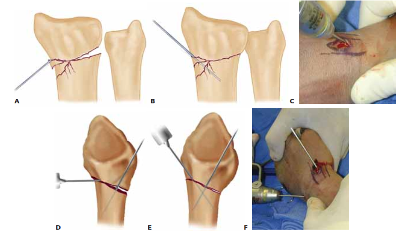
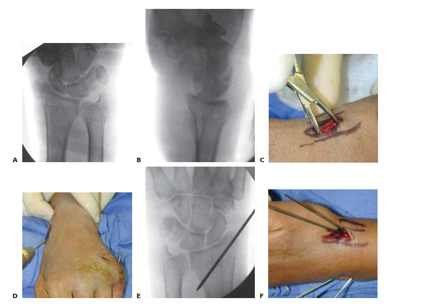
TECH FIG 2 • A. An incision is made over the radial styloid and a K-wire is manually inserted into the fracture site. B. The wire is levered distally to correct the radial inclination. C. The wire is advanced proximally, using power, into cortical bone. D. An incision is made over Lister’s tubercle, and a wire is inserted into the fracture site. E,F. The wire is levered distally to correct the dorsal angulation and advanced proximally using power into cortical bone.
AUTHOR’S PREFERRED TECHNIQUE FOR PERCUTANEOUS PINNING
