COMPLICATIONS OF FRACTURES
COMPLICATIONS OF FRACTURES
1 COMPLICATIONS OF MAJOR TRAUMA
Major tissue trauma activates cell defence mechanisms which combat infection, remove damaged tissue and facilitate tissue repair. These processes may be affected by systemic mediators which might cause an imbalance. This may be towards a generalised pro-inflammatory state (systemic inflammatory response syndrome or SIRS) accompanied by cell damage with increased cell wall permeability, or to suppression of inflammation (compensatory anti-inflammatory response syndrome or CARS) which may lead to a susceptibility to infection. Such imbalances are said to be the result either of particularly severe trauma or a particular individual response.3 The acute respiratory distress syndrome (ARDS) is regarded as being a local manifestation of SIRS, and in those who survive, the cause of multiple organ dysfunction (MODS). This may include cardiac, gastrointestinal, renal, hepatic, haematological and cerebral failure.
Hypoxia and acute respiratory insufficiency are common after trauma, and the causes include upper airway obstruction, chest injury (e.g. due to pneumothorax) and circulatory failure. Most respond to treatment of the underlying cause and the administration of oxygen, but if this fails other reasons, ARDS or the fat embolism syndrome (FES) must be suspected.
In both ARDS and FES there is no evidence of cardiac failure; chest radiographs show bilateral ‘snowstorm’ lungs, and there is disturbance of the PaO2/FiO2 ratio (the arterial oxygen concentration divided by the fractional inspired oxygen concentration). Where there has been a fat embolism the most distinctive features (which may appear 2 or 3 days after the injury) are:
Treatment:
(1) Aggressively carry out all routine resuscitative measures such as the administration of humidified oxygen and intravenous fluids. If indicated by the blood gas levels, proceed to endotracheal intubation and the use of a ventilator, with transfer to an intensive therapy unit. In rare cases a cardiopulmonary by-pass pump may be required. Where possible, active physiotherapy is advised. (2) If there is progressive deterioration in blood gas levels, surgical stabilisation of any femoral fracture is advocated. After such a procedure the patient should be left on a ventilator for not less than 24 hours; this may be discontinued when the blood gases improve, and there is an improvement in the tidal volume and the patient’s level of consciousness. (3) The administration of methylprednisolone in cases where the PaO2 is less than 60 mm Hg, whether discovered before the onset of frank symptoms or later, has been shown to have a beneficial effect. (A dose of 30 mg per kg body weight on admission and repeated after 4 hours has been recommended.)
2 COMPLICATIONS OF PROLONGED RECUMBENCY
Factors in risk assessment include:
Where the risks are judged to be more than trivial, the following may be considered:
Chemical prophylaxis: Where the risks are considered to be high, chemical prophylaxis should be considered. It would seem advantageous to continue this for 4–5 weeks, even although the patient has been allowed home in the interim. Careful consideration must be given as to when the medication should be commenced: the earlier it is started, the more effective it is – but too early, and haemorrhage may ensue. A number of agents, of varying efficacy, are in current use. These include:
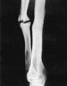
Non-Union